중증 근무력증의 분류 및 치료 지침
부산대학교 의학전문대학원 소아청소년과
이윤진
Submitted: 4 March, 2017 Revised: 25 March, 2017 Accepted: 25 March, 2017
Correspondence to Yun-Jin Lee, MD, PhD
Department of Pediatrics, Pusan National University Children’s Hospital, 20 Geumo-ro, Yangsan-si, Gyeongsangnam-do 50612, Korea
Tel: +82-55-360-2180, Fax: +82-55-360-2181 E-mail: [email protected]
Subgroup Classification and Therapeutic Guidance for Myasthenia Gravis
Myasthenia gravis (MG) is a chronic autoimmune disease of neuromuscular blockade, characterized by muscle weakness and fatigue, and is associated with the production of autoantibodies against the skeletal muscle acetylcholine receptor, muscle-specific kinase (MuSK), low-density lipoprotein receptor-related protein 4 (LRP4), and other muscle endplate proteins. In addition, MG may be classified according the location of the affected muscles (ocular vs. generalized), patient age at symptom onset, and thymic pathology. Subgroup classification based on serum antibodies and clinical features include early-onset, late-onset, thymoma-associated, MuSK and LRP4 antibody-negative, and ocular forms of MG, and can help with therapeutic decisions and prognosis. Pyridostigmine is the chosen symptomatic treatment. For patients who do not adequately respond to symptomatic therapy, corticosteroids, other immunomodulating agents, and thymectomy are the first-line immunosuppressive treatments. The treatment of MG is highly individualized and depends on the age of the patient, the type and severity of the disease, and the pace of progression.
Key Words: Myasthenia gravis, Child, Adult, Therapy, Thymectomy
Yun-Jin Lee, MD, PhD
Department of Pediatrics, Pusan National University Children’s Hospital, Pusan National University School of Medicine, Yangsan, Korea
Copyright © 2017 by The Korean Child Neurology Society
http://www.cns.or.kr
Introduction
Myasthenia gravis (MG) is an autoimmune disorder characterized by weakness and fatigability of skeletal muscles. Most patients who develop MG in adolescence or adulthood have autoantibodies that play a pathogenetically important role by attacking the acetylcholine receptor (AChR), fixing comple
ments, and reducing the number of AChRs over time1,2). These autoantibodies are believed to originate from hyperplastic germinal centers in the thymus, where myoid cells expressing AChR are gathered.
There are two clinical forms of MG: ocular and generalized. In the generalized type, weakness involves the ocular muscles and variable combinations of the bulbar, limb, and respiratory muscles. The definition of ocular MG is restricted to the ocular symptoms for 2 years without becoming generalized3). Juvenile MG (JMG) patients are subdivided according to the onset of the first symptoms as prepubertal (before the age of 12 years) and postpubertal (after the age of 12 years). Patients who have detectable antibodies to AChR or to the muscle
specific receptor tyrosine kinase (MuSK) are considered to have seropositive MG, whereas those lacking both AChR antibody (AChRAb) and MuSK antibody (MuSKAb) on standard assays are considered to have seronegative MG1,2). About half of the patients with the purely ocular type are seropositive, compared with approximately 90% of those with generalized disease. In contrast to adult MG, young children with JMG presenting generalized symptoms may have no or only slight elevated titer of specific antibodies, the diagnosis is often delayed4).
There is an ethnical difference in the presentation and prevalence of the disease. In Asian population, up to 50% of MG patients present with the first symptoms in childhood (age peak between 5 and 10 years, no difference in sex distribution). More than 70% of patients are limited to ocular symptoms and favorable course is common5,6). Among Caucasian prepubertal patients, presenting only with ocular symptoms, spontaneous remission is frequent and ranges from 15 to 34.7%7). In postpubertal children, female predominance is present5,6). The JMG population among African American shows differences compared to the Asian and European population, and demonstrated poorer response to thymectomy than Caucasian children and lower remission rates8). They reveal female pre
dominance in all age groups8).
Pathogenesis and subgroups
In most cases, MG is a condition mediated by autoantibodies against AChR1,2). However, the linkage between AChR antibodies and MG is not absolute. In addition, 10–20% of patients with MG have no measureable levels of autoantibodies against AChR. A proportion of these patients (40–70%) have antibodies directed against MuSK. Young children with JMG presenting generalized symptoms may have no or only slight elevated titer of specific antibodies4). The tests of these antibodies, expect for AChRAbs, are available only in specialized laboratories, and have not been performed in our country.
1. AChR-Ab subtypes
AChRAbs are highly specific for MG. They are directly pathogenic through crosslinking of AChRs, leading to the accelerated degradation of these receptors by complement binding and activation, and by inducing AChR conformational changes or blocking acethylcholine binding. No correlation has been shown between AChRAb level and disease severity.
However, in individual patients, the titers tend to decrease with successful immunotherapy and parallel clinical improvement9,10). Individual patients have a mix of immunologically different antibodies to AChR. This is partly due to the heterogeneity of the receptor. In addition, antibodies to the same receptor in a patient with MG can vary in their lightchain and subclass types11). This means that B lymphocytes producing AChR antibodies are also heterogeneous.
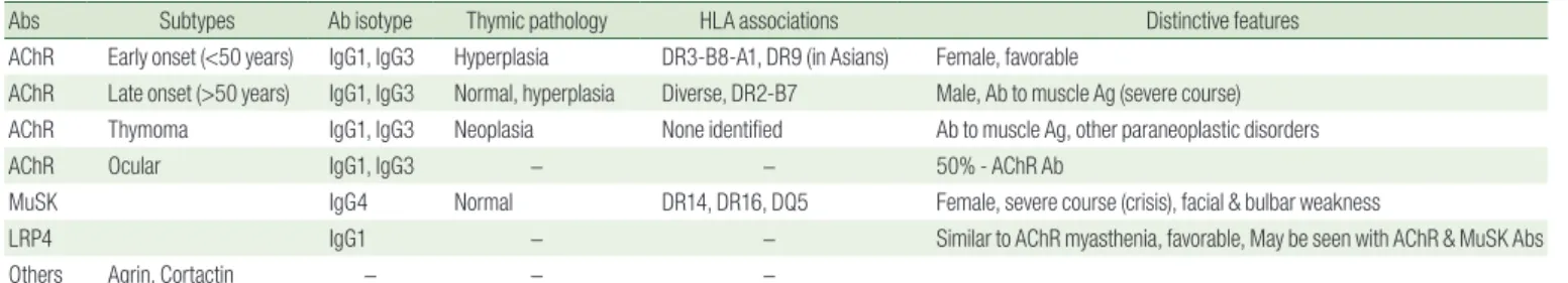
Table 1. Subgroup Characteristics and Antibodies of Myasthenia Gravis
Abs Subtypes Ab isotype Thymic pathology HLA associations Distinctive features
AChR Early onset (<50 years) IgG1, IgG3 Hyperplasia DR3-B8-A1, DR9 (in Asians) Female, favorable
AChR Late onset (>50 years) IgG1, IgG3 Normal, hyperplasia Diverse, DR2-B7 Male, Ab to muscle Ag (severe course) AChR Thymoma IgG1, IgG3 Neoplasia None identified Ab to muscle Ag, other paraneoplastic disorders
AChR Ocular IgG1, IgG3 − − 50% - AChR Ab
MuSK IgG4 Normal DR14, DR16, DQ5 Female, severe course (crisis), facial & bulbar weakness
LRP4 IgG1 − − Similar to AChR myasthenia, favorable, May be seen with AChR & MuSK Abs
Others Agrin, Cortactin − − −
Abs, antibodies; AChR, acethylcholine receptor; MuSK, muscle-specific tyrosine kinase; LRP4, lipoprotein receptor-related protein 4; HLA, human leukocyte antigen.
Fig. 1. Schematic view of the MuSK-LRP4-ColQ complex11)
. At the
postsynaptic membrane, MuSK is associated with LRP4. MuSK activation
through agrin-LRP4 binding triggers a signaling pathway, which includes
Dok7 recruitment, leading to AChR clustering. AChE binds through its ColQ
tail to perlecan and MuSK. MuSK, muscle-specific tyrosine kinase; LRP4,
lipoprotein receptor-related protein 4; AChR, acetylcholine receptor; AChE,
acetylcholinesterase; ColQ, collagen Q.
2. MuSK antibodies
MuSK autoantibodies have been found in 40–70% of Cauca
sian patients with AChRseronegative MG12). There are significant differences between MuSKAbpositive MG and AChRAb
positive MG in pathologic mechanism, clinical features, and response to pharmacologic treatments; onset at any age, female predominance, oculobulbar form, restricted myopathic form with prominent respiratory and/or proximal weakness, no thymic pathology (thymoma), and lower responsiveness to acetylcholinesterase inhibitors (Table 1)9,12). The MuSK protein is a tra nsmembra ne component of the postsynaptic neuromuscular junction. The nervederived proteoglycan agrin binds to lipoproteinrelated protein 4 (LRP4), leading to the activation of MuSK13). Activated MuSK interacts with the cytoplasmic adaptor protein Dok7, leading to recruitment of rapsyn to promote clustering of AChRs (Fig. 1)13,14).
3. Seronegative myasthenia
A subset (6–12%) of patients with MG who have no detectable antibodies directed to AChR or MuSK are classified as having seronegative MG15). Patients with seronegative MG are more likely to have purely ocular disease than those who are seropositive. Antibodies to other selfantigens have been detected in various reports on patients with seronegative MG (Table 1). (a) Antibodies to LRP4, the agrinbinding receptor of the MuSK complex (Fig. 1), inhibit AChR clustering in the membrane, and have been found in up to 50% of patients with seronegative MG16,17). (b) Agrin antibodies have been detected in a few patients with MG and AChR, MUSK, or LRP4 antibody18). (c) Cortactin is a protein that mediates clustering of AChR at the neuromuscular junction19). Titin and ryanodine receptor antibodies occur in some patients with AChRassociated MG.
These antibodies are present with a high frequency in thymomaassociated MG, with an intermediate frequency in lateonset MG, and very rarely in earlyonset and ocular MG20).
4. Thymus and the origin of autoimmunity in myasthenia Most patients with AChRAbpositive MG have thymic abnormalities including hyperplasia in 60–70% and thymoma in 10–12%1). The thymus contains a small number of “myoid” cells.
These cells are distinguished by striations and the presence of AChR on their surface. In addition, thymic epithelial cells produce unfolded AChR subunits that are hypothesized to prime helper T cells. These “autoimmunized” T cells then attack AChR on myoid cells and create infiltrating germinal centers in the hyperplastic thymus2).
Clinical features
MG is a relatively uncommon disorder with an annual incidence of approximately 7–23 new cases per million popula
tion21). There tends to be a bimodal distribution to the age of onset, with an early peak in the second and third decades (female predominance) and a late peak in the sixth to eighth decade (male predominance). Early in the disorder, the symp
toms may be transient in many patients and even spontaneously go into remission for weeks or longer. However, the manifesta
tions typically worsen and become more persistent. The maximum range of the symptoms was seen by 3 years from onset in 77%22). In postpubertal children, the clinical course is similar to adultonset MG. The patients often present with ocular symptoms at the onset; generalized weakness develops in up to 80% in the course of the disease4,5,7).
The cardinal feature of MG is fluctuating skeletal muscle weakness with diurnal alternation, often with true muscle fatigue23). More than 50% of patients present with ocular symptoms of ptosis and/or diplopia24). Ptosis usually aggravates after physical activity. Children may complain about double vision, particularly during prolonged reading or in the course of the day. About 50% of children with ocular symptoms develop systemic or bulbar muscle involvement within 2 years25,26). About 15% of patients present with bulbar symptoms, including dysarthria, dysphagia, and fatigable chewing. Imminent risk of aspiration may produce a “myasthenic crisis”27). Facial muscles are frequently involved and make the patient appear expres
sionless. Myasthenic crisis is a lifethreatening situation in which respiratory muscle weakness leads to respiratory insuffi ciency and pending respiratory failure.
Diagnostic approach
To diagnose JMG, the combination of a thorough history, repeated physical examination, and neurophysiological investigations, as well as serology tests are helpful clues.
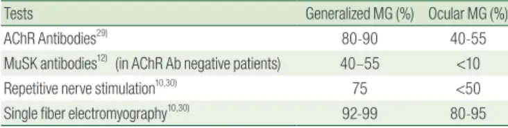
Postpubertal children may present with very indicative symptoms and have in up to 90% positive AChR antibodies in generalized MG28). The diagnosis in younger children constitutes a particular challenge as nonspecific symptoms may be present or the antibodies are only minimal elevated or even normal, also in case of severe generalized myasthenia. MG remains a clinical diagnosis. The approximate diagnostic sensitivity of the confirmatory laboratory tests in adult MG is shown in Table 2.
1. Serologic testing
Detection of AChRAb or MuSKAb, which is possible in approximately 90% of adult patients with generalized disease, provides the laboratory confirmation of MG10,29). In those with purely ocular myasthenia, the sensitivity of AChRAb testing is considerably lower (i.e., AChRAb is detectable in about half of the patients). In JMG patients with seronegative MG, most often young children with an ocular myasthenia, antibodies are not helpful because they may be negative in up to 50%8,25,26). MuSK antibodies are also rare in children and appear to be associated with a more severe disease, affecting mostly facial and bulbar weakness as well as frequent respiratory crises5,28). The detection of these antibodies is available only in specialized laboratories and could be initiated in seronegative JMG patients with distinct clinical symptoms and abnormal neurophysiological findings16). Here, larger number of patients and longterm followup are necessary to clarify the clinical and therapeutic implications of lowsensitivity AChRAb and LRP4Ab in pediatric population.
2. Electrophysiologic studies
Electrophysiologic tests usually allow the confirmation of diagnosis in patients with seronegative MG. Repetitive nerve stimulation (RNS) is the most frequently used test. Singlefiber electromyography is more technically demanding in children than RNS, and could be only performed in sedated children or in postpubertal patients with good compliance, Both the RNS test and singlefiber electromyography have a diagnostic sensitivity in generalized MG of about 75–95%10,30). However, the
sensitivity of RNS is much lower (15–45%) in patients with ocular myasthenia (Table 2).
3. Pharmacological testing
In edrophonium testing, edrophonium chloride (Tensilon), a shortacting cholinesterase inhibitor, is administered intra
venously (IV)29). An IV infusion should be started for rapid medication in the adverse events for the test. In children younger than 1 year, recommended dose is 0.15 mg/kg IV, preceded by a test dose of 0.01 mg/kg and then depending on effects increasing with 0.15 mg/kg steps to a maximum of 0.6 mg/kg. In children older than 1 year and weighing <34 kg, the dose is 0.5 mg IV initially, followed by subsequent 1 mg doses, repeated every minute to a maximum total dose of 5 mg. In adolescents with weighing >34 kg, the dose is 1–2 mg IV initially, then repeated every minute to a maximum total dose of 10 mg7). These doses may be given intramuscularly or subcutaneously.
Effects should be seen within 10 s and disappear within 120 s.
Adverse effects include nausea and emesis, headache, and bronchospasm. Atropine (0.01 mg/kg) should be prepared in a separate syringe before the administration of edrophonium, to block acute muscarinic effects of the cholinesterase inhibitor.
As a good alternative regimen, neostigmine methylsulfate (prostigmine) is administered intramuscularly at a dose of 0.04 mg/kg. If the result is negative or equivocal, another dose of 0.04 mg/kg may be administered 4 h after the first dose (typical dose, 0.5–1.5 mg). The peak effect is seen in 20–40 min. IV prostigmine is contraindicated because of the risk of cardiac arrhythmias, especially in young infants.
Treatment
Therapeutic standards for treatment of JMG have been adop
ted from adult patients31). Treatment options in JMG can be classified into three main categories: (a) symptomatic treatments Table 2. Approximate Sensitivity of the Confirmatory Tests for
Myasthenia Gravis in Adult
Tests Generalized MG (%) Ocular MG (%)
AChR Antibodies29) 80-90 40-55
MuSK antibodies12) (in AChR Ab negative patients) 40–55 <10
Repetitive nerve stimulation10,30) 75 <50
Single fiber electromyography10,30) 92-99 80-95
MG, myasthenia gravis; AChR, acethylcholine receptor; MuSK, muscle-specific kinase.
Table 3. Immune-modulating Agents Used in Myasthenia Gravis
Treatment Onset of action Time to maximal effect Initial dosing/frequency Monitor
Immunomodulator
Plasmapheresis 1-5 days 1-3 weeks 4-6 exchanges on alternate days BP, coagulation parameters
IVIG 3-10 days 1-3 weeks 1-2 g/kg (over 2-5 days) BP, renal/cardiac status
Prednisone 2-6 weeks 5-6 mo 0.5-1 mg/kg/d (maximum 30 mg/d), increase up to 2 mg/kg/d (maximum 60-80 mg/d) BP, glucose, bone density Azathioprine 6-12 months 1-2 years 0.5-1 mg/d, increase of 0.5 mg/kg/d every 4 weeks up to 2.5 mg/kg/d CBC, liver function
MMF 4-12 months 1-2 years 2.0-2.5 g/d in divided twice CBC
Cyclosporin 2-6 months 6-12 months 4-6 mg/kg/d in divided twice BP, renal function
Tacrolimus 1-2 months 6-12 months 3-5 mg/d BP, renal function, potassium
Surgery (thymectomy) 1-10 years 1-10 years
IVIG, intravenous immunoglobulin; MMF, mycophenolate mofetil; BP, blood pressure; CBC, complete blood count
(anticholinesterase agents), (b) chronic and rapid immunomo
dulating treatments, and (c) surgical treatment (thymectomy).
The time of onset of the clinical effect of each of these therapies for MG varies considerably (Table 3).
1. Symptomatic treatment
Acetylcholinesterase inhibitors are the firstline treatment for long time use owing to their safety. Pyridostigmine (Mestinon) is the usual choice and is administered orally32). In general, limb and bulbar symptoms (dysphagia, fatigable chewing, and dysar
thria) respond better to acetylcholinesterase inhibitors than the ocular manifestations (ptosis and diplopia). For adults and older adolescents, a common starting dose is 30 mg three times a day.
The dose is titrated according to its effect, increasing to usually 120 mg every 4 h while the patient is awake. Almost all adult patients require a total daily dose of ≤960 mg. For children and younger adolescents, the initial dose is 0.5–1 mg/kg every 4–6 h, up to a total daily dose of 5–7 mg/kg (maximum of 300 mg/
day)7,33). When a patient has significant persistent weakness
despite the use of pyridostigmine in sufficient doses, or when the adverse effects prohibit effective dosing, then immuno
therapy is generally warranted.
The most inconvenient muscarinic adverse effects include abdominal cramping and diarrhea. Others are increased saliva
tion and bronchial secretions, nausea, sweating, and bradycar
dia. The nicotinic adverse effects include fasciculations and muscle cramping.
2. Immunomodulation/Immunosuppression
The second therapeutic modality in MG is the administration of immunomodulating agents (Table 3). Prednisone and prednisolone are the firstline therapy in children with persisting symptoms, and are widely used as well as other agents, most commonly azathioprine, mycophenolate mofetil, and cyclo
sporine34). The administration of moderate or high doses of glucocorticoids leads to marked improvement or remission in about 70–75%. Prednisone is usually administered at high doses (0.75–1.0 mg/kg/day) for several months during the initial treatment of moderate or severe MG. In children, the starting dose is 0.5–1 mg/kg/day (maximum 30 mg/day), possibly increasing up to 2 mg/kg/day (maximum 60–80 mg/day) with often alternate day to minimize side effects. Several immuno
suppressive drugs are effective in MG (Table 3)34). To maximize the effects and minimize the adverse effects, a combination of immunosuppressive drugs is preferable for most patients.
Both plasmapheresis and intravenous immunoglobulin (IVIG) work quickly (over days) from initiation; however, the benefits
are only short term (weeks)35). These rapid immunotherapies are used most often in the following situations: (a) in the presence of myasthenic crisis, (b) preoperatively before thymectomy or other surgery, (c) as a “bridge” to sloweracting immunothe
rapies, (d) periodically to maintain remission in patients with MG that is not well controlled despite the use of chronic immuno
modulating drugs4).
A typical course of plasmapheresis consists of five exchanges every second day (3–5 L of plasma each) during 7–14 days. The beneficial clinical effect of plasmapheresis is usually seen within days; however, the benefit typically lasts only 3–6 weeks. IVIG is used in the same setting as plasmapheresis to quickly reverse an exacerbation of myasthenia35). The total dose of IVIG is 2 g/
kg, usually during 2–5 days (maximum dose 150 g). The effect of IVIG is seen typically in less than a week, and the benefit can last for 3–6 weeks. This regimen can be repeated every 4–8 weeks in patients who have failed to respond to other therapies.
3. Thymectomy
The benefit of thymectomy for patients with nonthymomatous MG is supported by the results of multicenter, assessorblinded trials. The benefit of thymectomy is not immediate. The 1year remission rate was <20%; however, during 7–10 years, the remission rate increased to up to 50%36). Remission rate were higher in children of generalized MG after thymectomy than the rate of spontaneous remission5,6,8,37). Thymectomy performed within the first year after onset is associated with higher remission rates37,38). Therefore, thymectomy is recommended as early as possible in case of generalized weakness.
Thymectomy is recommended by most centers for the following patients20): (a) those with thymomatous, generalized MG; (b) those aged <60 years with nonthymomatous, generali
zed AChRAbpositive MG; and (c) those aged <60 years with nonthymomatous, generalized MG and seronegative MG.
However, for patients with nonthymomatous, generalized MuSK
Abpositive MG, those with ocular MG, and for most patients aged ≥60 years, we suggest not performing thymectomy20,36).
Therapeutic approach
The treatment of MG is highly individualized and depends on the age of the patient, the type and severity of the disease, and the pace of progression.
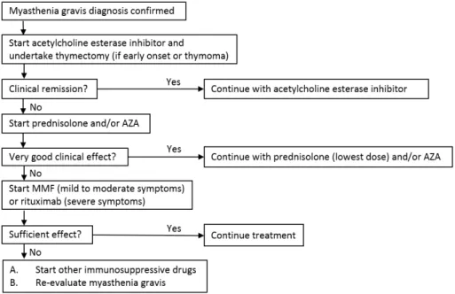
1. Generalized MG in adult (Fig. 2)
The initial step in most adult patients with mild or moderate
disease is symptomatic therapy in the form of pyridostigmine34). Those with severe disease, or rapidly worsening disease, should be treated much like patients with myasthenic crisis, by using rapid therapies (i.e., IVIG or plasmapheresis) followed by longer
acting immunotherapies such as glucocorticoids, azathioprine, mycophenolate mofetil, or cyclosporine. For those patients receiving pyridostigmine alone and whose symptoms are under good control or in remission, we simply follow their clinical course. Thymectomy should also be considered in all adult patients aged <60 years20).
An immunotherapeutic agent is required for patients who remain significantly symptomatic with pyridostigmine treatment, or who became symptomatic again after a temporary response to pyridostigmine34). In young adult patients, we favor the use of glucocorticoids. Often, a good response can be obtained with high doses initially, and that response can be maintained after tapering to low doses. For patients who cannot achieve a clinical improvement on prednisone, another immunomodulating agent is substituted. These include patients with relative contraindica
tions to glucocorticoids (such as diabetes or advanced age), liver disease (precludes azathioprine), renal disease (precludes cyclosporine), or leukopenia (problematic for both azathioprine and mycophenolate)34).
Thymectomy should be considered because of its potential longerterm benefit. Patients with thymoma clearly need surgical treatment. Although the need for thymectomy is less certain in patients with nonthymomatous tissue, there is evidence showing that thymectomy improves clinical outcomes.
2. Juvenile MG
Pyridostigmine is the firstline therapy in children. Steroids are generally used for severe disease that is unresponsive to anticholinesterase agents. Steroids retard bone growth, increase the risk of adult osteoporosis, and are especially problematic for chronic use in children32). Azathioprine, mycophenolate mofetil, and cyclosporine have been used successfully in JMG4,7); however, concerns about serious adverse effects, including impaired fertility and late development of malignancy, are of even greater concern in children than in adults.
As a longterm treatment for MG, thymectomy has been performed successfully and with low morbidity in children37). Thymectomy is a widely accepted option for peripubertal and postpubertal children with generalized MG who have positive AChRAbs or who are seronegative7,32,33,37). The rates of improve
ment and remission seen in an uncontrolled series seem to be similar to that reported in adults after thymectomy37). No significant deleterious consequences of removing the thymus in childhood have been reported4). However, children with pure ocular MG or MuSKpositive MG have not been shown to benefit from thymectomy. The role of thymectomy in prepubertal children remains controversial. This group has a higher incidence of spontaneous remission4,33).
요약
중증 근무력증은 근육의 빠른 피로와 약화를 특징으로 하는 만성 자가 면역 질환으로 골격근의 신경근 접합부의 차단이 원인이다. 근
Fig. 2. Treatment of generalized myasthenia gravis. AZA, azathioprine; MMF, mycophenolate mofetil.
신경 접합부에서 아세틸콜린 수용체에 부착하는 자가 항체로 인한 신 경 근육 전달 장애가 원인이다. 또한, MuSK, LRP4 및 그 외 운동종말 판 단백에 대한 자가 항체들이 관련되어 있다. 중증 근무력증은 침범 된 근부위 (안구형 및 전신형), 발생 연령 및 흉선의 병리 소견에 따라 분류될 수 있다. 혈청 항체 및 임상 증상에 따라 조기발병형, 후기발 병형, 흉선종, MuSK 혹은 LRP4 양성형, 혈청 항체 음성형, 및 안구형 의 소집단 분류가 가능해지고, 각 소집단별 치료 계획 수립과 예후 예 측을 용이하게 할 수 있다. Pyridostigmine 은 증상 치료를 위한 1차 선택 약물이다. 여기에 치료 반응이 부족한 경우에, 우선적인 면역억 제치료로써, 스테로이드, 다른 면역조절약물들, 그리고 흉선절제술을 고려하게 된다. 중증 근무력증의 치료는 환자 개인의 특성, 연령, 임상 증상 및 심한 정도 등에 따라 치료 형태를 결정하게 된다.
References
1) Drachman DB. Myasthenia gravis. N Engl J Med 1994;330:1797- 810.
2) Vincent A. Unravelling the pathogenesis of myasthenia gravis.
Nat Rev Immunol 2002;2:797-804.
3) Luchanok U, Kaminski HJ. Ocular myasthenia: diagnostic and treatment recommendations and the evidence base. Curr Opin Neurol 2008;21:8-15.
4) Andrews PI. Autoimmune myasthenia gravis in childhood.
Semin Neurol 2004;24:101-10.
5) Evoli A. Acquired myasthenia gravis in childhood. Curr Opin Neurol 2010;23:536-40.
6) Chiu HC, Vincent A, Newsom-Davis J, Hsieh KH, Hung T.
Myasthenia gravis: population differences in disease expression and acetylcholine receptor antibody titers between Chinese and Caucasians. Neurology 1987;37:1854-7.
7) Chiang LM, Darras BT, Kang PB. Juvenile myasthenia gravis.
Muscle Nerve 2009;39:423-31.
8) Andrews PI, Massey JM, Howard JF Jr, Sanders DB. Race, sex, and puberty influence onset, severity, and outcome in juvenile myasthenia gravis. Neurology 1994;44:1208-14.
9) Gilhus NE, Verschuuren JJ. Myasthenia gravis: subgroup classification and therapeutic strategies. Lancet Neurol 2015;14:1023-36
10) Lee KY, Kim SJ, Lee JH, Lee MH, Kim HS, Kim JK. Acetylcholine receptor antibody and clinical features in childhood onset myasthenia gravis. J Korean Child Neurol Soc 2005;13:210-9.
11) Vincent A, Whiting PJ, Schluep M, Heidenreich F, Lang B, Roberts A, et al. Antibody heterogeneity and specificity in myasthenia gravis. Ann N Y Acad Sci 1987;505:106-20.
12) McConville J, Farrugia ME, Beeson D, Kishore U, Metcalfe R, Newsom-Davis J, et al. Detection and characterization of MuSK antibodies in seronegative myasthenia gravis. Ann Neurol 2004;55:580-4.
13) Ghazanfari N, Fernandez KJ, Murata Y, Morsch M, Ngo ST,
Reddel SW, et al. Muscle specific kinase: organiser of synaptic membrane domains. Int J Biochem Cell Biol 2011;43:295-8.
14) Evoli A, Lindstrom J. Myasthenia gravis with antibodies to MuSK: another step toward solving mystery? Neurology 2011;77:1783-4.
15) Deymeer F, Gungor-Tuncer O, Yilmaz V, Parman Y, Serdaroglu P, Ozdemir C, et al. Clinical comparison of anti-MuSK- vs anti- AChR-positive and seronegative myasthenia gravis. Neurology 2007;68:609-11.
16) Pevzner A, Schoser B, Peters K, Cosma NC, Karakatsani A, Schalke B, et al. Anti-LRP4 autoantibodies in AChR- and MuSK- antibody-negative myasthenia gravis. J Neurol 2012;259:427-35.
17) Zhang B, Tzartos JS, Belimezi M, Ragheb S, Bealmear B, Lewis RA, et al. Autoantibodies to lipoprotein-related protein 4 in patients with double-seronegative myasthenia gravis. Arch Neurol 2012;69:445-51.
18) Gasperi C, Melms A, Schoser B, Zhang Y, Meltoranta J, Risson V, et al. Anti-agrin autoantibodies in myasthenia gravis. Neurology 2014;82:1976-83.
19) Madhavan R, Gong ZL, Ma JJ, Chan AW, Peng HB. The function of cortactin in the clustering of acetylcholine receptors at the vertebrate neuromuscular junction. PLoS One 2009;4:e8478.
20) Skeie GO, Apostolski S, Evoli A, Gilhus NE, Illa I, Harms L, et al.
Guidelines for treatment of autoimmune neuromuscular transmission disorders. Eur J Neurol 2010;17:893-902.
21) Breiner A, Widdifield J, Katzberg HD, Barnett C, Bril V, Tu K.
Epidemiology of myasthenia gravis in Ontario, Canada.
Neuromuscul Disord 2016;26:41-6.
22) Mantegazza R, Beghi E, Pareyson D, Antozzi C, Peluchetti D, Sghirlanzoni A, et al. A multicentre follow-up study of 1152 patients with myasthenia gravis in Italy. J Neurol 1990;237:339- 44.
23) Keesey JC. Clinical evaluation and management of myasthenia gravis. Muscle Nerve 2004;29:484-505.
24) Grob D, Brunner N, Namba T, Pagala M. Lifetime course of myasthenia gravis. Muscle Nerve 2008;37:141-9.
25) Lindner A, Schalke B, Toyka KV. Outcome in juvenile-onset myasthenia gravis: a retrospective study with long-term follow- up of 79 patients. J Neurol 1997;244:515-20.
26) Evoli A, Batocchi AP, Bartoccioni E, Lino MM, Minisci C, Tonali P. Juvenile myasthenia gravis with prepubertal onset.
Neuromuscul Disord 1998;8:561-7.
27) Sommer N, Melms A, Weller M, Dichgans J. Ocular myasthenia gravis. A critical review of clinical and pathophysiological aspects. Doc Ophthalmol 1993;84:309-33.
28) Finnis MF, Jayawant S. Juvenile myasthenia gravis: a paediatric perspective. Autoimmune Dis 2011;2011:404101.
29) Mahadeva B, Phillips LH 2nd, Juel VC. Autoimmune disorders of neuromuscular transmission. Semin Neurol 2008;28:212-27.
30) AAEM Quality Assurance Committee. American Association of Electrodiagnostic Medicine. Literature review of the usefulness
of repetitive nerve stimulation and single fiber EMG in the electrodiagnostic evaluation of patients with suspected myasthenia gravis or Lambert-Eaton myasthenic syndrome.
Muscle Nerve 2001;24:1239-47.
31) Della Marina A, Trippe H, Lutz S, Schara U. Juvenile myasthenia gravis: recommendations for diagnostic approaches and treatment. Neuropediatrics 2014;45:75-83.
32) Sanders DB, Wolfe GI, Benatar M, Evoli A, Gilhus NE, Illa I, et al.
International consensus guidance for management of myasthenia gravis: executive summary. Neurology 2016;87:419- 25.
33) Lim BC, Kang YJ, Hwang H, Chae JH, Kim KJ, Hwang YS.
Childhood myasthenia gravis: clinical features and response to steroid treatment. J Korean Child Neurol Soc 2003;11:120-7.
34) Díaz-Manera J, Rojas García R, Illa I. Treatment strategies for myasthenia gravis: an update. Expert Opin Pharmacother 2012;13:1873-83.
35) Illa I. IVIg in myasthenia gravis, Lambert Eaton myasthenic syndrome and inflammatory myopathies: current status. J Neurol 2005;252 Suppl 1:I14-8.
36) Saperstein DS, Barohn RJ. Management of myasthenia gravis.
Semin Neurol 2004;24:41-8.
37) Tracy MM, McRae W, Millichap JG. Graded response to thymectomy in children with myasthenia gravis. J Child Neurol 2009;24:454-9.
38) Hennessey IA, Long AM, Hughes I, Humphrey G. Thymectomy for inducing remission in juvenile myasthenia gravis. Pediatr Surg Int 2011;27:591-4.