Blood Res2015;50:254-67. bloodresearch.or.kr
264 Letters to the Editor
men and pelvis are essential to stage the disease accurately [12]. Currently, there is a controversy in the reported liter- ature regarding the accuracy of positron-emission tomog- raphy-CT scanning of MALTomas.
Radical parotidectomy is not indicated because of the associated morbidity, and because RT alone can secure local control and allow tissue preservation. Low dose RT (30 Gy) is extremely efficacious for local control of the disease, with local control rates ranging from 97% to 100%; 5-year progression free survival and overall survival are approx- imately 76% and 91%, respectively [13].
Regional and distant relapses are not common in gastric MALT lymphomas, but extragastric MALTomas tend to be more aggressive and may recur in the regional or distant lymph nodes and in other organs [14, 15]. According to Wenzel et al., patients with MALToma of the head and neck are at a relatively high risk for early dissemination and subsequent distant recurrence when only local therapies are applied. In the current case, there was no lymph node or other organ involvement.
Due to the high local control rate and low morbidity, together with the indolent biology of the disease, we con- clude that moderate-dose RT (25 to 30 Gy) is a safe and effectivetreatment option for stage I and II MALTomas in the parotid gland.
Babusha Kalra1, Pamela Alice Kingsley1, Preety Negi1, M Joseph John2, Kanwardeep Kwatra3, Uttam Braino George4
Departments of 1Radiotherapy, 2Clinical Hematology and Hemat Oncology, 3Pathology, 4Radiology, Christian Medical College and Hospital, Ludhiana, India
Correspondence to: Babusha Kalra Department of Radiotherapy, Christian Medical College
and Hospital, Brown Road, Ludhiana, Punjab 141008, India E-mail: kalrababusha@gmail.com
Received on Jan. 7, 2015; Revised on Oct. 26, 2015; Accepted on Nov. 5, 2015 http://dx.doi.org/10.5045/br.2015.50.4.262
AuthorsÊ Disclosures of Potential Conflicts of Interest No potential conflicts of interest relevant to this article were reported.
REFERENCES
1. Harris NL, Jaffe ES, Stein H, et al. A revised European-American classification of lymphoid neoplasms: a proposal from the International Lymphoma Study Group. Blood 1994;84:1361-92.
2. Thieblemont C, Berger F, Dumontet C, et al. Mucosa-associated lymphoid tissue lymphoma is a disseminated disease in one third of 158 patients analyzed. Blood 2000;95:802-6.
3. Balm AJ, Delaere P, Hilgers FJ, Somers R, Van Heerde P. Primary lymphoma of mucosa associated lymphoid tissue (MALT) in the parotid gland. Clin Otolaryngol Allied Sci 1993;18:528-32.
4. Mehle ME, Kraus DH, Wood BG, Tubbs R, Tucker HM, Lavertu
P. Lymphoma of the parotid gland. Laryngoscope 1993;103:
17-21.
5. Ciccone E, Truini M, Grossi CE. Lymphoid complement of the human salivary glands: function and pathology. Eur J Morphol 1998;36(Suppl):252-6.
6. Diss TC, Wotherspoon AC, Speight P, Pan L, Isaacson PG. B-cell monoclonality, Epstein Barr virus, and t(14;18) in myoepithelial sialadenitis and low-grade B-cell MALT lymphoma of the paro- tid gland. Am J Surg Pathol 1995;19:531-6.
7. Marioni G, Marchese-Ragona R, Marino F, et al. MALT-type lymphoma and Warthin's tumour presenting in the same parotid gland. Acta Otolaryngol 2004;124:318-23.
8. Rosenstiel DB, Carroll WR, Listinsky CM. MALT lymphoma pre- senting as a cystic salivary gland mass. Head Neck 2001;23:254-8.
9. Anacak Y, Miller RC, Constantinou N, et al. Primary muco- sa-associated lymphoid tissue lymphoma of the salivary glands:
a multicenter Rare Cancer Network study. Int J Radiat Oncol Biol Phys 2012;82:315-20.
10. Ferri C, Caracciolo F, Zignego AL, et al. Hepatitis C virus infection in patients with non-Hodgkin's lymphoma. Br J Haematol 1994;88:392-4.
11. Ando M, Matsuzaki M, Murofushi T. Mucosa-associated lym- phoid tissue lymphoma presented as diffuse swelling of the paro- tid gland. Am J Otolaryngol 2005;26:285-8.
12. Perry C, Herishanu Y, Metzer U, et al. Diagnostic accuracy of PET/CT in patients with extranodal marginal zone MALT lymphoma. Eur J Haematol 2007;79:205-9.
13. Tsai HK, Li S, Ng AK, Silver B, Stevenson MA, Mauch PM. Role of radiation therapy in the treatment of stage I/II mucosa-asso- ciated lymphoid tissue lymphoma. Ann Oncol 2007;18:672-8.
14. Wenzel C, Fiebiger W, Dieckmann K, Formanek M, Chott A, Raderer M. Extranodal marginal zone B-cell lymphoma of muco- sa-associated lymphoid tissue of the head and neck area: high rate of disease recurrence following local therapy. Cancer 2003;97:
2236-41.
15. Zinzani PL, Magagnoli M, Ascani S, et al. Nongastrointestinal mucosa-associated lymphoid tissue (MALT) lymphomas: clinical and therapeutic features of 24 localized patients. Ann Oncol 1997;8:883-6.
Sickle cell- thalassemia with concomitant hemophilia A: a rare presentation
TO THE EDITOR: Sickle cell- thalassemia (HbS- tha- lassemia) is a sickling disorder of red blood cells in varying severity, which results from compound heterozygosity for sickle cell trait and thalassemia trait. In India, the fre- quency of the S gene reaches as high as 40%, particularly in the tribal groups, whereas the incidence of the thalasse- mia gene is around 3–4% in the general population [1].
Hence, the occurrence of HbS- thalassemia due to in-
bloodresearch.or.kr Blood Res 2015;50:254-67.
Letters to the Editor 265
Fig. 1. Photomicrograph of peripheral blood film showing anisopoi-
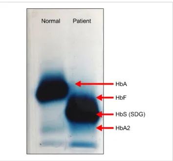
kilocytosis with sickle-shaped cells, target cells, and nucleated RBCs. Fig. 2. Hb electrophoresis at alkaline pH showing a faint band in the A2 region, a prominent band in the SDG region, and HbF.
heritance of both defects is expected to be seen. The preva- lence of sickle cell- thalassemia in India has been reported as <1% in various studies [2]. HbS- thalassemia was first described by Silvestroni and Bianco [3] in 1944 as a micro- drepanocytic disease. Serjeant et al. [4] studied the manifes- tations of this disease. It is characterized by hepatosple- nomegaly, chronic anemia, recurrent attacks of pyogenic infections, avascular necrosis of the bones, and subarachnoid hemorrhage. Hemophilia A is an X-linked recessive disease with a prevalence of 1 in 10,000 male births [5]. It has been estimated that in India 1,300 children with hemophilia are born each year. The chance of both these disorders being present together is extremely rare (1 in 250,000).
Here we report an interesting case that not only shows the coinheritance of both these disorders but also the manner in which the presence of one has impacted the manifestation of the other.
CASE
A 19-year-old male presented with diffuse abdominal pain. There was no history of vomiting, loose stools, or bleeding. He had a history of being admitted previously with the same complaint and had received a transfusion of three units of red blood cells (RBCs) in the past. On examination, the patient had pallor and icterus, and the spleen was palpable 2 cm below the left costal margin.
Blood tests revealed hemoglobin (Hb) of 7.8 g/dL (13–17 g/dL), RBC count 3.15×10¹²/L (4.5–5.5×10¹²/L), mean corpus- cular volume (MCV) 76.0 fL (83–101 fL), mean corpuscular hemoglobin (MCH) 25.0 pg (27–32 pg), and mean corpus- cular hemoglobin concentration (MCHC) 32.0 g/dL (31.5–
34.5 g/dL). The white blood cell (WBC) count was 9.8×10⁹/L (4–11×109/L) and the platelet count was 160×109/L (150–
450×109/L). The peripheral blood film revealed moderate anisopoikilocytosis with microcytes, target cells, and a few tear drop cells and sickle-shaped cells along with 10 nucleat-
ed RBCs (NRBCs)/100 WBCs (Fig. 1). His sickle cell test result was positive. Hb electrophoresis showed a band in the SDG region along with a faint band in the A2 region (Fig. 2). High-performance liquid chromatography (HPLC) showed HbS 81.4%, HbF 5.5%, and HbA2 6.2%. Mutation study analysis was performed using the amplification re- fractory mutation system-polymerase chain reaction (ARMS- PCR) assay, which revealed c.15G>A mutation. The patient was diagnosed as compound heterozygous for HbS-
thalassemia.
After 5 months, the patient presented with excessive bleeding for 6 days following a dental extraction. A coagu- lation workup was performed. The patient’s prothrombin time (PT) and international normalized ratio (INR) were 11.8 seconds (10.4–14.1 sec) and 1.12. The activated partial thromboplastin time (aPTT) was 64.2 seconds (23.0–31.05 sec). Thrombin time was 14.0 seconds (14–19 sec). Factor VIII assay was performed, and the level was found to be
<1%. The level for factor IX was 60%. Screening for com- mon thrombophilia markers was performed to rule out the coinheritance of any prothrombotic factor, which could be responsible for the mild phenotype of hemophilia in this case. The investigations revealed protein C 76.1%
(normal range, 70–140%), protein S 95.8% (normal range, 70–140%), and antithrombin 126.7% (70–140%). Tests for factor V Leiden and prothrombin G20210A mutations were negative. In the literature, it has been shown that the pres- ence of prothrombotic risk factors can influence the onset of the first symptomatic bleeding in children with previously undiagnosed hemophilia A [6].
Plasma fibrinogen and von Willebrand factor were found to be normal. The patient received local tranexamic acid and 2 units of fresh frozen plasma for 2 days, following
Blood Res2015;50:254-67. bloodresearch.or.kr
266 Letters to the Editor
which the bleeding stopped. The patient’s family also under- went screening. His father has beta thalassemia trait and his mother has sickle cell trait. His sister was found to be a beta thalassemia carrier. The coagulation workup showed no abnormalities in any family member. However, his maternal uncle had expired in an accident. The patient has followed up with us, and experienced one episode of epistaxis and pain crisis in the interim. It was planned to initiate treatment with hydroxyurea owing to the increased episodes of pain crisis.
DISCUSSION
Coinheritance of thalassemia and hemophilia A is an uncommon association and coinheritance with sickle tha- lassemia is still rarer. HbS- thalassemia is divided into sickle cell-+ thalassemia and sickle cell-° thalassemia, which have, respectively, reduced or no amounts of HbA present. The clinical and hematologic features in HbS-
thalassemia are quite variable. The clinical severity largely depends upon the nature of the thalassemia mutations.
HbS- thalassemias are classified as HbS-° thalassemia, hav- ing an absence of HbA and with a severe clinical course similar to SS disease, and HbS-+ thalassemia, usually asso- ciated with 20–30% of HbA and with a milder clinical course [7].
Among various mutations, IVS 1-5 (G→C), a severe + thalassemia allele, was found to be the commonest, followed by codon 15 (G→A), codon 30 (G→C), and codon 8/9 (+G), which are severe thalassemia alleles. In the Indian pop- ulation, the commonest thalassemia mutation is seen in 30–80% of heterozygotes, while the majority of the remain- ing thalassemia alleles are of the type [8].
Joints are vulnerable to hemorrhage in hemophilia be- cause of low levels of thromboplastin in synovial tissue [9]. In addition, in any male child presenting with recurrent episodes of prolonged bleeding, occurring spontaneously or following injury or surgical procedures, hemophilia should be suspected [10]. In our case, the patient presented with bleeding following a dental extraction.
Colah et al. [11] reported an interesting consanguineous family from Western India with a combination of thalasse- mia and hemophilia A. Their first child (a male) was diag- nosed with -thalassemia major at 8 months of age and was subsequently transfused every month. At age 2, his gums bled for 5 days after a fall. The coagulation data showed prolonged aPTT with factor VIII assay <1%. The patient was thus diagnosed as suffering from severe hemophilia A with -thalassemia major.
We found two other reports in the literature in which there was coinheritance of thalassemia with bleeding disorders. In one study, there was a report of two sisters with multiple sclerosis, lamellar ichthyosis, -thalassemia minor, and a quantitative deficit of factor VIII-von Willebrand complex [12], whereas the second was a report of a female presenting with Wilson’s disease with con- comitant thalassemia and factor V deficiency [13].
There is also strong evidence for the presence of a hyper- coagulable state in both thalassemia and sickle cell anemia due to platelet activation and the generation of intravascular thrombi [14]. Low plasma levels of protein C, protein S, and antithrombin; elevated plasma levels of thrombin-an- tithrombin (TAT) complexes, prothrombin fragment 1+2 (F1+2), D-dimer complexes, and circulating antipho- spholipid antibodies; platelet activation during vaso-occlu- sive crises; abnormal external exposure of phosphatidylser- ine (PS) and adherence of sickle erythrocytes to the vascular endothelium; reduced nitric oxide levels in the presence of hemolytic anemia; and increased tissue factor expression have been detected in sickle cell patients [15]. In our case, although the patient had a factor VIII level of <1%, bleeding complications did not occur owing to the hypercoagulable state attributed to coinheritance of sickle thalassemia, resulting in a thrombohemorrhagic balance.
To the best of our knowledge, no case describing the combination of sickle cell- thalassemia with hemophilia A has been reported prior to now. The rarity of the co- inheritance of these two disorders and the alterations in presentation, along with the chances of missing the diagnosis of a bleeding disorder with a hypercoagulable state, prompt- ed us to report this case.
Pratibha Dhiman1, Rahul Chaudhary2, Krishna Sudha2
1Department of Hematology, Institute of Liver and Biliary Sciences, 2Department of Pathology, ESI Hospital, New Delhi, India Correspondence to: Pratibha Dhiman Department of Hematology, Institute of Liver and Biliary
Sciences, Sector D1, Vasant kunj, New Delhi 110070, India E-mail: dhimandrpratibha@yahoo.com
Received on Feb. 2, 2015; Revised on Apr. 23, 2015; Accepted on Nov. 13, 2015 http://dx.doi.org/10.5045/br.2015.50.4.264
AuthorsÊ Disclosures of Potential Conflicts of Interest No potential conflicts of interest relevant to this article were reported.
REFERENCES
1. Mukherjee MB, Nadkarni AH, Gorakshakar AC, Ghosh K, Mohanty D, Colah RB. Clinical, hematologic and molecular vari- ability of sickle cell- thalassemia in western India. Indian J Hum Genet 2010;16:154-8.
2. Mohanty D, Colah RB, Gorakshakar AC, et al. Prevalence of
-thalassemia and other haemoglobinopathies in six cities in India: a multicentre study. J Community Genet 2013;4:33-42.
3. Silvestroni E, Bianco I. Microdrepanocito-anemia in un soggetto di razza Bianca. Boll A Accad Med Roma 1944;70:347.
4. Serjeant GR, Ashcroft MT, Serjeant BE, Milner PF. The clinical features of sickle-cell- thalassaemia in Jamaica. Br J Haematol 1973;24:19-30.
bloodresearch.or.kr Blood Res 2015;50:254-67.
Letters to the Editor 267
5. Mosher DF. Disorders of blood coagulation. In: Wyangaarden JB, Smith LH, Bennett JC, eds. Cecil textbook of medicine.
Philadelphia, PA: WB Saunders, 1992:1004-6.
6. Escuriola Ettingshausen C, Halimeh S, Kurnik K, et al.
Symptomatic onset of severe hemophilia A in childhood is de- pendent on the presence of prothrombotic risk factors. Thromb Haemost 2001;85:218-20.
7. Weatherall DJ, Clegg JB. The thalassaemia syndromes. 4th ed.
Oxford, UK: Blackwell Science Ltd, 2001:395.
8. Kulozik AE, Bail S, Kar BC, Serjeant BE, Serjeant GE. Sickle cell-beta+ thalassaemia in Orissa State, India. Br J Haematol 1991;77:215-20.
9. Gregg-Smith SJ, Pattison RM, Dodd CA, Giangrande PL, Duthie RB. Septic arthritis in haemophilia. J Bone Joint Surg Br 1993;75:368-70.
10. Hoyer LW. Hemophilia A. N Engl J Med 1994;330:38-47.
11. Colah RB, Shetty SD, Surve RR, et al. Prenatal diagnosis in a family at risk for beta-thalassemia and hemophilia A: an uncommon association. Hemoglobin 2004;28:343-6.
12. Capra R, Mattioli F, Kalman B, Marcianò N, Berenzi A, Benetti A. Two sisters with multiple sclerosis, lamellar ichthyosis, beta thalassaemia minor and a deficiency of factor VIII. J Neurol 1993;240:336-8.
13. Giannini E, Fasoli A, Botta F, Testa R. Wilson's disease with con- comitant beta thalassaemia and factor V deficiency. Ital J Gastroenterol Hepatol 1998;30:633-5.
14. Eldor A, Rachmilewitz EA. The hypercoagulable state in thalassemia. Blood 2002;99:36-43.
15. Ataga KI. Hypercoagulability and thrombotic complications in hemolytic anemias. Haematologica 2009;94:1481-4.
