https://doi.org/10.5624/isd.2017.47.2.109
Introduction
Recurrent caries under restorations is the main reason for restorative failure and replacement in the primary den- tition.1,2 Improper or delayed diagnosis of carious lesions may lead to significant loss of hard tissue and may result in unsuccessful treatment outcomes. A visual examina- tion should be combined with a probing and radiographic examination to improve the detection of recurrent caries.
Radiography is the most common and useful method for diagnosing recurrent caries under restorations, and bite- wing radiography is usually preferred by clinicians.3-5 Despite being the most preferred radiographic technique, bitewing radiography has several drawbacks, such as distortion, superimposition, and poor beam angulation.6 Therefore, it may fail to provide adequate information re- garding the diagnosis, location, and extension of recurrent caries under different restorations.7 Moreover, the radi- opaque appearance of restorations may conceal the cari- ous lesions.8 Cone-beam computed tomography(CBCT) was introduced in order to overcome the shortcomings of 2-dimensional radiographic techniques.9 CBCT enables the acquisition of 3-dimensional information regarding
Effects of various cone-beam computed tomography settings on the detection of recurrent caries under restorations in extracted primary teeth
Kıvanç Kamburoğlu1,*, Gül Sönmez1, Zeynep Serap Berktaş1, Hakan Kurt1, Doĝukan Özen2
1Department of Dentomaxillofacial Radiology, Faculty of Dentistry, Ankara University, Ankara, Turkey
2Department of Biostatistics, Faculty of Veterinary Medicine, Ankara University, Ankara, Turkey
ABSTRACT
Purpose: The aim of this study was to assess the ex vivo diagnostic ability of 9 different cone-beam computed tomography(CBCT) settings in the detection of recurrent caries under amalgam restorations in primary teeth.
Materials and Methods: Fifty-two primary teeth were used. Twenty-six teeth had dentine caries and 26 teeth did not have dentine caries. Black class II cavities were prepared and restored with amalgam. In the 26 carious teeth, recurrent caries were left under restorations. The other 26 intact teeth that did not have caries served as controls.
Teeth were imaged using a 100×90-mm field of view and a 0.2-mm voxel size with 9 different CBCT settings. Four observers assessed the images using a 5-point scale. Kappa values were calculated to assess observer agreement.
CBCT settings were compared with the gold standard using a receiver operating characteristic analysis. The area under the curve(AUC) values for each setting were compared using the chi-square test, with a significance level of α=.05.
Results: Intraobserver kappa values ranged from 0.366 to 0.664 for observer 1, from 0.311 to 0.447 for observer 2, from 0.597 to 1.000 for observer 3, and from 0.869 to 1 for observer 4. Furthermore, interobserver kappa values among the observers ranged from 0.133 to 0.814 for the first reading and from 0.197 to 0.805 for the second reading. The highest AUC values were found for setting 5(0.5916) and setting 3(0.5886), and were not found to be statistically significant(P>.05).
Conclusion: Variations in tube voltage and tube current did not affect the detection of recurrent caries under amalgam restorations in primary teeth.(Imaging Sci Dent 2017; 47: 109-15)
KEY WORDS: Cone-Beam Computed Tomography; Tooth, Deciduous; Dental Caries; Diagnosis
Copyright ⓒ 2017 by Korean Academy of Oral and Maxillofacial Radiology
This is an Open Access article distributed under the terms of the Creative Commons Attribution Non-Commercial License(http://creativecommons.org/licenses/by-nc/3.0) which permits unrestricted non-commercial use, distribution, and reproduction in any medium, provided the original work is properly cited.
Imaging Science in Dentistry·pISSN 2233-7822 eISSN 2233-7830 Received April 5, 2017; Revised May 9, 2017; Accept May 13, 2017
*Correspondence to : Prof. Kıvanç Kamburoğlu
Department of Dentomaxillofacial Radiology, Dentistry Faculty, Ankara University, 06500, Beşevler, Çankaya, Ankara, Turkey
Tel) 90-312-2965632, Fax) 90-312-2123954, E-mail) [email protected]
dentomaxillofacial structures in a single scan, including 180°-360° rotation around the structures using a cone- shaped X-ray beam.10 Due to the high radiation exposure, CBCT must not be used routinely for the detection of car- ies, particularly among children. However, CBCT imaging can be conducted for various other reasons in children, such as evaluating impaction, eruption, and pathology.5-11 Therefore, available CBCT images taken for different rea- sons in children may also be used to diagnose recurrent caries during routine image interpretation.
Beam-hardening artifacts and streak artifacts are thought to be limiting factors in the detection of recurrent caries under restorations when assessing CBCT images.5 Beam- hardening artifacts occur adjacent to high density struc- tures and are seen as dark bands, and streak artifacts oc- cur due to radiation from a metallic object and are seen as linear hyper-densities extending along the width of the field.12 The presence of beam-hardening artifacts and streak artifacts has the potential to completely affect the quality of the CBCT image and, thus, its diagnostic abil- ity. The potential difference of an X-ray beam source, which is referred to as the tube voltage and is measured in peak kilovoltage(kVp), and the tube current, which is measured in milliamps(mA), are factors that may deter- mine the quantity and quality of the X-ray beam.13 The tube voltage is thought to be the most important factor af- fecting image quality and diagnostic ability. High-contrast radiographs obtained with low tube voltage have been rec- ommended for detecting caries using intraoral radiogra- phy.14 A previous study revealed that a tube voltage of 50 kVp was better than 65kVp and 70kVp when used with a phosphor-plate intraoral radiography system for detecting proximal caries in primary teeth.15 However, on the con- trary, another study established that there was no signifi- cant difference between 60kVp and 90kVp for the diag- nosis of artificial peri-implant defects and carious lesions in permanent teeth using intraoral radiography16. To the best of our knowledge, no studies have compared differ- ent CBCT setting parameters in detecting recurrent caries under amalgam restorations at the primary dentition.
In routine dental practice, exposure settings of CBCT systems are generally determined by manufacturer recom- mendations. Nearly all commercially available CBCT systems have pre-set exposure modes for different clinical needs and also allow for the manual selection of exposure settings. It is difficult to diagnose recurrent caries under amalgam restorations in primary teeth due to the com- pression of structures in intraoral radiography and the oc- currence of metal artifacts in CBCT. Therefore, it is clini-
cally useful to assess the performance of CBCT systems using different imaging settings for the visibility of recur- rent caries lesions under restorative materials. The aim of this study was to assess the ex vivo diagnostic ability of 9 different CBCT settings in the detection of recurrent car- ies under amalgam restorations in primary teeth.
Materials and Methods
A total of 52 extracted or exfoliated primary mandibu- lar first molars and second molars were included. Twenty- six teeth had dentine caries and 26 teeth did not have dentine caries. The presence of caries was determined by visual and intraoral radiographic examination prior to cavity preparation. Teeth were immersed in 2% sodium hypochlorite for 20 minutes and stored in distilled water.
In all 52 teeth, Black class II cavities were prepared and then amalgam restorations were placed. In the 26 carious teeth, recurrent caries were left under amalgam resto- rations. The other 26 intact teeth that did not have caries but had been restored with amalgam restorations served as controls. Cavex Avalloy(Cavex Holland BV, Haarlem, Netherlands) was utilized with zinc phosphate liner for amalgam restorations(Adhesor Cement, SpofaDental, Jicin, Czech Republic). Each group of teeth was stained by a researcher with 2 different-colored nail varnishes.
Teeth were randomly mixed prior to the acquisition of images to ensure random selection.
Image acquisition
For the imaging procedures, teeth were placed in the mandibular molar sockets of a child mandible within a dry skull. The dry skull and the mandible were covered with 2cm of red wax in order to simulate soft tissue.
All teeth were randomly placed in the alveolar sock- ets toge ther in groups of 4(first and second mandibu- lar deci duous molars on the left and right hemi-man- dibles). After the teeth were placed, they were imaged using a 100×90-mm field of view(FOV), a 0.2-mm vox- el size, and 9 different settings of a CBCT system(Plan- meca ProMax 3D ProFace, Planmeca, Helsinki, Finland), as shown in Figure 1 and Table 1. No artifact reduction was used.
Image interpretation
Four observers, including 2 dentomaxillofacial radiol- ogy specialty students(observer 1 and observer 2) and 2 dentomaxillofacial radiology specialists(observer 3 and observer 4), separately reconstructed the multi-planar
reformatted images in a dimly lit room. They used the software provided by the CBCT system(Planmeca Rome- xis, Planmeca, Helsinki, Finland) on a medical diagnostic monitor(NEC, Tokyo, Japan) that had a 21.3-inch screen and 2048 ×1536 resolution.12 The observers prepared panoramic and cross-sectional views with a 1-mm section interval and 1-mm thickness. A total of 10 images that were not included in the study were used for a calibra-
tion session prior to the interpretation sessions. No time restriction was placed on the observers, and evaluations of each image were repeated 1 week after the initial view- ings.
For each tooth, the cross-sectional images were asse- ssed for the presence or absence of recurrent caries under restorations and were scored using a 5-point scale as fol- lows: 1, caries are definitely present; 2, caries are pro- bably present; 3, uncertain or unable to tell if caries are present; 4, caries are probably not present; and 5, caries are definitely not present. Histological validation of the status of the caries was performed by serially sectioning each tooth mesiodistally in parallel to the long axis of the crown using an Accutom-50(Struers, Ballerup, Den- mark). Both sides of each section were examined under a stereomicroscope(×10)(Stemi 2000, Carl Zeiss, Jena, Germany). Teeth were recorded as either sound or having a carious lesion, which was defined as a demineralized white or yellowish-brown discolored area in the enamel or dentine. Histological status was determined by con- sensus of the researchers. Histological examination of the
Table 1. Nine different cone-beam computed tomography imag- ing settings used in this study
Setting Tube voltage
(kVp) Tube current
(mA) Exposure time (s)
Setting 1 60 1 12.1
Setting 2 60 6.3 12
Setting 3 60 12.5 12
Setting 4 78 1 12.1
Setting 5 78 6.3 12
Setting 6 78 12.5 12
Setting 7 96 1 11.9
Setting 8 96 6.3 12
Setting 9 96 12.5 11.9
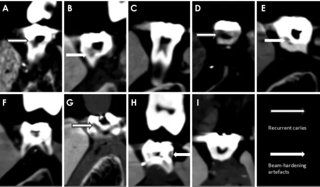
Fig. 1. Cross-sectional cone beam computed tomography images obtained by Planmeca Promax 3D Max. A. Setting with 60kVp and 1 mA amalgam restoration with recurrent caries. B. Setting with 60kVp and 6.3mA amalgam restoration with beam hardening artifact. C.
Setting with 60kVp and 12.5mA amalgam restoration without recurrent caries. D. Setting with 78kVp and 1mA amalgam restoration with recurrent caries. E. Setting with 78kVp and 6.3mA amalgam restoration with beam hardening artifact. F. Setting with 60kVp and 12.5mA amalgam restoration without recurrent caries. G. Setting with 96kVp and 1mA amalgam restoration with recurrent caries. H. Setting with 96 kVp and 6.3mA amalgam restoration with beam hardening artifact. I. Setting with 96kVp and 12.5mA amalgam restoration without recurrent caries.
A B C D E
F G H I
52 tooth surfaces confirmed our first examination and re- vealed that 26 surfaces had dentine caries and 26 surfaces did not have caries.
Statistical analysis
Weighted kappa coefficients were calculated to assess the intraobserver and interobserver agreement for each set- ting. Kappa values were interpreted according to the fol- lowing criteria: 0.10, no agreement; 0.11-0.40, poor agree- ment; 0.41-0.60, moderate agreement; 0.61-0.80, strong agreement; and 0.81-1.00, excellent agreement.17 Scores obtained for the different CBCT settings were compared with the gold standard using receiver operating charac- teristic(ROC) curves to evaluate the ability of observers to differentiate between teeth with recurrent caries and without recurrent caries. The area under the curve(AUC) values were calculated with standard errors and 95% con- fidence intervals, and AUC values for each setting were compared using the chi-square test and a significance level of α=.05. The effects of the observer, the reading, and the image setting on diagnosis were calculated using 3-way repeated analysis of variance(ANOVA). All cal- culations were carried out using Stata 12/MP4 statistical software(StataCorp LP., College Station, TX, USA).
Results
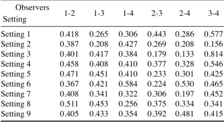
Table 2 shows the intraobserver kappa coefficients cal- culated for each observer for their first and second read- ings. The intraobserver kappa coefficients ranged from 0.366 to 0.664 for observer 1, from 0.311 to 0.447 for observer 2, from 0.597 to 1.000 for observer 3, and from 0.869 to 1 for observer 4. This suggested poor and moder- ate intraobserver agreement for observer 1 and observer 2, respectively, and strong and excellent intraobserver agree- ment for observer 3 and observer 4, respectively. Table 3 and Table 4 show the interobserver kappa coefficients for the first and second readings by the different CBCT imag- ing settings, respectively. Interobserver kappa coefficients among observers ranged from 0.133 to 0.814 for the first reading and from 0.197 to 0.805 for the second reading.
In general, we found poor and moderate interobserver agreements for both readings. The AUC values for differ- ent settings for each observer and reading were also cal- culated. No statistically significant difference was found among different CBCT settings used for the detection of recurrent caries under amalgam restorations(P>.05).
However, when the data were analyzed without taking into account intraobserver and interobserver dependency
Table 3. Interobserver kappa coefficients among observers for the first reading
Observers 1-2 1-3 1-4 2-3 2-4 3-4 Setting
Setting 1 0.418 0.265 0.306 0.443 0.286 0.577 Setting 2 0.387 0.208 0.427 0.269 0.208 0.156 Setting 3 0.401 0.417 0.384 0.179 0.133 0.814 Setting 4 0.458 0.408 0.410 0.377 0.328 0.546 Setting 5 0.471 0.451 0.410 0.233 0.301 0.425 Setting 6 0.367 0.421 0.584 0.224 0.530 0.465 Setting 7 0.408 0.341 0.322 0.306 0.197 0.452 Setting 8 0.511 0.453 0.256 0.375 0.334 0.341 Setting 9 0.405 0.433 0.354 0.392 0.481 0.415
Table 4. Interobserver kappa coefficients among observers for the second reading
Observers 1-2 1-3 1-4 2-3 2-4 3-4 Setting
Setting 1 0.531 0.517 0.490 0.673 0.492 0.455 Setting 2 0.535 0.523 0.350 0.590 0.256 0.406 Setting 3 0.590 0.609 0.570 0.572 0.487 0.805 Setting 4 0.564 0.511 0.393 0.525 0.579 0.613 Setting 5 0.429 0.476 0.474 0.292 0.372 0.535 Setting 6 0.440 0.445 0.626 0.214 0.371 0.487 Setting 7 0.213 0.231 0.394 0.259 0.197 0.236 Setting 8 0.445 0.495 0.409 0.235 0.238 0.458 Setting 9 0.401 0.418 0.418 0.292 0.436 0.264
Table 5. Area under the curve(AUC), standard error, and 95% CIs for different settings
Setting AUC SE LB(95% CI) UB(95% CI)
Setting 1 0.5485 0.0272 0.49509 0.60187 Setting 2 0.5465 0.0270 0.49362 0.59946 Setting 3 0.5886 0.0262 0.53731 0.63993 Setting 4 0.5832 0.0269 0.53052 0.63596 Setting 5 0.5916 0.0266 0.53944 0.64376 Setting 6 0.5634 0.0270 0.51051 0.61632 Setting 7 0.5530 0.0271 0.49987 0.60611 Setting 8 0.5798 0.0268 0.52727 0.63226 Setting 9 0.5719 0.0271 0.51888 0.62490 LB: lower bound, UB: upper bound
Table 2. Intraobserver kappa coefficients between the first and second readings for 4 observers
Setting Observer 1 Observer 2 Observer 3 Observer 4
Setting 1 0.382 0.390 0.616 0.890
Setting 2 0.366 0.447 0.744 0.869
Setting 3 0.445 0.312 1.000 0.987
Setting 4 0.532 0.324 0.770 0.986
Setting 5 0.447 0.415 0.705 0.900
Setting 6 0.540 0.329 0.710 0.986
Setting 7 0.664 0.311 0.597 0.941
Setting 8 0.659 0.319 0.784 1.000
Setting 9 0.495 0.321 0.695 0.941
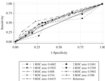
in general, the highest AUC values were found for setting 5 and setting 3, though they were not found to be statis- tically significant(P>.05)(Table 5). Figure 2 shows the ROC curves for observer 3 for the first reading of all set- tings. According to ANOVA, there was a significant main effect of observers(P<.05) and a non-significant main effect of reading and image setting on diagnosis(P>.05).
Discussion
The aim of this study was to evaluate different CBCT settings and determine the best setting for the detection of recurrent caries under amalgam restorations in prima- ry teeth. No significant difference among various set- tings was found, despite setting 5(78kVp, 6.3mA, and an average exposure time of 12.0 seconds) and setting 3 (60kVp, 12.5mA, and an average exposure time of 12.0 seconds) showing the highest AUC values. Our results suggested that variations in the tube current and the tube voltage did not affect the detection of recurrent caries un- der amalgam restorations. In addition, the low AUC val- ues, most of which were lower than 0.60 for all observ- ers, indicated that CBCT may not be the ideal imaging technique for the detection of recurrent caries in primary teeth. However, dentists are often asked to assess caries under amalgam restorations when CBCT images have been taken for other reasons, particularly among children, as amalgams are still preferred in most regions of the world.
Potential limitations to the CBCT system should be taken into account when assessing recurrent caries under resto- rations. Furthermore, observer performance and diagnos-
tic accuracy were not affected by using different CBCT settings through changing the tube voltage, tube current, or exposure time. Observer consistency increased with experience, as indicated by the contrast between observ- er 2, who was a new specialty student and had the lowest intraobserver and interobserver agreement values, and ob- server 4, who showed the highest intraobserver agreement values for all readings and was the most experienced spe- cialist. Variations within and among observers were due to difficulties in interpreting the CBCT images for diag- nosing recurrent caries. In a previous study, the highest AUC for images evaluated on a variety of display types was found for an image assessed on a medical diagnostic monitor that used dedicated software.12 Therefore, in this study, the same monitor and dedicated Planmeca software were used, which enabled the observers to scroll through images and easily inspect each section for caries or arti- facts that resembled caries.
Dental caries are among one of the most common health problems. It is, therefore, crucial to assess available diag- nostic methods in order to make an accurate and early diag- nosis.18 Detecting recurrent caries under amalgam resto- rations can be a complicated process when beam harden- ing artifacts mimic caries, or vice versa. Furthermore, de- tecting recurrent caries under restorations in primary teeth is of paramount importance, as they improve quicker than carious lesions in permanent teeth.19 Artificial carious lesions are easier to diagnose than real carious lesions.
However, their appearance may not reflect actual clinical conditions. Therefore, we used real carious lesions under restorations that were clinically, radiographically, and his- tologically validated.
Despite the negative effect of metal artifacts on the qual- ity and interpretability of CBCT images, a previous study found greater, yet statistically non-significant(P>.05) AUC values for CBCT images than for intraoral images in detecting recurrent buccal caries under restorations.5 In a study where the diagnostic abilities of visual inspection, film, charge-coupled device, photostimulable phosphor plate, and CBCT images were assessed in detecting prox- imal caries, experienced observers found higher AUC values for CBCT images, though the difference among the modalities was not statistically significant.20 The au- thors of the aforementioned study used images with a 0.3-mm voxel size, whereas we used a 0.2-mm voxel size, which may have improved our diagnostic ability.
In addition, Kulczyk et al.21 examined the effects of the presence of amalgam fillings in detecting proximal car- ies and concluded that CBCT images should not be used
Sensitivity
1.00 0.75 0.50 0.25 0.00
0.00 0.25 0.50 0.75 1.00 1-Specificity
1 ROC area: 0.4682 2 ROC area: 0.5481 3 ROC area: 0.5925 4 ROC area: 0.5769 5 ROC area: 0.608 6 ROC area: 0.5902 7 ROC area: 0.534 8 ROC area: 0.588
9 ROC area: 0.6435 Reference
Fig. 2. Receiver operating characteristic curves for observer 3 for the first reading for all settings.
as a diagnostic modality for detecting caries if amalgam fillings are present. That study included CBCT images of 102 extracted permanent teeth that were obtained with a 9-inch FOV, a 0.25-mm voxel size, an automatically ad- justed tube current, a constant tube voltage of 110kVp, and a 36-second scanning time, and, similar to our find- ings, showed that the presence of metallic artifacts made it difficult to detect recurrent caries in CBCT images.
Alth ough CBCT imaging provides a 3-dimensional visu- alization of recurrent caries, the presence of metallic arti- facts from amalgam filling material represents a potential limita tion to this imaging technique. Another predominant limitation of the CBCT system, motion artifacts, was not an issue in the ex vivo setting of our study.
To our knowledge, no previous study has compared the use of different CBCT setting parameters to detect recur- rent caries under amalgam restorations at the primary den- tition. However, a recent study assessed the effects of variations in tube current and FOV on the detection of a vertical root fracture in teeth with intracanal posts and found that decreasing the tube current resulted in a higher efficacy in detecting a vertical root fracture.22 According to another study, differences in exposure parameters did not affect the diagnostic ability of observers to detect a vertical root fracture.23
The main objective of our study was to evaluate the effects of changes in tube voltage and tube current set- tings on the detection of recurrent caries under amalgam restorations in primary teeth. We therefore did not atte- mpt to reduce artifacts through settings offered by the CBCT system in order to keep our findings unbiased.
Unlike intraoral images, CBCT images make it possible to view restorations and carious lesions in axial, coro- nal, and cross-sectional views. However, patients receive higher radiation doses with CBCT than with intraoral radiography, which must be taken into consideration. The average effective doses for a 10-year-old phantom and an adolescent phantom were found to be 116 microsieverts (μSv) and 79μSv, respectively. It was also calculated that the percentage attributable lifetime mortality risks were 0.002% and 0.001% for a 10-year-old and an adolescent patient, respectively, which are considerably higher than the risk to an adult having received the same doses. It is, therefore, crucial that dental CBCT examinations on children must be fully justified over conventional techni- ques.24 Radiation exposure was not an issue in our ex vivo research. However, we used CBCT with a medium FOV and a 0.2-mm voxel size, as these settings are pre- ferred for most diagnostic tasks. Ultimately, our findings
may not apply to CBCT systems and images obtained us- ing different settings.
In conclusion, there were no statistically significant dif- ferences among different CBCT settings used for the de- tection of recurrent caries under amalgam restorations in primary teeth. Our results suggest that variations in tube voltage and tube current did not affect the detection of recurrent caries under amalgam restorations in primary teeth.
References
1. American Academy of Pediatric Dentistry. Guideline on car- ies-risk assessment and management for infants, children, and adolescents. Pediatr Dent 2013; 35: E157-64.
2. Mjör IA, Toffenetti F. Secondary caries: a literature review with case reports. Quintessence Int 2000; 31: 165-79.
3. Okida RC, Mandarino F, Sundfeld RH, de Alexandre RS, Sundefeld ML. In vitro-evaluation of secondary caries forma- tion around restoration. Bull Tokyo Dent Coll 2008; 49: 121- 4. Ando M, González-Cabezas C, Isaacs RL, Eckert GJ, Stook-8.
ey GK. Evaluation of several techniques for the detection of secondary caries adjacent to amalgam restorations. Caries Res 2004; 38: 350-6.
5. Murat S, Kamburoğlu K, Isayev A, Kurşun S, Yüksel S. Vis- ibility of artificial buccal recurrent caries under restorations using different radiographic techniques. Oper Dent 2013; 38:
197-207.
6. Kamburoğlu K, Ilker Cebeci AR, Gröndahl HG. Effectiveness of limited cone-beam computed tomography in the detection of horizontal root fracture. Dent Traumatol 2009; 25: 256-61.
7. Mialhe FL, Pereira AC, Meneghim Mde C, Ambrosano GM, Pardi V. The relative diagnostic yields of clinical, FOTI and radiographic examinations for the detection of approximal caries in youngsters. Indian J Dent Res 2009; 20: 136-40.
8. Tveit AB, Espelid I. Class II amalgams: interobserver vari- ations in replacement decisions and diagnosis of caries and crevices. Int Dent J 1992; 42: 12-8.
9. Tyndall DA, Rathore S. Cone-beam CT diagnostic applica- tions: caries, periodontal bone assessment, and endodontic applications. Dent Clin North Am 2008; 52: 825-41.
10. White SC. Cone-beam imaging in dentistry. Health Phys 2008; 95: 628-37.
11. Kamburoğlu K, Kurt H, Kolsuz E, Öztaş B, Tatar I, Çelik HH.
Occlusal caries depth measurements obtained by five different imaging modalities. J Digit Imaging 2011; 24: 804-13.
12. Baltacıoĝlu İH, Eren H, Yavuz Y, Kamburoğlu K. Diagnostic accuracy of different display types in detection of recurrent caries under restorations by using CBCT. Dentomaxillofac Radiol 2016; 45: 20160099.
13. Kau CH, Bozic M, English J, Lee R, Bussa H, Ellis RK. Cone- beam computed tomography of the maxillofacial region-an update. Int J Med Robot 2009; 5: 366-80.
14. Svenson B, Welander U, Anneroth G, Söderfeldt B. Exposure
parameters and their effects on diagnostic accuracy. Oral Surg Oral Med Oral Pathol 1994; 78: 544-50.
15. Sogur E, Baksı BG, Orhan K, Paksoy SC, Dogan S, Erdal YS, et al. Effect of tube potential and image receptor on the de- tection of natural proximal caries in primary teeth. Clin Oral Investig 2011; 15: 901-7.
16. Kaeppler G, Dietz K, Reinert S. Influence of tube potential setting and dose on the visibility of lesions in intraoral radiog- raphy. Dentomaxillofac Radiol 2007; 36: 75-9.
17. Landis JR, Koch GG. The measurement of observer agree- ment for categorical data. Biometrics 1977; 33: 159-74.
18. Yamaguchi K, Miyazaki M, Takamizawa T, Inage H, Moore BK. Effect of CPP-ACP paste on mechanical properties of bovine enamel as determined by an ultrasonic device. J Dent 2006; 34: 230-6.
19. Wilson PR, Beynon AD. Mineralization differences between human deciduous and permanent enamel measured by quanti- tative microradiography. Arch Oral Biol 1989; 34: 85-8.
20. Senel B, Kamburoglu K, Uçok O, Yüksel SP, Ozen T, Avsever
H. Diagnostic accuracy of different imaging modalities in de- tection of proximal caries. Dentomaxillofac Radiol 2010; 39:
501-11.
21. Kulczyk T, Dyszkiewicz Konwińska M, Owecka M, Krzyżos- taniak J, Surdacka A. The influence of amalgam fillings on the detection of approximal caries by cone beam CT: in vitro study. Dentomaxillofac Radiol 2014; 43: 20130342.
22. Safi Y, Hosseinpour S, Aziz A, Bamedi M, Malekashtari M, Vasegh Z. Effect of amperage and field of view on detection of vertical root fracture in teeth with intracanal posts. Iran En- dod J 2016; 11: 202-7.
23. Pinto MG, Rabelo KA, Sousa Melo SL, Campos PS, Oliveira LS, Bento PM, et al. Influence of exposure parameters on the detection of simulated root fractures in the presence of various intracanal materials. Int Endod J 2017; 50: 586-94.
24. Theodorakou C, Walker A, Horner K, Pauwels R, Bogaerts R, Jacobs R, et al. Estimation of paediatric organ and effec- tive doses from dental cone beam CT using anthropomorphic phantoms. Br J Radiol 2012; 85: 153-60.