대한소화기학회지 1999;34:774 - 783
9)
접수: 1999년 3월 18일, 승인: 1999년 6월 20일
연락처: 정영화, 138-736, 서울시 송파구 풍납동 388-1, 서울중앙병원 소화기내과 Tel: (02) 2224-3184, Fax: (02) 476-0824
※ 본 논문의 요지는 1998년 제37차 대한소화기학회 추계학술대회에서 발표되었음.
간세포암 및 주위 간조직 내 T ransforming Growth Factor- β1 mRNA 발현
울산대학교 의과대학 서울중앙병원 내과, 일반외과*
송병철・정영화・양수현・김정아・홍인란・이재균 정 성 애・이 영 주*・이 승 규*・이 영 상・서 동 진
E x p re s s i o n o f Tr a n s fo r m i n g Gr o w t h F a c t o r -β1 m R N A i n H e p a t o c e ll u a r Ca r c i n o m a a n d S u rr o u n d i n g Li v e r
Byung-Che ol Son g, M.D., Youn g-Hw a Ch ung, M.D., Soo Hyu n Yang, M.D., J un g A Kim , In Ran Hong, J ae Kyoon Le e , M.D., Sun g Ae J u ng, M.D., Youn g-J oo Le e , M.D.*,
S u n g G y u L e e , M .D .*, Yu n g S a n g Le e , M.D. a n d D o n g J i n S u h , M.D.
Departments of Internal Medicine and General Surgery*,
University of Ulsan College of Medicine, Asan Medical Center, Seoul, Korea
Background/Aims: Transforming growth factor-β1 (TGF-β1) has a central role in hepatic fibrosis
and is also related to the development and progression of hepatocellular carcinoma (HCC). However the effects of TGF-β1 overexpression in HCC remains unclear. This study was designed to assess the role of TGF-β1 in HCC. Methods: We semiquantitated TGF-β1 mRNA by competitive reverse transcription polymerase chain reaction from HCC and surrounding liver (SL) of 21 patients. We analyzed the relationship between TGF-β1 mRNA expression and clinicopathological characteristics to assess the role of TGF-β1 in HCC. Results: TGF-β1 mRNA was overexpressed in HCC than in SL (median 2.1×10⁴ vs 5.1×10³ copies/μg of RNA, p=0.01). TGF-β1 mRNA was overexpressed in all cases of well differentiated HCC. However, in the cases of poorly defferentiated HCC, the overexpression was observed in 57% of the patients (p=0.06). The overexpression occurred more frequently in single nodular type (87%) than in multinodular type (33%, p=0.03). It also occurred in 100% of cases of tumor less than 3 cm, in 71% of cases of tumor sized between 3 and 5 cm, an in 50% of cases of tumor bigger than 5 cm (p=0.06). In addition, expression of TGF-β1 mRNA in HCC was inversely correlated with the level of serum α-fetoprotein (r=-0.47, p=0.03). Conclusions:TGF-β1 mRNA is generally overexpressed in HCC, suggesting that it may have important roles in the development of HCC. Our data also indicates that TGF-β1 mRNA expression may play more roles in small, single nodular and well differentiated HCC. (Kor J Gastroenterol 1999;34:774 - 783)
Key Words: Hepatocellular carcinoma, TGF-β1 mRNA
송병철 외 10인. 간세포암 내 Transforming Growth Factor-β1 mRNA 발현 775
서 론
간세포암의 발생 및 성장에는 여러 인자들이 복합 적으로 관여하며 특히 만성 간질환 환자에서 암 유전 자 혹은 암억제유전자의 변이와 함께 각종 성장인자 들의 과발현이 중요한 역할을 한다고 알려져 있다.1 간세포암의 발생 혹은 성장과 연관성이 시사되는 성장인자들은 transforming growth factor-α(TGF- α),2-5 transforming growth factor-β1 (TGF-β1),6-9 insulin like growth factor-II,10,11 fibroblast growth factor-212,13 및 hepatocyte growth factor 등14,15이다.
TGF-β1은 25 kD의 homodimeric polypeptide로16 만 성 간질환에서는 간조직의 백혈구 및 간내 간질세포 등에서 발현되어 세포외기질의 주성분인 collagen과 fibronectin 등의 합성을 촉진하고 이들의 분해를 억 제함으로써 간섬유화의 중심적 역할을 한다.17-19 TGF-β1은 세포의 증식과 분화에도 관여하는데,20 시험관 내에서 정상 간세포의 증식을 억제하고21 백 서에서 부분간절제술에 의한 간세포 재생 과정에서 DNA 합성이 정점에 이른 후에 증가하여 간세포의 과증식을 억제하며,22 부분간절제 후 TGF-β1을 투 여하면 초기의 간재생이 억제된다고 알려져 있다.23 또한 시험관 내에서 TGF-β1은 간세포암 세포주의 증식을 억제한다고 알려져 있으나,24-26 생체 내에서 는 오히려 간세포암조직에 과발현되어6 간세포암의 성장을 촉진하는 것으로 알려져27,28 간세포암의 발 생 혹은 진행에 긍정적으로 작용할 가능성이 시사되 어 왔다. 또한 간세포암 환자의 종양조직 내 TGF-β1 mRNA 역시 주위 간조직보다 과발현되고7,8 혈중과 요중에도 TGF-β1이 증가된다는 보고 등29-32도 이러 한 가설을 뒷받침하고 있다. 그러나 과발현된 TGF- β1이 간세포암의 발생과 진행에 어떻게 관여하는 지에 대한 연구는 미흡하다. 따라서 본 연구에서는 간세포암조직은 물론 동일 환자의 주위 간조직에서 TGF-β1 mRNA의 발현 양을 정량하고 이를 임상적 및 종양학적 특성들과 비교함으로써 간세포암의 발생 과 진행에서 TGF-β1의 역할을 분석하고자 하였다.
대상 및 방법
1. 대 상
간세포암으로 수술받은 21예를 대상으로 하여 간 세포암과 주위 간조직에서 TGF-βl mRNA의 양을 경쟁적 역전사 중합효소연쇄반응(competitive reverse transcription polymerase chain reaction, competitive RT-PCR)33으로 측정하고, TGF-β1 mRNA의 발현 양상과 간세포암의 임상적, 종양학적 특성들과의 연 관성을 분석하였다.
2. 방 법
1) 간조직에서 m RNA 추출
간세포암조직과 주위 간조직으로부터 acid guani- dium thiocyanate-phenol-chloroform을 이용한 single- step method34를 응용하여 만든 RNA-STAT 60TM (TEL-TEST B, INC., TX, USA)으로 전체 세포내 RNA를 추출하였다. 방법을 요약하면, RNA-STAT- 60TM 1 mL에 간조직 약 100 mg을 넣고 균질화하여 실온에 5분 방치한 후 200 μL의 chloroform을 넣고 15초간 vortex하여 잘 섞은 후 3분간 실온에 재방치 하였다. 4℃에서 12,000 g으로 10분간 원심분리한 후 상층액을 취하여 500 μL의 isopropanol과 혼합하 고 실온에서 10분간 방치한 후 12,000 g으로 4℃에 서 10분간 원심분리하여 RNA pellet을 얻었다. RNA pellet은 75% ethanol 1 mL를 첨가하여 vortex한 후 7,500 g으로 4℃에서 5분 동안 원심분리하였고 이 과정을 2회 반복하여 세척하였다. 실온에서 건조한 RNA는 0.1% diethyl-pyrocarbonate (DEPC) 처리한 deionized distilled-water (DDW) 20 μL에 용해시킨 후 분광광도계를 이용하여 260 nm 파장에서 흡광도 를 측정하였고, A260/A280 비가 1.8 이상인 표본을 실험에 이용하였다. 또한 전체 RNA를 1% 한천겔에 서 MOPS (4-Morpholinepropane sulfonic acid) 완충 액으로 전기영동하여 RNA 적합성을 재확인하였다.
776 The Korean Journal of Gastroenterology : Vol. 34, No. 6, 1999
2) 경쟁적 역전사 중합효소연쇄반응을 이용한 T GF - β1 m RN A 의 정량
(1) TGF-β1 mRNA에 대한 competitor
TGF-β1 mRNA에 대한 competitor는 TGF-β1 mRNA을 포함하여 11개의 사이토카인 mRNAs에 대해 경쟁적으로 반응할 수 있는 각각의 priming site 를 갖게 설계된 plasmid pHCQ35을 이용하였다.
(2) cDNA의 합성
8개의 시험관에 4 μL의 0.1% DEPC-DDW를 넣 고, 첫 시험관에 competitor 1 μL (108 RNA copies μg/μL)를 넣어 5-1, 5-2, ---, 5-8까지 희석한 후 조직에 서 추출한 RNA 1 μL (1 g)를 혼합하여 총 5 μL를 만 든 후 65℃에서 10분 반응시키고 얼음 위에서 3분 동안 냉각시켜 원형(template)으로 이용하였다. 여기 에 5× RT buffer 4 μL, 0.1% DEPC-DDW 9 μL, oligo (dT)15 1 μL (50 ng), dNTP 0.5 μL (100 μM) AMV reverse transcriptase (Promega, Madison, WI) 0.2 μL (2 U), RNasin (Promega, Madison, WI) 0.3 μL (12 U)를 혼합하여 총 20 μL를 만든 후, 42℃에 서 60분 반응시키고 95℃에서 5분 간 반응시켜 역전 사 효소를 비활성화시킨 후 얼음 위에서 냉각시켜 cDNA를 만들었다. 합성된 cDNA는 0.1% DEPC- DDW 20 μL를 첨가하여 2배로 희석하여 40 μL를 만 들었고 이 중 4 μL를 RT-PCR의 원형으로 사용하였다
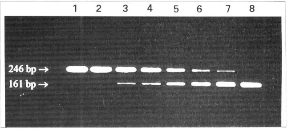
(3) 경쟁적 역전사 중합효소연쇄반응 및 정량분석 증폭에 이용된 시발체의 염기서열과 위치는Table 1과 같고 TGF-β1 mRNA와 competitor에 대한 증폭 산물의 크기는 각각 161 bp와 246 bp이다.
합성된 cDNA 4 μL, 10× PCR buffer 4 μL, dNTP 0.5 μL, 5' -primer 0.5 μL (10 pmol), 0.1% DEPC- DDW 31 μL를 혼합하여 40 μL를 만들고 mineral oil
50 μL를 첨가하였다. PCR은 automated thermal cycler (Perkin Elmer, Norwalk, USA)를 이용하였고
‘hot-start' PCR법을 적용하였는데, 94℃에서 4분간 DNA를 변성시키고 65℃에서 15분간(이 과정에서 10분이 지나면 10× PCR buffer 1 μL, 3' -primer 10 pmol, Taq polymerase 1U, MgCl2 1.5 mM, 0.1%
DEPC-DDW 5.3 μL를 혼합한 10 μL를 첨가) annealing 하였다. 이어서 94℃ 45초, 60℃ 45초, 72℃ 2분으로 구성되는 cycle을 35회 시행한 후, 마지막에 72℃에 서 7분간 extension하였다. PCR산물은 10 μL 취하 여 6×DNA loading buffer (0.25% bromophenol blue, 0.25% xylene cyanol, 30% glycerol)에 혼합하 여 2% 한천겔에서 120 V로 40분간 전기영동한 후 ethidium bromide로 염색하여 자외선조사기로 확인 하였다(Fig. 1). 최종적으로 TGF-β1 mRNA와 competitor의 증폭산물의 밀도를 영상밀도계 (Imaging densitometer GS-670, BioRad, Hurcules, CA)를 이용하여 측정한 후 log 밀도비로 표준 그래 프를 그렸고 표준 그래프의 R2값이 0.95 이상인 경 우를 분석에 사용하였다. 증폭산물의 log 밀도비가 1이 되는 지점을 TGF-β1 mRNA의 양으로 정하였 다(Fig. 2).
3) 통계 분석
TGF-β1 mRNA 양은 중앙값(범위, 최소값-최대 값)을, 그 이외의 연속 변수는 평균±표준편차로 표 시하였다. 간세포암과 주위 간조직 내 TGF-β1 mRNA양의 비교는 Wilcoxon signed rank test, 빈도 분석은 chi square test 혹은 Fisher' s exact test 그리 고 두 변수 간의 연관성은 Spearman' s rank cor- relation coefficient로 각각의 유의성을 검정하였다.
Table 1. Primers used for the Competitive RT-PCR
Primers Nucleotide position Sequences 5′primer
3′primer
1678-1697 1819-1838
5'-GCCCTGGACACCAACTATTGCT-3'
5'-AGGCTCCAAATGTAGGGGCAGG-3'
Song, et al. Transforming Growth Factor-β1 mRNA in Hepatocelluar Carcinoma 777
결 과
1. 간세포암 환자의 임상적 특징
대상 21예 중 남자는 16예, 여자는 5예였으며, 평 균 나이는 52±8세(39-65세)였다. 원인별로는HBsAg 양성인 환자가 17예(81%)로 대부분이었으며, anti-
HCV 양성은 1예(5%), anti-HBc 만 양성인 경우는 3 예(14%)였다. 혈청 α-fetoprotein (AFP)은 20 ng/mL 미만인 경우가 8예(38%), 20-400 ng/mL인 경우는 2 예(10%) 그리고 400 ng/mL 이상인 경우가 11예 (52%)였다. 종양의 크기는 평균 6.3±4.5 cm (1.7- 17.0 cm)로, 장경이 3 cm 미만이 6예, 3-5 cm이 7예 그리고 5 cm 이상인 경우가 8예였다. 종양의 형태는 Fig. 1. Products of competitive RT-PCR. The amounts of TGF-β1 mRNA was determined
where the density of competitor RNA transcripts was equal to those of TGF-β1 mRNA transcripts. Upper lane (246 bp), RT-PCR products of competitor; lane 1, 5-1 (5×10⁷ copies/
μg RNA); lane 2, 5-2; lane 3, 5-3; lane 4, 5-4; lane 5, 5-5; lane 6, 5-6; lane 7, 5-7; lane 8, 5-8; lower lane (161 bp), those of TGF-β1 mRNA.
Fig. 2. The intensities of PCR bands were measured using image densitometer. The log ratio of densities (competitor/TGF-β1 mRNA) was plotted against the log reciprocal of the amount of competitor of TGF-β1 mRNA.
778 대한소화기학회지 : 제 34 권 제 6 호 1999
단결절형이 15예, 다결절형이 6예였다. Edmondson- Steiner36에 의한 세포의 분화도는 Grade I이 2예, Grade II는 5예, Grade III가 13예 그리고 Grade IV 는 1예였다. 주위 간조직은 16예에서 만성 간염의 소견을, 5예에서 간경변증 소견을 보였다(Table 2).
2. 간세포암과 주위 간조직에서 T GF - β1 m RN A 발현양상
간세포암과 주위 간조직에서 TGF-β1 mRNA의 중앙값은 각각 2.1×10⁴ (9.2×10²-1.9×10⁶), 5.1×
103 copies/μg RNA (2.1×10²-3.6×10⁴)로 간세포암 조직 내에서 주위 간조직에 비해 일반적으로 높게 발현되었다(p=0.01)(Fig. 3). TGF-β1 mRNA 발현 양 은 21예 중 15예에서 간세포암조직 내에서 주위 간 조직보다 높게 발현되었으나 6예에서는 오히려 주 위 간조직에서 증가되어 있었다. 종양의 분화가 좋 은(Grade I 또는 Grade II) 7예 모두에서 간세포암조 직 내 TGF-β1 mRNA 발현이 높았던 반면, 분화가 나쁜(Grade III 또는 Grade IV) 예에서는 14예 중 8 예(57%)에서만 발현이 높았다(p=0.06). 또한 단결절 형 15예 중 13예(87%)에서, 다결절형 6예 중 2예 (33%)에서 간세포암조직 내 발현이 높았다(p=0.03).
종양의 장경이 3 cm 미만인 경우는 6예 모두에서
Fig. 3. Expression of TGF-β1 mRNA in HCC and surrounding liver (SL) in 15 cases (○). On the other hand, in 6 cases (●), TGF-β1 mRNA was much more expressed in the SL than in HCC (p=0.01). Median value of TGF-β1 mRNA was 5.1×10³ vs. 2.1×10⁴ copies/μg of total RNA in SL and HCC respectively.
Table 2. Clinicopathological Characteristics of Pa- tients with HCC
Age (years) Sex (M/F) Etiology
HBV/HCV/NBNC*
AFP (ng/mL) 20/20-400/ 400
Pathology of surrounding liver CH/LC
Tumor size (cm) 3/3-5/ 5 Tumor type
SN/MN Histologic grade
Grade Ⅰ/Ⅱ/Ⅲ/Ⅳ
52±1.7 16/5 17/1/3 8/2/11 16/5 6/7/8 15/6 2/5/13/1 HBV, hepatiti B virus; HCV, hepatitis C virus;
AFP, α-fetoprotein; CH, chronic hepatitis; LC, liver cirrhosis; HCC, hepatocellular carcinoma; SN, sin- gle nodular type; MN, multinodular type.
* NBNC means HBsAg(-), anti-HCV(-) but anti- HBc(+).
송병철 외 10인. 간세포암 내 Transforming Growth Factor-β1 mRNA 발현 779
(100%), 3-5 cm인 경우는 7예 중 5예(71%), 그리고 5 cm 이상인 경우는 8예 중 4예(50%)에서 간세포암 조직 내 발현이 증가하여, 종양의 크기가 커질수록 TGF-β1 mRNA 발현이 감소하는 경향을 보였다 (p=0.06)(Table 3).
한편 간세포암조직 내 TGF-β1 mRNA의 발현 정 도는 혈청 AFP치와 역상관관계를 나타냈다(r=0.47,
p=0.03)(Fig. 4). 그러나 TGF-β1 mRNA 발현과 나 이, 성별, 혈청 트란스아미나제, 알부민, 빌리루빈, 담관 침범 그리고 미세혈관 침범 여부와는 상호 연 관성이 없었다.
고 찰
간세포암의 발생 과정을 이해하는 데 있어서 암 유전자와 종양억제유전자의 변이뿐만이 아니라 성 장인자들의 역할에 대한 관심이 증가하고 있다.
TGF-β1은 정상 간세포의 증식을 억제하는24,37 성장 인자임에도 불구하고 간세포암조직 내에서 과발현 되어 오히려 간세포암의 성장을 촉진한다는 것은27,28 매우 흥미로운 사실이다.
본 연구에서 간세포암조직에서 TGF-β1 mRNA를 정량한 결과 주위 간조직에 비하여 일반적으로 증가 된다는 사실을 확인할 수 있었다. 간세포암 환자에 서 혈중과29 요중에31,32 TGF-β1이 증가하고 수술 또 는 적절한 경간동맥색전술 후에 감소함이29,32 보고되 었는데, 본 연구 결과로 보아 간세포암 환자들의 혈중 과 요중에 증가하는 TGF-β1은 간세포암조직 내의 전사단계에서 과발현됨을 확인할 수 있었다.
Table 3. Comparison of TGF-β1 mRNA Expression in HCC and Surrounding Liver according to the Characteristics of HCC Variables
Variables HCC SL (%) HCC≤SL (%) p value
Histologic grade
Ⅰ or Ⅱ
Ⅲ or Ⅳ Tumor type
Single nodular Multinodular Tumor size
3 cm 3-5 cm 5 cm
Microvascular invasion (-)
(+)
7 (100) 8 (57) 13 (87)
2 (33) 6 (100) 5 (71) 4 (50) 11 (73)
4 (67)
0 (0) 6 (43) 2 (13) 4 (67) 0 (0) 2 (29) 4 (50) 4 (27) 2 (33)
0.06
0.03
0.06
1.0
HCC, Hepatocellular carcinoma; SL, Surrounding liver. HCC>SL means that the level of TGF-β1 mRNA expression is higher in HCC than in SL.
Fig. 4. Correlation between TGF-β1 mRNA and serum AFP. TGF-β1 mRNA in HCC and serum AFP were inversely correlated (r=-0.47, p=0.03).
780 The Korean Journal of Gastroenterology : Vol. 34, No. 6, 1999
간세포암 환자에서 과발현된 TGF-β1이 세포 증 식을 억제하지 않고 오히려 간암세포의 증식을 촉 진하는 기전으로는 몇 가지 가능성들이 제시되고 있다. 첫째, TGF-β1의 작용에 필수적인 TGF-β1 수 용체가 정상 간세포에는 세포막에 발현되는 데 비 해 간암세포에는 주로 세포질 내에 존재하기 때문 에 TGF-β1의 증식억제작용을 피할 수 있다는 것이 다.38 둘째, epidermal growth factor (EGF) 수용체의 발현을 유도하여 TGF-α 혹은 EGF의 작용을 통해 종양이 성장할 것이라는 가설도 있다.28,39 셋째, 종 양 및 주위 조직의 세포외기질과 integrin,27 type IV collagenase 등40의 합성을 촉진하고, 혈관 형성을 촉 진하며,41 종양 주위의 정상 간세포의 apoptosis를 유 발하여42,43 주위 환경을 간세포암 성장에 유리한 환 경으로 만들고, 넷째, TGF-β1이 숙주의 면역능을 억제하여 종양의 성장과 진행을 도와주는 것으로 알려져 있다.44,45
본 연구에서는 TGF-β1 mRNA 발현이 종양의 특 성에 따라 다양하게 나타남을 관찰할 수 있었다. 즉 분화가 좋은 간세포암에서는 TGF-β1 mRNA 발현 이 모든 환자에서 주위 간조직보다 증가되어 있었던 반면에, 분화가 나쁜 환자에서는 57%만이 주위 간 조직보다 증가해 있었다. 간세포암의 형태에 있어서 는 단결절형에서 다결절형보다 간세포암조직 내의 TGF-β1 mRNA 발현이 증가되어 있었고, 종양의 크 기가 작을수록 주위 간조직보다 TGF-β1 mRNA 발 현이 증가되는 경향을 보였다. 따라서 간세포암의 발생과 진행에 있어서, TGF-β1은 분화가 좋고 단결 절형이면서 크기가 작은 간세포암의 발생과 진행에 보다 많은 기여를 할 것으로 생각된다. 그러나 분화 가 나쁘거나 다결절형이거나 종양의 크기가 큰 경우 에는 상대적으로 주위 간조직보다 TGF-β1 mRNA 의 과발현이 없어 TGF-β1보다는 다른 성장인자 혹 은 유전자 변이 등이 관여할 것으로 생각된다. 분화 가 나쁘고 종양의 크기가 큰 간세포암에서 p53 유전 자 변이가 흔히 동반된다는 보고들46-48은 종양학적 특성에 따라 유전자 변이와 성장인자의 역할이 다를 수 있음을 뒷받침하고 있다.
본 연구에서는 혈청 AFP 농도가 간세포암조직 내 의 TGF-β1 mRNA 양과 반비례함을 확인할 수 있었
다. 이는 TGF-β1이 AFP과는 다른 기전으로 간세포 암에서 증가함을 시사하며 시험관 내 연구에서 TGF-β1이 AFP의 유전자의 촉진자(promotor)를 억 제한다는 보고49와 일치하는 소견이다.
결론적으로 TGF-β1은 간세포암조직 내에서 전사 단계의 과발현에 의하여 증가되며, 특히 분화가 좋 고 단결절형이며 종양의 크기가 작은 간세포암에서 과발현되어 이들의 발생과 진행에 관여하리라 생각 된다.
요 약
목적: 만성 간질환 환자에서 암유전자 혹은 억제 유전자의 변이와 함께 각종 성장인자들의 과발현이 간세포암의 발생 및 진행에 중요한 역할을 하고 있 음이 보고되고 있다. TGF-β1은 만성 간질환 환자의 간섬유화에 중심적 역할을 할 뿐만 아니라 간세포암 의 발생과 성장에도 관여한다고 알려져 있다. 그러 나 TGF-β1이 간세포암의 발생 혹은 진행에 어떻게 기여하는지에 관한 연구는 미흡하다. 본 연구는 TGF-β1 mRNA 발현 양을 정량하고 이를 임상적 및 종양학적 특성들과 비교함으로써 간세포암에서 TGF- β1의 역할을 분석하고자 하였다. 대상 및 방법: 수 술받은 21예의 간세포암 및 주위 간조직에서 경쟁적 역전사 중합효소연쇄반응을 이용하여 TGF-β1 mRNA 를 정량하고 이들의 발현 정도와 간세포암의 종양학 적 특성 및 혈청학적검사 소견들과의 연관성을 분석 하였다. 결과: TGF-β1 mRNA는 간세포암조직 내에 서 주위 간조직에 비해 높게 발현되었다(중앙값; 2.1
×104 vs. 5.1×10³ RNA copies/μg RNA) (p=0.01).
TGF-β1 mRNA는 분화가 좋은(Grade I 혹는 Grade II) 7예 모두에서 간세포암조직 내에서 과발현된 반 면, 분화가 나쁜(Grade III 또는 IV) 경우 14예 중 8 예(57%)에서만 과발현되었다(p=0.06). 또한 TGF-βl mRNA의 과발현 빈도는, 단결절형에서 다결절형에 비해 높았다(87% vs 33%)(p=0.03). 직경 3 cm 미만 (n=6), 3-5 cm(n=7), 5 cm 이상인(n=8) 종양의 각각 100%, 71%, 50%에서 과발현되어 종양이 커질수록 과발현의 빈도가 감소하는 경향을 보였다(p=0.06).
한편 간세포암조직 내 TGF-β1 mRNA의 발현 정도
Song, et al. Transforming Growth Factor-β1 mRNA in Hepatocelluar Carcinoma 781
는 혈청 AFF치에 반비례하였다(r=0.47)(p=0.03). 그 러나 TGF-β1 mRNA의 발현 정도와 혈청 트란스아 미나제, 알부민 및 빌리루빈과 현미경적인 미세혈관 혹은 담도 침범 여부와는 상호 연관성이 없었다.
결론: 일반적으로 TGF-β1은 간세포암조직 내에서 전사단계에 과발현되며, 종양의 특성에 따라 간세포 암의 발생 혹은 진행 과정에 다양하게 기여하는 것 으로 생각된다. TGF-β1은 분화가 좋고 단결절형이 면서 크기가 작은 간세포암의 발생 혹은 진행에 기 여할 것으로 생각된다.
색인단어: 간세포암, TGF-β1 mRNA
감사의 글
본 연구에 이용한 plasmid pHCQ1을 제공해 주신 서울중앙병원 소화기내과 양석균 선생님께 감사를 드립니다.
참 고 문 헌
1. Moradopour D, Wands JR. Hepatic oncogenesis. In Zakim D, Boyer TD, ed. Hepatology. Volume II. 3rd ed. Philadelphia: WB Saunders, 1996:1490-1512.
2. Jhappan C, Stahle C, Harkins RN, Fausto N, Smith GH, Merlino GT. TGF-α overexpression in trans genic mice induces liver neoplasia and abnorma development of the mammary gland and pancreas Cell 1990;61:1137-1146.
3. Kira S, Nakanishi T, Suemori S, Kitamoto M Watanabe Y, Kajiyama G. Expression of transform ing growth factor alpha and epidermal growth facto receptor in human hepatocellular carcinoma. Liver 1997;17:177-182.
4. Schaff Z, Hsia CC, Sarosi I, Tabor E. Overexpres sion of transforming growth factor-α in hepatocel lular carcinoma and focal nodular hyperplasia from European patients. Hum Pathol 1994;25:644-51.
5. Chung Y-H, Kim JA, An GS, et al. Semiquantitative analysis of transforming growth factor alpha mRNA in liver tissues of patients with chronic viral hepa
titis and hepatocellular carcinoma. Gastroenterology 1997;112(abstr):1254A.
6. Nakatsukasa H, Evarts RP, Hsia CC, Mardsen E Thorgeirsson SS. Expression of transforming growth factor-β1 during chemical hepatocarcinogenesis in the rat. Lab Invest 1991;65:511-517.
7. Derynck R, Goeddel DV, Ullrich A, et al. Synthesi of messenger RNAs for transforming growth factor α ad β and the epidermal growth factor receptor b human tumor. Cancer Res 1987;47:707-712.
8. Ito N, Kawata S, Tamura S, et al. Elevated levels o transforming growth factor-β messenger RNA and its polypeptide in human hepatocellular carcinoma Cancer Res 1991;51:4080-4083.
9. Kim HK, Chung Y-H, Kim JA, Yang SH, Lee YS Suh DJ. Expression of transforming growth factor- beta1 in chronic hepatitis and hepatocellular carci noma associated with HCV infection. Hepatology 1998;28:244A.
10. Ueno T, Takahashi K, Matsuguchi T, Ikejiri K Endo H, Yamamoto M. Reactivation of rat insulin like growth factor II gene during hepatocarcino genesis. Carcinogenesis 1988;9:1779-1783.
11. Cariani E, Lasserre C, Seurin D, et al. Differentia expression of insulin-like growth factor II mRNA in human primary liver cancer, benign liver tumor, and liver cirrhosis. Cancer Res 1988;48:6844-6849.
12. Mise M, Arii S, Higashituji H, et al. Clinical sign ficance of vascular endothelial growth factor and basic fibroblast growth factor gene expression in liver tumor. Hepatology 1996;23:455-464.
13. Kin M, Sata M, Ueno T, et al. Basic fibroblas growth factor regulates proliferation and motility of human hepatoma cells by an autocrine mechanism. J Hepatol 1997;27:677-687.
14. Bottaro DP, Rubin JS, Faletto DL, et al. Identi fication of the hepatocyte growth factor receptor as the c-met proto-oncogene product. Science 1991;251:
802-804.
15. Jeffers M, Rong S, Woude GF. Hepatocyte growth factor/scatter factor-Met signaling in tumorigenicity and invasion/metastasis. J Mol Med 1996;74:505-
782 대한소화기학회지 : 제 34 권 제 6 호 1999
513.
16. Derynck R, Jarrett JA, Chen EY, et al. Human transforming growth factor-β complementary DNA sequence and expression in normal and transformed cells. Nature 1985;316:701-705.
17. Friedman SL. The cellular basis of hepatic fibrosis mechanism and treatment strategies. N Engl J Med 1993;328:1828-1835.
18. 송일한, 정영화, 김정아 등. 백서에서 구역 선택적 간 세포 손상에 의한 transforming growth factor β1의 발현양상에 관한 연구. 대한소화기학회지 1998;32:
196-210.
19. Jung SA, Chung Y-H, Park NH, et al. Experimenta model of hepatic fibrosis induced by periporta necrosis with allylalcohol. Digestion 1998;59(abstr):
535A.
20. Norgaard P, Hougaard S, Poulsen HS, Spang-Thom sen M. Transforming growth factor beta and cancer Cancer Treat Rev 1995;21:367-403.
21. Carr BI, Hayashi I, Branum EL, Moses HL. Inhi bition of DNA synthesis in rat hepatocytes by platelet-derived type β transforming growth factor.
Cancer Res 1986;46:2330-2334.
22. Braun L, Mead JE, Panzica M, Mikumo R, Bell GI Fauso N. Transforming growth factor β mRNA in creases during liver regeneration: a possible para crine mechanism of growth regulation. Proc Nat Acad Sci USA 1988;85:1539-1543.
23. Russell WE, Coffey R Jr, Ouellette AJ, Moses HL Type β transforming growth factor reversibly inhi bits the early proliferative response to partial hepa tectomy in the rat. Proc Natl Acad Sci USA 1988;85:5126-5130.
24. Wollenberg GK, Semple E, Quinn BA, Hayes MA Inhibition of proliferation of normal, preneoplastic, and neoplastic rat hepatocytes by transforming growth factor-β. Cancer Res 1987;47:6595-6599.
25. Lin JK, Chou CK. In vitro apoptosis in the huma hepatoma cell line induced by transforming growth factor β1. Cancer Res 1992;52:385-388.
26. Fukuda K, Kojiro M, Chiu JF. Induction of apo ptosis by transforming growth factor-β1 in the ra
hepatoma cell line McA-RH7777: a possible asso- ciation with tissue transglutaminase expression.
Hepatology 1993;18:945-953.
27. Factor VM, Kao CY, Santoni-Rugiu E, Woitach JT Jensen MR, Thorgeirsson SS. Constitutive expres sion of mature transforming growth factor β1 in th liver accelerates hepatocarcinogenesis in transgenic mice. Cancer Res 1997;57: 2089-2095.
28. Arrick BA, Lopez AR, Elfman F, Ebner R, Damsky CH, Derynck R. Altered metabolic and adhesive properties and increased tumorigenesis associated with increased expression of transforming growth factor β1. J Cell Biol 1992;118:715-726.
29. Shirai Y, Kawata S, Tamura S, et al. Plasma trans forming growth factor-β1 in patients with hepato cellular carcinoma. Cancer 1994;73:2275-2279.
30. Shirai Y, Kawata S, Ito N, et al. Elevated levels o plasma transforming growth factor-β in patients with hepatocellular carcinoma. Jpn J Cancer Res 1992;83: 676-679.
31. Tsai JF, Jeng JF, Chuang LY, et al. Elevated urinar transforming growth factor-β1 level as a tumor marker and predictor of poor survival in cirrhotic hepatocellular carcinoma. Br J Cancer 1997;76:244- 250.
32. Tsai JF, Jeng JF, Chuang LY, et al. Urinary transforming growth factor-β1 in relation to serum α-fetoprotein in hepatocellular carcinoma. Scand J Gastroenterol 1997;32:254-260.
33. Wang AM, Doyle MV, Mark DF. Quantitation o mRNA by the polymerase chain reaction. Proc Nat Acad Sci USA 1989;86:9717-9721.
34. Chomczynski P, Sacchi N. Single-step method of RNA isolation by acid guanidium thiocyanate- phenol-chloroform extraction. Anal Biochem 1987;
162:156-159.
35. Jung HC, Eckmann L, Yang S-K, et al. A distinc array of proinflammatory cytokines is expressed in human colon epithelial cells in reponse to bacteria invasion. J Clin Invst 1995;95:55-65.
36. Edmondson HA, Steiner PE. Primary carcinoma of the liver: a study of 100 cases among 48,900
송병철 외 10인. 간세포암 내 Transforming Growth Factor-β1 mRNA 발현 783
necropsies. Cancer 1954;7:462-503.
37. Oberhammer FA, Pavelka M, Sharma S, et al Induction of apoptosis in cultured hepatocyte and in regressing liver by transforming growth factor-β1.
Proc Natl Acad USA 1992;89:5408-5412.
38. Bedossa P, Peltier E, Terris B, Franco D, Poynard T Transforming growth factor-beta1 (TGF-β1) and TGF-β1 receptors in normal, cirrhotic, and neoplas tic human liver. Hepatology 1995;21:760-766.
39. Assoian RK, Frolik CA, Roberts AB, Miller DM Sporn MB. Transforming growth factor-β controls receptor levels for epidermal growth factor on NRK fibroblasts. Cell 1984;36:35-41.
40. Samuel SK, Hurta RA, Kondaiah P, et al. Autocrin induction of tumor protease production and invasion by a methallothionein-regulated TGF-β1 (Ser223, 225). EMBO J 1992;11:1599-1605.
41. Ueki N, Nakazato M, Ohkawa T, et al. Excessive production of transforming growth-factor β1 can play an important role in the development o tumorigenesis by its action for angiogenesis: validity of neutralizing antibodies to block tumor growth Biochem Biophys Acta 1992;1137:189-196.
42. Braun L, Gruppuso P, Mikumo R, Fausto N Transforming growth factor β1 in liver carcino genesis: messenger RNA expression and growth effects. Cell Growth Differ 1990;1:103-111.
43. Gressner AM, Lahme B, Mannherz HG, Polzar B TGF-β-mediated hepatocellular apoptosis by rat and human hepatoma cells and primary rat hepatocyte.
Hepatol 1997;26:1079-1092.
44. Torre-Amione G, Beauchamp RD, Koeppen H, et al A highly immunogenic tumor transfected with a murine transforming growth factor type β1 cDNA escapes immune surveillance. Proc Natl Acad Sc USA 1990;87:1486-1490.
45. Li XF, Takiuchi H, Zou JP, et al. Transforming growth factor-β (TGF-β)-mediated immunosup- pression in the tumor-bearing states: enhanced production of TGF-β and a progressive increase in TGF-β susceptibility of anti-tumor CD4+ T cel function. Jpn J Cancer Res 1993;84:315-325.
46. Youn KH, Chung Y-H, Hong IR, et al. Correlation of p53 mutations and microvascular invasions of hepatocelluar carcinoma. Hepatology 1998;28(abstr):
243A.
47. Oda T, Tsuda H, Scarpa A, Sakamoto M, Hirohash S. p53 gene mutation spectrum in hepatocellular carcinoma. Cancer Res 1992;52:6358-6364.
48. Hsu HC, Tseng HJ, Lai PL, Lee PH, Peng SY Expression of p53 gene expression in 184 unifoca hepatocellular carcinoma: association with tumor growth and invasiveness. Cancer Res 1993;53:4691- 4694.
49. Nakao K, Nakata K, Mitsuoka S, et al. Transforming growth factor β1 differentially regulates α-fetopro tein and albumin in HuH-7 human hepatoma cells Biochem Biophys Res Commun 1991;174:1294- 1299.