Ileocolic Intussusception
Accompanied with Inflamed Appendix: 2 Case Reports
염증성 충수돌기를 동반한 회결장 장중첩증: 2예 보고
Hyung Ju Lee, MD1 , Sook Min Hwang, MD1* , Young Joo Won, MD1 , Ji Young Woo, MD1 , Kon Hee Lee, MD2 , Min Eui Hong, MD3
Departments of 1Radiology, 2Pediatrics, and 3Pathology, Kangnam Sacred Heart Hospital, Hallym University College of Medicine, Seoul, Korea
Intussusception and acute appendicitis are common emergency conditions in children. They should be promptly differentiated in pediatric patients presenting with suggestive symptoms.
However, both diseases may occur simultaneously. Herein, we present two cases of intussus- ception of the appendix accompanied with appendicitis.
Index terms Intussusception; Appendicitis; Ultrasonography; Tomography, X-Ray Computed
INTRODUCTION
Intussusception and acute appendicitis are common conditions with similar clinical manifestations, such as abdominal pain, vomiting, and/or irritability. Both require ur- gent attention and careful examination in children because the more the diagnosis is delayed, the greater the likelihood that severe complications, including peritonitis and possible death, will occur (1, 2). It is important to promptly differentiate these two con- ditions in pediatric patients presenting suggestive symptoms. In ileocolic intussuscep- tion, the appendix tends to be part of the intussusception (3). In rare cases, however, pathologic appendix and intussusception can coexist (3, 4). In this report, we present two cases of ileocolic intussusception with the pathologic appendix.
CASE REPORT CASE 1
A 35-month-old girl visited the emergency department with abdominal pain. She had a history of recurrent intussusception, treated by pneumatic reduction 6 months ago.
She complained abdominal pain and vomiting, which began 2 days ago. The pain was
Received May 20, 2020 Revised July 20, 2020 Accepted August 2, 2020
*Corresponding author Sook Min Hwang, MD Department of Radiology, Kangnam Sacred
Heart Hospital, Hallym University College of Medicine,
1 Singil-ro, Yeongdeungpo-gu, Seoul 07441, Korea.
Tel 82-2-829-5155 Fax 82-2-829-5241
E-mail [email protected] This is an Open Access article distributed under the terms of the Creative Commons Attribu- tion Non-Commercial License (https://creativecommons.org/
licenses/by-nc/4.0) which permits unrestricted non-commercial use, distribution, and reproduc- tion in any medium, provided the original work is properly cited.
ORCID iDs Hyung Ju Lee https://
orcid.org/0000-0002-6466-4012 Sook Min Hwang
https://
orcid.org/0000-0003-1755-4846 Young Joo Won
https://
orcid.org/0000-0003-2834-9688 Ji Young Woo
https://
orcid.org/0000-0002-6200-0159 Kon Hee Lee
https://
orcid.org/0000-0002-9013-8606 Min Eui Hong
https://
orcid.org/0000-0002-4409-4286
https://doi.org/10.3348/jksr.2020.0100 709 colicky and cyclic. In the emergency department, her vital signs were stable (body tempera- ture, 37.2°C; pulse rate, 98 beats/min; respiratory rate, 20 breaths/min). On physical examina- tion, there was tenderness in the right lower quadrant of abdomen and auscultation indicat- ing a hypoactive bowel. There was no rebound tenderness or palpable mass in the abdomen.
Abdominal radiography and laboratory tests were performed. Abdominal radiography showed oval-shaped haziness in the right upper quadrant and localized bowel gas in the left upper quadrant without distension. Laboratory investigations revealed as followings: hemo- globin, 12.6 g/dL; hematocrit, 37.6%; platelet count, 439000/mm3; white blood cell count, 12360/mm3; neutrophils, 63.3%; monocytes, 4.9%; and lymphocytes, 28.1%, and elevated high-sensitivity C-reactive protein level (6.4 mg/L; normal range, 0–1 mg/L). We suspected in- tussusception and performed abdominal ultrasonography (US), which showed a multicen- tric, bowel-in-bowel mass appearance in the right lower quadrant, consistent with intussus- ception (Fig. 1A).
Pneumatic reduction was performed without complications. After 6 hours, the patient complained cyclic abdominal pain and US was repeated. The second ultrasonographic ex- amination revealed recurrent intussusception and pneumatic reduction was successfully performed. After 12 hours, the patient complained abdominal pain again and on US recur- rent intussusception was depicted. However, the third pneumatic reduction was failed. After- ward, contrast-enhanced abdominal and pelvic CT was performed, which showed ileocolic intussusception and wall thickening of the appendix, which was partially trapped within the intussusception (Fig. 1B).
In emergency surgery, the appendix was found to be inflamed and partially trapped within the intussusception. Thus, open appendectomy and manual reduction of the intussusception were performed. The surgically resected appendix appeared edematous and enlarged. Histo- logic examination showed multifocal inflammatory infiltration (Fig. 1C). There was no fecalith within the inflamed appendix on gross examination. Five days after surgery, the patient was discharged without specific complications.
CASE 2
A 35-month-old girl visited emergency department with vomiting and cyclic abdominal pain started one day ago. She was on medication due to acute nasopharyngitis diagnosed in a local pediatric hospital with symptoms of cough and rhinorrhea three days ago. In the emer- gency department, her vital signs were stable (body temperature, 36.8°C; pulse rate, 130 beats/min; respiratory rate, 30 breaths/min). On physical examination, we noted tenderness and abdominal guarding in the right lower quadrant. We found no rebound tenderness or palpable mass in the abdomen.
Abdominal radiography and laboratory tests were performed. Abdominal radiography showed paucity of bowel gas. Laboratory tests revealed as followings: hemoglobin, 13.4 g/dL;
hematocrit, 38.8%; platelet count, 245000/mm3; white blood cell count, 9290/mm3; neutrophils, 77.9%; monocytes, 2.6%; and lymphocytes, 16.4%, and normal C-reactive protein level.
Initially, acute appendicitis was suspected and contrast enhanced abdominal CT was per- formed. On CT, bowel in bowel appearance in the right upper to lower abdomen was seen, suggesting ileocolic intussusception. In addition, enlarged appendix was noted with promi-
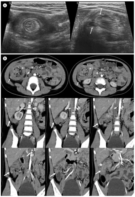
Fig. 1. A 35-month-old girl presenting with recurrent intussusception with acute appendicitis.
A. Initial ultrasonographic images. The transverse image shows a multicentric, hypo- and hyperechoic, bowel-in-bowel mass appearance. The longitudinal image showing the multilayered bowel, echogenic mesentery fat, and two lymph nodes (arrows) within the intussusception.
B. Axial and coronal contrast-enhanced CT images show the appendix (arrows) partially trapped within the intussusception (asterisks). Prominent appendiceal wall enhancement and periappendiceal fluid collection are observed.
A
B
https://doi.org/10.3348/jksr.2020.0100 711
Fig. 1. A 35-month-old girl present- ing with recurrent intussusception with acute appendicitis.
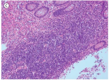
C. Microscopically, there is transmu- ral neutrophil infiltration at the ap- pendix (hematoxylin and eosin stain,
× 200).
Fig. 2. A 35-month-old girl presenting with ileocolic intussusception with inflamed appendicitis.
Axial and coronal contrast-enhanced CT images show the appendix (arrows) partially trapped within the intus- susception (asterisks). Enlargement of the appendix with prominent appendiceal wall enhancement and peri- appendiceal fat stranding are also noted.
C
nent appendiceal wall enhancement. There was also periappendiceal inflammation. (Fig. 2).
Ileocolic intussusception was treated through pneumatic reduction. The reduction was suc- cessful without complication. After reduction the patient had conservative treatment includ- ing pain control and Nil Per Os (NPO) under the close observation. The patient had not com- plained recurrent abdominal pain and discharged after five days.
DISCUSSION
Intussusception and acute appendicitis are two common and important conditions requir- ing adequate investigation at the proper time. They manifest with common symptoms, such as abdominal pain, vomiting, and/or irritability. Depending on the child's age at the time of diagnosis, differential diagnosis can be made; a child affected by intussusception tends to be younger than a child affected by acute appendicitis (4). Furthermore, there are some charac- teristic symptoms helpful for differentiation, such as currant jelly-like stool and cyclic irrita- bility in intussusception and right lower quadrant pain in acute appendicitis. In many cases, if one condition is suspected, then the other may be overlooked (4).
Intussusception occurs when one segment of bowel and its associated mesentery enter into an adjacent segment, and it is one of the most common causes of intestinal obstruction in children under 3 years of age (3, 4). Generally, the cause of intussusception is idiopathic and most cases (75–90%) in children are ileocolic or ileocecal (3, 4). It is rare in children younger than 4 years of age to have a lead point as the cause of intussusception (4). When a lead point is present, the most common causes are Meckel’s diverticulum and Henoch-Schönlein pur- pura (3-5). The various other causes include lymphoma, duplication, hemangioma, and in- testinal polyps (5-7).
In our cases, patients were younger than 4 years of age (both are 35 months), thus we ini- tially thought cases were idiopathic. In our first case, intussusception recurred twice and on the third recurrence, pneumatic reduction failed. After the failure of pneumatic reduction, we performed CT to determine any complications and to find a possible lead point that was not identified on previous US. On CT, we found the inflamed appendix partially entrapped within the intussusception. Our second case also showed intussusception with inflamed ap- pendix on initial CT. Intussusception with appendicitis, such as in our cases, is rare in both pediatric and adult patients and has been described in only a few case reports in the English literature (3, 4, 8).
Appendicitis is a common and major surgical emergency in childhood. Appendicitis is caused by obstruction of the lumen by fecalith or lymphoid hyperplasia (8, 9). When intus- susception and appendicitis coexist, as in our cases, it is confusing whether the inflamed ap- pendix is a lead point of the intussusception or the intussusception caused strangulation and inflammation of the appendix. In our cases inflamed appendix was not invaginated but par- tially entrapped into intussusception on CT. We presumed luminal obstruction by intussus- ception might be the cause of luminal obstruction of appendix. For the first case, multiple trial of pneumatic reduction was failed and operative reduction was done. During operative treatment for intussusception, appendectomy was also performed. However, for the second case, pneumatic reduction succeeded and symptoms were resolved. After reduction, the lu-
https://doi.org/10.3348/jksr.2020.0100 713 minal obstruction was removed and appendiceal inflammation might be improved. After three years of follow up periods, the second patient did not complain any symptoms suggest- ing acute appendicitis.
Although rare, the appendix can be a lead point for intussusception due to anatomical and pathologic factors. Anatomical factors include a fully mobile appendix, narrow meso- appendix, and poorly fixed or high caecum, whereas pathologic conditions include appen- dicular inflammation, calcified fecalith, polyp, tubulovillous adenoma, mucocele, mucinous cystadenoma, mucinous cystadenocarcinoma, carcinoid tumor, and mucosa-associated lym- phoma (3).
As the rarity of coexistence of intussusception and appendicitis, there is no established management yet. Treatment may vary depending on the patient's condition, and further re- search is needed. It is necessary to keep in mind that inflamed appendix and ileocolic intus- susception can coexist and various conditions involving appendix can be a lead point as well.
Author Contributions
Conceptualization, H.S.M.; data curation, L.H.J., W.Y.J., H.M.E.; investigation, H.S.M., L.H.J., W.Y.J., L.K.H., H.M.E.; methodology, H.S.M.; project administration, H.S.M.; supervision, H.S.M.; writing—
original draft, L.H.J., W.Y.J.; and writing—review & editing, W.J.Y., L.K.H.
Conflicts of Interest
The authors have no potential conflicts of interest to disclose.
Funding None
REFERENCES
1. McCollough M, Sharieff GQ. Abdominal surgical emergencies in infants and young children. Emerg Med Clin North Am 2003;21:909-935
2. Jiang J, Jiang B, Parashar U, Nguyen T, Bines J, Patel MM. Childhood intussusception: a literature review.
PLoS One 2013;8:e68482
3. Joshi SB, E H, Kinhal V, Kola SK, K SV. Intussusception in children with a pathological appendix acting as a
“lead point”-a series of 3 cases. J Clin Diagn Res 2015;9:PD03-04
4. Kee HM, Park JY, Yi DY, Lim IS. A case of intussusception with acute appendicitis. Pediatr Gastroenterol Hep- atol Nutr 2015;18:134-137
5. Navarro O, Daneman A. Intussusception. Part 3: diagnosis and management of those with an identifiable or predisposing cause and those that reduce spontaneously. Pediatr Radiol 2004;34:305-312; quiz 369 6. Guo WL, Hu ZC, Tan YL, Sheng M, Wang J. Risk factors for recurrent intussusception in children: a retrospec-
tive cohort study. BMJ Open 2017;7:e018604
7. Ksia A, Mosbahi S, Brahim MB, Sahnoun L, Haggui B, Youssef SB, et al. Recurrent intussusception in chil- dren and infants. Afr J Paediatr Surg 2013;10:299-301
8. Raja R, Sreeramulu PN, Srinivasan D, Singh R. A rare case: ileo-ileal intussusception with acute appendici- tis. Int Surg J 2018;5:2642-2645
9. Marjon L, Hull N, Thomas K. Concurrent acute appendicitis and ileocolic intussusception in a 1-year-old child. Radiol Case Rep 2018;13:655-657
염증성 충수돌기를 동반한 회결장 장중첩증: 2예 보고
이형주1 · 황숙민1* · 원영주1 · 우지영1 · 이건희2 · 홍민의3
소아 환자에서 장중첩증과 급성 충수돌기염은 흔한 응급 질환이다. 의심되는 증상이 있을 때 두 질환을 감별하는 것은 중요하다. 하지만, 드물게 두 질환이 한꺼번에 발생하는 경우도 있 다. 이에, 우리는 병적인 충수돌기를 동반한 장중첩증을 보였던 두 증례를 소개한다.
한림대학교 의과대학 강남성심병원 1영상의학과, 2소아청소년과, 3병리과