서 론
비만세포는 알레르기 반응을 일으키는 주요 효력 세포이며 전형 적으로는 알레르겐 특이 IgE 항체와의 교차연결에 의해 활성화된 다. 하지만, IgE 면역반응이 아닌 비특이적인 자극에 의해서도 활성 화될 수 있다.1 역사적으로 비만세포는 1878년 Paul Ehrlich에 의해 처음으로 기술되었으며 세포질에 존재하는 큰 과립이 특이하게 염 색되는 것을 발견하였다. 그는 이런 과립들이 주변 조직에 영양을 공급시킨다고 판단을 해서 Mastzellen (독일어로 ‘fatten'이라는 뜻) 으로 명명했다.2 현재는 비만세포가 호흡기, 소화기, 그리고 비뇨생 식기, 진피, 혈관을 둘러싸고 있는 조직에 위치하면서 인체에서 면 역 계통 유지 및 방어에 중요한 역할을 하는 것으로 알려져 있다.1-3 한편, 비만세포 활성에 따른 질환은 비만세포의 비정상적인 증식
및 침윤, 과도한 매개물질 분비로 발생할 수 있고 인체의 모든 기관 을 침범할 수 있으며 피부, 소화기, 심혈관, 호흡기, 그리고 신경계 등을 포함하는 이상 소견과 증상을 일으킬 수 있다.4 이번 종설에 서는 비만세포의 생화학적인 특징, 비만세포 활성 관련 질환, 그리 고 치료적인 접근에 대해 최근 경향을 중심으로 다루어 보도록 하 겠다.
비만세포의 특징과 활성
1. 비만세포의 생물학적인 특성
비만세포는 골수와 비장에 존재하는 미분화 CD34+ 줄기 세포 (pluripotent stem cell)에서 기원하고 골수성 경로를 따라 분화한 다. 비만세포의 주요 성장 인자는 줄기 세포 인자(stem cell factor)
Allergy Asthma Respir Dis 5(5):248-255, September 2017 https://doi.org/10.4168/aard.2017.5.5.248 REVIEW
Correspondence to: Hee-Kyoo Kim https://orcid.org/0000-0003-0692-1639 Department of Internal Medicine, Kosin University College of Medicine, 262 Gamcheon-ro, Seo-gu, Busan 49267, Korea
Tel: +82-51-990-6249, Fax: +82-51-990-6249, E-mail: naum67@naver.com Received: May 3, 2017 Revised: May 22, 2017 Accepted: June 23, 2017
© 2017 The Korean Academy of Pediatric Allergy and Respiratory Disease The Korean Academy of Asthma, Allergy and Clinical Immunology This is an Open Access article distributed under the terms of the Creative Commons Attribution Non-Commercial License
알레르기 염증에서의 비만세포 역할과 비만세포 관련 질환
김희규
고신대학교 의과대학 내과학교실
The roles of mast cells in allergic inflammation and mast cell-related disorders
Hee-Kyoo Kim
Department of Internal Medicine, Kosin University College of Medicine, Busan, Korea
Mast cells, which are major effector cells in allergic reactions, are found in the perivascular spaces of most tissues and contain pro-in- flammatory and vasoactive mediators. These mediators are released after IgE receptor cross-linking induced by allergens or other stimuli, including anaphylatoxins (C3a and C5a), aggregated IgG, certain drugs, venoms, and physical stimuli (pressure and tempera- ture changes), as well as cytokines and neuropeptides. The excess release of these mediators can cause variable allergic symptoms and signs, such as bronchospasm, itching, flushing, nausea, vomiting, diarrhea, abdominal pain, vascular instability, and anaphylaxis.
Furthermore, mast cell disorders may involve either excessive proliferation of mast cells or abnormal mast cell reactivity. Mast cell disorders can be broadly divided into 3 types: primary, secondary, and idiopathic. All of these disorders present with signs and symp- toms of mast cell activation and differ in severity and involvement of various organ systems. The best characterized primary disorder is mastocytosis. Systemic and cutaneous forms of the disease are well described. Secondary disorders include typical allergic dis- eases and some types of urticarial diseases. In this article, the biochemical characteristics of mast cells and the role of mast cells in al- lergic inflammation, as well as the classification, diagnosis, and management of mast cell-related disorders, will be reviewed.
(Allergy Asthma Respir Dis 2017;5:248-255)
Keywords: Mast cell, Allergy and immunology, Mastocytosis
2017-03-16 https://crossmark-cdn.crossref.org/widget/v2.0/logos/CROSSMARK_Color_square.svg
이며 이와 결합하는 비만세포의 표면 수용체는 tyrosine kinase kit (CD117)로 불린다. 미성숙 세포 또는 전구 세포는 여러 케모카인에 의해 표적기관으로 이동하게 되며 각 조직으로 이동한 후 주위 미 세환경의 영향을 받으며 성숙하게 된다. 일단 조직으로 이동한 비 만세포는 혈액을 통해 재순환하지 않는다.2-4
비만세포의 일차적인 역할은 선천면역과 획득면역의 주요 작동 반응을 담당하여 일부 세균이나 바이러스, 그리고 기생충에 대한 방어에 관여한다. 그 기전으로는 미생물 생성물에 대한 사이토카 인 분비, 보체 활성, 탈과립에 의한 세포 내 물질 분비, 그리고 다른 염증 작동 세포의 모집 등을 통해 이루어진다. 또한, 아섬유세포 증 식 및 콜라겐 합성 등을 자극하여 상처 회복과 반흔 형성을 통해 조 직재생에도 관여하는 것으로 알려져 있다.2
한편, 병적으로 활성화될 경우 각종 알레르기 질환을 비롯하여 비만세포증, 죽상동맥경화, 자가면역질환, 그리고 화상부위 염증 반응 악화 등을 유발할 수 있다.4
2. 비만세포 형태 및 주요 활성 매개물질
성숙한 비만세포는 정상적으로는 원형 또는 방추형의 단핵 형태 를 취하고 있다. 세포질 내에는 수많은 과립이 존재하고 있으며 내 부에 있는 히스타민이나 단백분해효소는 toluidin blue, alcian blue, aniline과 같은 염기성 청색 염색물질에 결합한 뒤 변색성(meta- chromatic) 세포질 염색으로 발현된다. 또한, IgE나 kit 등에 대한 세포막 수용체나 과립 단백분해효소(특히 tryptase)에 대한 단클 론성 항체도 비만세포를 확인하는 데 도움이 된다.
비만세포는 세포 내 단백분해효소 내용에 따라 2가지로 나뉜다.
전통적으로는 트립신 분해효소(tryptase)만 포함하고 있는 세포 (tryptase-positive mast cells, MCT)와 트립신 분해효소를 포함해 서 chymase, carboxypeptidase A, cathepsin G 등을 포함하고 있는
세포(tryptase- and chymase-positive mast cells, MCTC)로 나눈다 (Table 1). 전자는 상하기도 상피 등의 점막에 분포하며 후자는 결 합 조직, 피부, 기도 평활근 등에 분포하는 것으로 알려져 있다.5,6
비만세포가 활성화되면 다양한 염증 매개 물질을 생성 및 분비 한다. 이러한 매개 물질은 이미 생성된 매체(preformed), 새롭게 생 성된 매체(newly-synthesized mediators), 그리고 사이토카인(cy- tokine)과 케모카인(chemokine) 등 크게 3가지로 나뉜다. 이미 생 성된 매체는 세포 내 과립에 저장되어 있다가 자극에 의해 세포 외 부 환경으로 급속하게(수초에서 수분 이내) 분비된다. 히스타민, 단 백분해효소, chymase 그리고 proteoglycan 등이 대표적인 물질이 다. 새로 생성되는 매체는 세포막의 인지질을 이용해서 아라키돈 산이 생성되어 다양한 후속 물질들(eicosanoids)이 합성된다. 비만 세포에서 만들어진 다기능성 사이토카인과 케모카인들은 다른 염 증세포들을 모집하고 활성화시켜 염증을 증폭시킨다. 일부 tumor necrosis factor-alpha (TNF-α)는 과립 내에 저장되어 있다가 염증 초기에 분비되기도 한다. 대표적인 Th2 사이토카인으로 interleu- kin (IL)-4와 IL-5가 비만세포 활성 시기에 분비된다.5,6
3. 비만세포 활성 및 탈과립 과정(Fig. 1) 1) IgE 매개 활성
고전적인 비만세포 활성화는 high-affinity IgE receptor (FcεRI) 이라는 고친화성 IgE 수용체를 통해 일어난다. 이 수용체에 결합되 어 있는 IgE들이 알레르겐과 같은 항원에 결합되면 교차 연결을 통 해 활성화가 일어나며 이것은 탈과립의 강력한 자극원이 된다. Fcε RI은 하나의 α 및 β사슬과 두 개의 γ사슬로 구성되어 있는 4분자체 구조물이다. 이 수용체는 IL-4와 IL-13에 의해 세포막의 발현이 증 가된다. 또한, 혈중 IgE 농도가 높아지면 수용체의 수도 증가한다.
교차 연결된 FcεRI는 β 및 γ사슬의 세포 내 영역인 ITAM (im- munoreceptor tyrosine-based activation motif)의 인산화를 유도 하고 Lyn이라는 tyrosine kinase를 활성화시키고 이는 또 다른 ty- rosine kinase인 Syk의 활성화를 통해 mitogen-activated protein kinases 경로와 protein kinase C (PKC) 및 Ca의존성 경로를 활성 화시킨다. PKC 활성화는 myosin 경쇄(light chain)를 포함한 다양 한 단백질을 인산화 시켜 비만세포의 탈과립 및 세포 외 유출에 관 여하는 것으로 알려져 있다.6,7
2) 비IgE 매개 활성
IgE 비의존성 비만세포 활성 과정에 관여하는 물질로 protease, cytokine (stem cell factor, TNF-α, interferon-γ 등), complement, adenosine, Toll-like receptor 리간드, neuropeptide, 고장성 물질 등 이 있다. 또한, 비만세포 내 SHIP (src homology 2-containing inosi- tol phosphatase)와 같이 음성 조절(negative regulation)을 시키는 물질의 결핍으로 비만세포 활성 유발 역치를 낮출 수도 있다.8,9 Table 1. Characteristics of human mast cell subtypes according to protease
contents
Characteristic MCT MCTC
Protease content Tryptase Tryptase, chymase Proteoglycan content Heparin Heparin
Common location Epithelium Lamina propria, connective tissue, skin, airway smooth muscle Putative primary role Host defense Tissue repair
Relative LTC4 release High Skin: low Relative PGD2 release High Skin: high Cytokine profile IL-4, IL-13: low IL-4, IL-13: high
IL-5, IL-6: high
Activated by antigen Yes Yes
Activated by substance P No Yes
Responds to C5a No Yes
MCT, tryptase-only mast cell; MCTC, tryptase and chymase-containing mast cell; LTC4, leukotriene C4; PGD2, prostaglandin D2.
4. 비만세포와 선천면역
1990년 중반 한 연구에서 비만세포가 탈과립 없이 LPS에 대해 사 이토카인을 생산할 수 있다는 결과로 숙주 방어에도 일차적인 역 할을 할 수 있음을 알게 되었다. 이후 비만세포는 병원체에 대해 초 기 TNF 생산에 관여하며 톨유사수용체(Toll-like receptor), 보체, pathogen-associated mannose-binding protein, 면역글로불린 결 합 단백 등의 자극에 의해 세포가 활성화되어 숙주 방어에 필요한 주요 효력 세포를 모집하는 선천 면역의 기능을 담당하고 있는 것 으로 보인다.10 최근에는 IL-33가 비만세포의 ST2 수용체 자극을 통 해 비만세포의 주요 염증 사이토카인 생성, 활성화 및 증식, 그리고 알레르기 질환의 악화에 관여하는 것으로 밝혀져 그 경로 조절 과 정에 관심을 가지고 있다.2,11
주요 알레르기 질환에서의 비만세포의 역할
비만세포는 병태생리와 관련해서 천식과 여러 알레르기 질환 발 생에 주요 염증세포다. 이들 질환은 주로 IgE 매개된 반응으로 비만 세포의 역할은 병소 부위에서 즉시형 반응이 특징이다.12,13 최근에
는 후기 반응이나 만성 알레르기 반응에도 연관되어 있다는 증거 들이 있어 알레르기 염증 전반에 관여하고 있음을 알 수 있다.14 다 음은 주요 질환별로 비만세포의 역할에 대해 소개하고자 한다.
1. 천식
아토피 천식 환자에게 원인 알레르겐 노출 시 거의 대부분에서 10–20분 사이에 폐활량의 급속한 감소가 관찰되며 2시간에 걸쳐 서서히 회복되는데 이를 조기 천식 반응(early asthmatic reaction) 이라 하고 약 50%의 환자에서 4–6시간쯤에 폐활량이 다시 감소하 게 되는데 이를 후기 천식 반응(late asthmatic reaction)이라 한다.15 조기 반응에서 비만세포가 관여하는 증거로는 IgE 연관 매개물질 의 분비 속도, 기관지 내 국소 알레르겐 유발검사에서 기관폐포세 척액(bronchoalveolar lavage)의 비만세포 기원 트립신 분해 효소의 수분 내 급격한 증가,16 비만세포 탈과립 저해에도 관여하는 살부타 몰 노출 시 히스타민 수치의 감소,17 그리고 항IgE약제(omalizum- ab)를 3–4개월 투여하였을 때 조기 천식 반응의 현저한 감소18 등이 있다. 한편, 후기 천식 반응에서 기도 수축에 관여하는 비만세포의 역할을 규명하기는 다소 어려운 면이 있다. 왜냐하면, 관여 매개물 Fig. 1. IgE and non-IgE mediated mast cell activation and mediators. FcεRI, Fc epsilon RI (high-affinity IgE receptor); TLR, Toll-like receptor; GPCRs, G protein coupled re- ceptors; ATP, adenosine triphosphate; DAG, diacylglycerol; PKC, protein kinase C; PGD2, prostaglandin D2; PGE2, prostaglandin E2; LTB4, leukotriene B4; LTC4, leukotriene C4.
Food, aeroallergens, drugs, latex,
insect bites/stings etc.
Chemicals, drugs, food (additives), infection physical factors (cold, heat...), exercise, emotions, etc.
질이 대식 세포나 호산구 등에서 유래할 수 있기 때문이다. 하지만, 비만세포에서 합성된 염증 유발 leukotriene C4와 prostaglandin D2, 염증 사이토카인 등이 주변 염증세포를 활성화시키거나 모집 시키는 역할을 하여 기도의 지속적인 알레르기 염증을 유지시키는 역할을 하는 것으로 생각되고 있다.19
전형적인 알레르기 면역반응을 통하지 않고 비만세포의 탈과립 에 의해 천식 반응이 나타날 수 있는데 운동유발 천식이나 비알레 르기성 천식이 그 예가 될 수 있다. 찬공기 노출에 의한 물리적인 변 화, 운동에 의한 국소조직의 삼투압 변화는 비만세포의 탈과립을 유발할 수 있으나 이외의 기전에 의한 기도 수축도 관여할 것으로 추정하고 있다.20
만성 천식의 기도 염증과 기도 개형에도 비만세포가 관여하는 것 으로 알려져 있다. 트립신분해효소는 섬유아세포의 증식을 촉진시 켜 1형 콜라겐 합성을 자극할 수 있으며 히스타민과 함께 기도 평활 근 증식을 유도한다. 또한, 비만세포는 호산구와 다른 염증세포를 모집하거나 활성화시켜 염증, 조직 손상, 섬유화 그리고 개형에 관여 한다. 재미있는 사실은 폐기능이 정상인 무증상의 천식 환자에게도 히스타민이 기관폐포 세척액에서 관찰되고 있어 비만세포의 탈과 립이 소량이지만 지속적으로 이루어지고 있음을 제시하고 있다.21
비아토피성(내인성) 천식은 임상적으로는 중증 호산구성 특징 을 보이며 면역학적으로 아토피성 천식과 차이가 나지만 기본적인 비만세포 역할은 유사하다는 의견이 지배적이다. 그 개념에 대한 첫 번째 증거는 기관지 점막 내에서 비아토피성 천식에서도 비만세 포의 FcεRI표현이 동일하게 증가되어 있고 점막에 국한된 IgE의 합 성이 증가되었다는 보고가 있다.22,23 두 번째는 Th2 사이토카인인 IL-4, IL-5의 증가가 비아토피성 천식에서도 증가되어 있다.24 이는 IgE합성을 유도시키며 비만세포의 FcεRI표현도 증가시키게 된다.25 이러한 국소 알레르기성 반응을 유도하는 원인으로 전신 감작 없 이 확인되지 않은 외부 항원, 감염, 내부 항원 등이 제시되고 있다.
그 중에서 포도상구균(Staphylococcus aureus)에서 유래하는 균체 외독소(enterotoxin)가 초항원(superantigen)으로 작동하여 면역 자극원이 될 수 있다. 이 독소는 항원 제시 세포에 의한 항원 제시 과정 없이 T세포를 자극하고 이후 B세포의 항체 종류 변환(class- switching)을 통해 초항원에 대한 특이 IgE 생성을 유도할 수 있 다.26 외부 항원 없이 IgE 신호를 활성화시키는 또 한 가지 가능한 기 전은 FcεRI 자가항체(autoantibodies)의 생성이다. 또 다른 연구에 서는 상피세포 경로를 통해 알레르기 염증에 영향을 주는 IgG 자 가항체가 내인성 천식에서 발견되었다는 보고가 있다.27
2. 알레르기비염
계절성과 통년성 알레르기비염 모두에서 활성화된 비만세포 증 가에 대한 보고가 있으며 CD34+, 트립신분해효소 음성반응 비만 세포 증가에 대한 증거는 병소부위로 전구세포의 이동이 많다는
것을 제시한다. 비만세포 중에서도 점막형 세포(MCT)가 증가되어 있고 해당세포에서 IgE염색 비율이 증가되어 있었다는 증거가 있 다.28 알레르기비염에서도 점막형 세포에서 염증 사이토카인인 IL- 4, IL-5 등의 분비를 증가시켰고 국소 스테로이드에 의해 억제되었 다. 실험에서도 알레르기 비염의 후기 반응이 항IgE 치료를 하였을 때 현저하게 감소되어 즉시형 알레르기 염증 반응 전반에 비만세포 가 관여하고 있음을 보여주었다.29
3. 두드러기
두드러기는 유병 기간에 따라 급성과 만성으로 나눌 수 있으나 분류체계가 복잡하고 다양한 원인이 관여하고 있다. 대부분의 두 드러기는 비만세포의 탈과립과 연관이 있다고 보여지며, 말초 비만 세포 배양과 관련된 흥미로운 한 연구에서 두드러기 환자가 정상인 보다 비만세포의 기저 히스타민 분비가 더 높았다는 보고가 있다.30 또한, 만성 자발성 두드러기의 약 30%에서는 FcεRI 또는 IgE에 대 해 순환형 자가항체가 발견되고 있다.31 하지만, 이러한 경우 피부 이외 인체의 다른 부위에서는 비만세포 활성이 없어 만성두드러기 에서의 자가항체 존재 의미에 대해 더 연구가 필요하다.
4. 아토피피부염
아토피피부염의 병태생리에서 IgE의 역할은 아직 명확하지 않다.
그럼에도 불구하고 아토피피부염에서 비만세포는 주변 환경을 감 지하여 면역병태에 관여할 것으로 제시되고 있다. 그 한 예로서 정 상인의 피부에는 전 비만세포 중 조직형 비만세포(MCTC)가 90%
이상이지만 아토피피부염에서는 점막형 비만세포가 증가되어 있었 다.32 최근 한 동물 연구에서 비만세포가 피부장벽기능의 조절에 관 여하고 있음을 제시하고 있어 질환 발생에 한 축이 될 수 있겠다.33
5. 아나필락시스
전신 아나필락시스는 생명을 위협하는 중대하면서도 위급한 알 레르기 반응이다. 전형적으로는 IgE 매개된 반응으로 음식물을 비 롯하여 약물, 벌독, 운동 등에 의하지만 원인을 밝히기 힘든 경우도 있다. 한편, IgE가 매개되지 않는 비면역학적인 반응을 아나필락시 스양 반응이라고 부른다.3,9,34 최근 한 동물 실험에서 G 단백질 연결 수용체(G-protein-coupled receptor)의 일종인 Mrgprb2라는 비만 세포 특이 수용체가 발견되어 이는 염증 유발 펩타이드 또는 약물 에 의한 비IgE 매개반응을 중개하는 것으로 밝혀졌다.35
비만세포에 있는 트립신분해효소는 두 가지 형태 즉 α형과 β형이 있는데 전자는 평상시 분비 물질이라면 후자는 알레르기 염증반 응에서 급격하게 분비되는 물질이다.36 히스타민과 함께 β형 트립신 분해효소는 비만세포에서 함께 분비되나 증상 발생 후 히스타민은 5분 이내에 혈중 농도가 최고에 이르며 트립신분해효소는 15–120 분 사이에 최고 수준에 도달한다. 이는 트립신분해효소가 히스타
민보다 고분자이며 헤파린과 오랫동안 결합되어 있음으로 조직에 서 혈중으로 확산이 히스타민보다 느려지기 때문이다.37
최근에는 platelet-activating factor (PAF)가 아나필락시스 발생 과 비만세포의 활성에 관여함을 알게 되어 관심을 가지고 있다. 이 는 비만세포뿐만 아니라 호염기구, 호산구, 단핵구 등에서도 생성 된다. 생물학적인 작용으로 기관지 수축, 혈관 투과도 및 부종을 종 등 각종 염증 반응에 관여한다. 주목할만한 사항은 PAF가 다른 생 물학적 표식자에 비해 아나필락시스 반응의 중증도와 상관관계가 높다는 사실이다.38
비만세포증 및 비만세포 관련 질환
비만세포 질환은 비만세포의 활성화 및 매개 물질 분비에 의한 반복적인 증상을 보이는 형태를 보이며 비만세포의 증가 여부, 원인 물질 여부 등에 따라 일차성, 이차성 그리고 특발성으로 나뉜다. 39,40
1. 비만세포증
비만세포증은 한 가지 이상의 조직에 과도한 비만세포가 축척되 는 특징을 가진 질환이다. 비만세포증은 크게 피부국한형와 전신형 으로 나뉘며 각각은 임상적 형태, 병리 그리고 예후에 따라 몇 가지 아형으로 분류할 수 있다. 비만세포증은 드문 질환이며 성별로는 소아에서는 남아가 성인에서는 여성에서 다소 빈도가 높게 보고되 고 있다. 발생 연령은 신생아부터 성인에 이르기까지 가능하다.41
비만세포증은 피부 가려움증, 피부홍조, 오심, 구토, 설사, 복통 그리고 혈관 불안정성(vascular instability) 등의 다양한 임상적인 특징을 보일 수 있다. 특징적인 병리 특징은 피부, 위장관, 골수, 간, 비장, 그리고 임파선에서 비만세포의 과증식이 특징이며 흔히 혈액 학적인 이상과 동반된다. 비만세포의 비정상적인 증식은 성장 인자 중 하나인 stem cell factor에 반응하는 비만세포 수용체 KIT (CD117)의 변이와 관련이 있다.39-42
1) 피부국한형 비만세포증(cutaneous mastocytosis)
성인보다 소아 연령대에서 흔하다. 가장 흔한 증상은 가려움이다.
특징적인 피부 소견으로 주로 다발성의 과색소성 반점 또는 구진성
의 피부병변이 관찰되며 그 부위를 문지르거나 긁으면 두드러기 형 태를 보인다(Darier's Sign). 이는 주요 진단적인 기준에도 속한다. 소 아 환자들은 청소년이 되면서 소실 또는 호전되는 경우가 많다. 임 상상에 따라 색소성 담마진(urticaria pigmentosa/maculopapular cutaneous mastocytosis), 미만형(diffuse cutaneous mastocytosis), 그리고 피부 비만세포종(cutaneous mastocytoma), 총 3가지 아형 으로 분류될 수 있으며 그 중 색소성 담마진이 가장 흔하다. 일반적 으로 피부국한형은 전신 증상은 잘 나타나지 않으나 때때로 위장관 경련, 아나필락시스와 같은 증상이 발생할 수 있다. 이런 증상은 육 체적인 자극, 스트레스, 그리고 운동 등에 의해 촉발될 수 있다.
약 60%–80% 환자들에게서 기능획득(gain-of-function) KIT 변 이(mutation)와 관련이 있다. 소아에서는 간비대, 림프비대, 또는 말 초혈액 이상이 없다면 전신 비만세포증을 진단하기 위해 골수 검 사를 시행할 필요는 없다.
진단은 임상적인 증상과 피부조직검사로 할 수 있으며 전형적인 피부의 반점과 다리에 징후 양성인 경우 임상 진단이 가능하다. 피 부조직검사상 방추형 모양과 이염색과립을 가지고 있는 비만세포 침윤이 있으나 다른 염증세포들의 축적이 없는 것이 전형적인 특징 이다.43
2) 전신 비만세포증(systemic mastocytosis)
전신 비만세포증 환자들은 흔히 두드러기, 혈관부종, 안면 홍조, 삽화적인 저혈압, 설사, 두통 등을 경험하며, 이러한 증상은 술, 아 스피린, 곤충교상, 감염, 또는 조영제 노출에 의해 유발될 수 있다.
하지만, 비만세포증 환자들에서 세균, 진균 또는 바이러스 감염이 증가되지는 않는다. 약 80% 환자에게서 색소성 담마진이 동반된다.
주요 검사 소견으로 증상 발생이 없는 상태에서도 혈청 총 트립 신 분해 효소의 증가(정상 <11.4 ng/mL)가 있을 경우 강하게 의심 할 수 있다. 전신 비만세포증 환자에서는 >20 ng/mL가 전형적이 라고 볼 수 있다. 소변에서 히스타민 대사 물질(11 β-prosta- glandinF2α 또는 N-methyl histamine)증가를 보일 수 있다. 성인 90% 이상에서 c-kit 유전자에서 exon 17번의 점 돌연변이(point mutation, D816V)를 가지고 있다.39-41
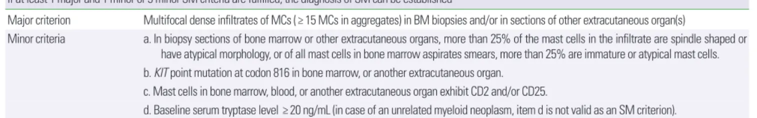
진단 기준은 World Health Organization 분류에 따라 1차 기준
Table 2. Diagnostic criteria of SM by World Health Organization
If at least 1 major and 1 minor or 3 minor SM criteria are fulfilled, the diagnosis of SM can be established
Major criterion Multifocal dense infiltrates of MCs (≥ 15 MCs in aggregates) in BM biopsies and/or in sections of other extracutaneous organ(s)
Minor criteria a. In biopsy sections of bone marrow or other extracutaneous organs, more than 25% of the mast cells in the infiltrate are spindle shaped or have atypical morphology, or of all mast cells in bone marrow aspirates smears, more than 25% are immature or atypical mast cells.
b. KIT point mutation at codon 816 in bone marrow, or another extracutaneous organ.
c. Mast cells in bone marrow, blood, or another extracutaneous organ exhibit CD2 and/or CD25.
d. Baseline serum tryptase level ≥ 20 ng/mL (in case of an unrelated myeloid neoplasm, item d is not valid as an SM criterion).
SM, systemic mastocytosis; MC, mast cell; BM, bone marrow.
1개 및 2차 기준 1개를 만족하거나 2차 기준 3개 이상을 만족하면 전신 비만세포증으로 진단할 수 있다(Table 2). 아형으로는 indo- lent systemic mastocytosis (ISM), smoldering systemic mastocy- tosis (SSM), systemic mastocytosis with an associated hematologic (non-MC lineage) neoplasm (SM-AHN), aggressive systemic mas- tocytosis (ASM), and mast cell leukemia (MCL)로 나뉜다.44
감별해야 할 질환으로 단순 두드러기, 홍조의 경우 카르시노이드 증후군, 갈색세포종, 다른 원인에 의한 아나필락시스, 그리고 소화 기 증상으로는 염증성 장질환, 자극성 장 증후군, 흡수 장애 증후 군, 졸린거-엘리슨 증후군 및 골수 증식성 질환 등이 있다.
3) 진단적인 접근
비만세포증이 의심되는 모든 환자에서 총 혈청 트립신 분해효소 를 측정해야 한다. 증상이 있을 때 그 수치가 올라가 있다면 증상이 소실된 후 적어도 24시간 이내에 재측정을 해서 올라가 있는지를 확인해야 한다. 그렇지 않다면 단순 아나필락시스일 가능성이 높
다.39,44 일부 연구에서는 혈장 헤파린 농도 측정이 전신 비만세포 활
성 질환 확인에 도움을 줄 수 있다는 보고도 있어 보조적인 수단이 될 수 있다.45
4) 국내 사례 보고
피부 국한형의 경우 소아의 경우 보고가 드물지 않으나46 성인에 서는 적으며 대부분 색소성 담마진 형태이다.47,48 전신형은 4예가 보 고되어 있으며 대부분 백혈병이나 악성 종양과 동반되어 있다.49,50 이외에 위장관에 국한된 비만세포증 1예가 보고되어 있는데 만성 설사로 내원한 70세 남자에서 위장점막 조직 검사를 통해 진단이 되었다.51
5) 예후
예후는 피부국한형 비만세포증 환자가 가장 좋으며 전신형 중에 서는 비활동성(ISM) 아형이 그 뒤를 따른다. 하지만, 비비만세포 계 통성(non-MC lineage) 질환, 공격성(ASM) 비만세포증 및 비만세 포 백혈병(MCL)은 예후가 좋지 않다.39,52-54
2. 단일클론형 비만세포 활성화 증후군
임상 증상은 전신성 비만세포증과 유사하다. 하지만, 색소성 담마 진이 없으며 기저 트립신 분해효소가 정상이거나 약간 증가된 소견 을 보인다. 골수검사에서 c-KIT 점변이(주로 D816V) 및 CD25의 과 도한 표현을 관찰할 수 있으나 비만세포의 다발성 응집은 보이지 않 는다. 전신 비만세포증 진단의 2차 기준은 1–2개 정도 만족한다.40,41
3. 이차성 및 특발성 비만세포 관련 질환
이차성 비만세포 활성 질환은 비만세포의 양이나 기능은 정상이
며 알레르겐, 자가항체, 약물, 또는 보체 활성 물질에 의해 비만세포 가 반응하는 질환이며 IgE 매개 알레르기 질환, 일부 두드러기 등 이 이에 속한다. 특발성은 원인을 밝힐 수 없을 때 붙여지며 특발성 두드러기, 특발성 혈관부종, 특발성 아나필락시스 등이 이 범주에 속한다. 새로운 유전 결핍이 밝혀지면 일차성 비만세포 질환으로 분류될 가능성도 있다.39-41
비만세포 활성 질환의 치료
비만세포 활성을 유발하는 물질이나 환경을 회피하는 것이 일차 적으로 필요하다. 급성 아나필락시스가 발생하면 즉시 에피네프린 을 근주한다. 이어 항히스타민제 및 당류코르티코이드를 정주한다.
증상완화, 비만세포 활성 저해 또는 증식 억제에 작용하는 치료가 아래와 같이 시도되고 있다.
1. 증상 조절 치료
흔한 증상에 대한 완화 치료제로는 비진정 지속성 H1 수용체 길 항제(로라타딘, 세티리진, 펙소페나딘 등)가 추천된다. Cyprohep- tadine은 H1 및 seretonin 수용체 복합 길항체로 설사, 홍조 그리고 두통이 있는 환자에게 고려될 수 있다. H2 수용체 길항제인 fa- motidine이나 ranitidine은 속쓰림, 오심, 복부 경련, 그리고 설사 등의 소화기 증상이 있는 환자들에게 도움이 될 수 있다. 류코트리 엔 조절제는 H1 및 H2수용체 길항제와 병합 사용 시 도움이 될 수 있다. 아세틸살리실릭산은 홍조와 혈관부종이 있는 일부 환자에서 부작용이 없다면 유용할 수 있다. 삼환계 항우울제는 H1 수용체 길 항과 비만세포 활성을 저해할 수 있는 성질이 있다. 솔라렌(pso- ralen) 투여와 동시에 UV (ultraviolet) A 또는 협대역 UVB를 조사 하는 광화학요법은 소양감 감소 및 일시적인 피부 병변을 줄이는데 도움이 될 수 있다. 국소 스테로이드는 일반적으로 권고되지 않는 다.39,43,44,55
2. Anti-IgE 치료를 포함한 비만세포 활성 저해 약물
Ketotifen과 disodium cromoglycate는 비만세포를 활성화 차단 하는 효과가 있다.53,56 항 IgE 단클론성 항체는 천식, 만성 두드러기 에 주로 사용하지만,57,58 재발성 아나필락시스 삽화를 방지할 수 있 었던 증례들이 보고되고 있다.59,60
3. 비만세포 증식 및 생존 방해 약물
미만세포 항상성에 중심 역할을 하는 줄기세포인자(stem cell factor) 수용체인 kit의 저해제인 imatinib이 KIT D816V변이가 없 는 전신 비만세포증 환자 소수에서 비만세포를 감소시켰다는 보고 가 있다.61 또한, 최근 중증 난치성 천식에서 imatinib이 기도과민성, 비만세포 수, 그리고 트립신 분해효소 분비를 감소시킨 보고가 있
으며 이는 비만세포가 중증 천식의 병태생물학적 과정에 관여하고 있음을 제시하는 증거이기도 하다.62 또한 PKC412라는 multiki- nase inhibitor가 in vitro 실험에서 IgE 의존 비만세포의 활성뿐만 아니라 성장과 생존을 억제시켰다.63 골수 이식 시도 및 몇 가지 표 적 치료제도 개발되고 있으나 아직은 그 성과가 미미하다.44,52,55
결 론
비만세포는 기본적으로 조직의 항상성 및 손상된 조직의 수복에 관여한다. 하지만, 비정상적인 활성에서는 다양한 생물화학적인 물 질을 분비하여 알레르기 증상을 일으키는 병태생리와 관련이 있 다. 과거에는 비만세포가 조기 염증반응에 관여할 것으로 생각하였 지만 현재는 후기반응 및 만성경과에도 깊이 관여하고 있다는 증거 들이 있어 알레르기 염증 전반에 대한 비만세포의 역할이 강조되 고 있다. 한편, 비만세포 활성 질환이 드물지 않을 것으로 보이나 아 직 그 인식이 부족하여 전형적인 알레르기 질환 이외에는 진단되는 경우가 적은 편이다. 특히 중증의 알레르기 증상을 보이며 다발성 전신 증상을 보일 때 비만세포증에 대한 의심을 해 볼 필요가 있다.
향후 과제로는 비만세포 활성에 대한 보다 실용적이고 신뢰할만 한 생물학적 표식자를 발견할 필요가 있으며 중증 질환에 대한 효 과적인 치료제가 개발되어야 하겠다.
REFERENCES
1. Rivera J, Fierro NA, Olivera A, Suzuki R. New insights on mast cell acti- vation via the high affinity receptor for IgE. Adv Immunol 2008;98:85- 2. Moon TC, St Laurent CD, Morris KE, Marcet C, Yoshimura T, Sekar Y, et 120.
al. Advances in mast cell biology: new understanding of heterogeneity and function. Mucosal Immunol 2010;3:111-28.
3. Blank U, Falcone FH, Nilsson G. The history of mast cell and basophil re- search - some lessons learnt from the last century. Allergy 2013;68:1093- 4. Boyce JA. The biology of the mast cell. Allergy Asthma Proc 2004;25:27-101.
5. Kawakami T, Galli SJ. Regulation of mast-cell and basophil function and 30.
survival by IgE. Nat Rev Immunol 2002;2:773-86.
6. Stone KD, Prussin C, Metcalfe DD. IgE, mast cells, basophils, and eosino- phils. J Allergy Clin Immunol 2010;125(2 Suppl 2):S73-80.
7. Theoharides TC, Alysandratos KD, Angelidou A, Delivanis DA, Sis- manopoulos N, Zhang B, et al. Mast cells and inflammation. Biochim Biophys Acta 2012;1822:21-33.
8. Boyce JA. Mast cells: beyond IgE. J Allergy Clin Immunol 2003;111:24- 9. Yu Y, Blokhuis BR, Garssen J, Redegeld FA. Non-IgE mediated mast cell 32.
activation. Eur J Pharmacol 2016;778:33-43.
10. Marshall JS, Jawdat DM. Mast cells in innate immunity. J Allergy Clin Immunol 2004;114:21-7.
11. Saluja R, Khan M, Church MK, Maurer M. The role of IL-33 and mast cells in allergy and inflammation. Clin Transl Allergy 2015;5:33.
12. Modena BD, Dazy K, White AA. Emerging concepts: mast cell involve- ment in allergic diseases. Transl Res 2016;174:98-121.
13. Amin K. The role of mast cells in allergic inflammation. Respir Med 2012;106:9-14.
14. Serrano-Candelas E, Martinez-Aranguren R, Valero A, Bartra J, Gasta- minza G, Goikoetxea MJ, et al. Comparable actions of omalizumab on mast cells and basophils. Clin Exp Allergy 2016;46:92-102.
15. Robinson DS. The role of the mast cell in asthma: induction of airway hy- perresponsiveness by interaction with smooth muscle? J Allergy Clin Immunol 2004;114:58-65.
16. Wenzel SE, Fowler AA 3rd, Schwartz LB. Activation of pulmonary mast cells by bronchoalveolar allergen challenge. In vivo release of histamine and tryptase in atopic subjects with and without asthma. Am Rev Respir Dis 1988;137:1002-8.
17. Church MK, Hiroi J. Inhibition of IgE-dependent histamine release from human dispersed lung mast cells by anti-allergic drugs and salbutamol.
Br J Pharmacol 1987;90:421-9.
18. Boulet LP, Chapman KR, Côté J, Kalra S, Bhagat R, Swystun VA, et al. In- hibitory effects of an anti-IgE antibody E25 on allergen-induced early asthmatic response. Am J Respir Crit Care Med 1997;155:1835-40.
19. Liu MC, Hubbard WC, Proud D, Stealey BA, Galli SJ, Kagey-Sobotka A, et al. Immediate and late inflammatory responses to ragweed antigen challenge of the peripheral airways in allergic asthmatics. Cellular, medi- ator, and permeability changes. Am Rev Respir Dis 1991;144:51-8.
20. Bradding P, Feather IH, Howarth PH, Mueller R, Roberts JA, Britten K, et al. Interleukin 4 is localized to and released by human mast cells. J Exp Med 1992;176:1381-6.
21. Ying S, Durham SR, Corrigan CJ, Hamid Q, Kay AB. Phenotype of cells expressing mRNA for TH2-type (interleukin 4 and interleukin 5) and TH1-type (interleukin 2 and interferon gamma) cytokines in bronchoal- veolar lavage and bronchial biopsies from atopic asthmatic and normal control subjects. Am J Respir Cell Mol Biol 1995;12:477-87.
22. Humbert M, Grant JA, Taborda-Barata L, Durham SR, Pfister R, Menz G, et al. High-affinity IgE receptor (FcepsilonRI)-bearing cells in bronchial biopsies from atopic and nonatopic asthma. Am J Respir Crit Care Med 1996;153(6 Pt 1):1931-7.
23. Ying S, Humbert M, Meng Q, Pfister R, Menz G, Gould HJ, et al. Local expression of epsilon germline gene transcripts and RNA for the epsilon heavy chain of IgE in the bronchial mucosa in atopic and nonatopic asth- ma. J Allergy Clin Immunol 2001;107:686-92.
24. Humbert M, Durham SR, Ying S, Kimmitt P, Barkans J, Assoufi B, et al.
IL-4 and IL-5 mRNA and protein in bronchial biopsies from patients with atopic and nonatopic asthma: evidence against "intrinsic" asthma being a distinct immunopathologic entity. Am J Respir Crit Care Med 1996;154:1497-504.
25. Del Prete G, Maggi E, Parronchi P, Chrétien I, Tiri A, Macchia D, et al.
IL-4 is an essential factor for the IgE synthesis induced in vitro by human T cell clones and their supernatants. J Immunol 1988;140:4193-8.
26. Barnes PJ. Intrinsic asthma: not so different from allergic asthma but driven by superantigens? Clin Exp Allergy 2009;39:1145-51.
27. Kwon B, Lee HA, Choi GS, Ye YM, Nahm DH, Park HS. Increased IgG antibody-induced cytotoxicity against airway epithelial cells in patients with nonallergic asthma. J Clin Immunol 2009;29:517-23.
28. Bentley AM, Jacobson MR, Cumberworth V, Barkans JR, Moqbel R, Schwartz LB, et al. Immunohistology of the nasal mucosa in seasonal al- lergic rhinitis: increases in activated eosinophils and epithelial mast cells.
J Allergy Clin Immunol 1992;89:877-83.
29. Hanf G, Noga O, O'Connor A, Kunkel G. Omalizumab inhibits allergen challenge-induced nasal response. Eur Respir J 2004;23:414-8.
30. Saini SS, Paterniti M, Vasagar K, Gibbons SP Jr, Sterba PM, Vonakis BM.
Cultured peripheral blood mast cells from chronic idiopathic urticaria patients spontaneously degranulate upon IgE sensitization: relationship to expression of Syk and SHIP-2. Clin Immunol 2009;132:342-8.
31. Sabroe RA, Greaves MW. Chronic idiopathic urticaria with functional autoantibodies: 12 years on. Br J Dermatol 2006;154:813-9.
32. Järvikallio A, Naukkarinen A, Harvima IT, Aalto ML, Horsmanheimo M.
Quantitative analysis of tryptase- and chymase-containing mast cells in atopic dermatitis and nummular eczema. Br J Dermatol 1997;136:871-7.
33. Sehra S, Serezani APM, Ocaña JA, Travers JB, Kaplan MH. Mast cells regulate epidermal barrier function and the development of allergic skin inflammation. J Invest Dermatol 2016;136:1429-37.
34. Schwartz LB. Diagnostic value of tryptase in anaphylaxis and mastocyto- sis. Immunol Allergy Clin North Am 2006;26:451-63.
35. McNeil BD, Pundir P, Meeker S, Han L, Undem BJ, Kulka M, et al. Identi- fication of a mast-cell-specific receptor crucial for pseudo-allergic drug reactions. Nature 2015;519:237-41.
36. Schwartz LB, Sakai K, Bradford TR, Ren S, Zweiman B, Worobec AS, et al. The alpha form of human tryptase is the predominant type present in blood at baseline in normal subjects and is elevated in those with system- ic mastocytosis. J Clin Invest 1995;96:2702-10.
37. Schwartz LB, Yunginger JW, Miller J, Bokhari R, Dull D. Time course of appearance and disappearance of human mast cell tryptase in the circu- lation after anaphylaxis. J Clin Invest 1989;83:1551-5.
38. Pałgan K, Bartuzi Z. Platelet activating factor in allergies. Int J Immuno- pathol Pharmacol 2015;28:584-9.
39. Theoharides TC, Valent P, Akin C. Mast cells, mastocytosis, and related disorders. N Engl J Med 2015;373:1885-6.
40. Valent P. Mast cell activation syndromes: definition and classification. Al- lergy 2013;68:417-24.
41. Valent P, Akin C, Arock M, Brockow K, Butterfield JH, Carter MC, et al.
Definitions, criteria and global classification of mast cell disorders with special reference to mast cell activation syndromes: a consensus proposal.
Int Arch Allergy Immunol 2012;157:215-25.
42. Frieri M, Patel R, Celestin J. Mast cell activation syndrome: a review. Curr Allergy Asthma Rep 2013;13:27-32.
43. Hartmann K, Escribano L, Grattan C, Brockow K, Carter MC, Alvarez- Twose I, et al. Cutaneous manifestations in patients with mastocytosis:
Consensus report of the European Competence Network on Mastocyto- sis; the American Academy of Allergy, Asthma & Immunology; and the European Academy of Allergology and Clinical Immunology. J Allergy Clin Immunol 2016;137:35-45.
44. Valent P, Akin C, Metcalfe DD. Mastocytosis: 2016 updated WHO classi- fication and novel emerging treatment concepts. Blood 2017;129:1420-7.
45. Vysniauskaite M, Hertfelder HJ, Oldenburg J, Dreßen P, Brettner S, Homann J, et al. Determination of plasma heparin level improves identi- fication of systemic mast cell activation disease. PLoS One 2015;10:
e0124912.
46. Chang SE, Kang SK, Jee MS, Choi JH, Sung KJ, Moon KC, et al. Clinico- pathological study of 30 Cases of Cutaneous Mastocytosis. Korean J Der- matol 2002;40:501-5.
47. Park CH, Kim KM, Kim HG, Son SW, Kim SH, Park HW, et al. A case of cutaneous mastocytosis with urticaria developed after hot bath. Korean J Asthma Allergy Clin Immunol 2005;25:328-32.
48. Lee EH, Kim MR, Kang TW, Kim SC. Diffuse cutaneous mastocytosis with generalized bullae. Ann Dermatol 2010;22:77-80.
49. Youk J, Koh Y, Kim JW, Kim DY, Park H, Jung WJ, et al. A scientific treat- ment approach for acute mast cell leukemia: using a strategy based on next-generation sequencing data. Blood Res 2016;51:17-22.
50. Bae MH, Kim HK, Park CJ, Seo EJ, Park SH, Cho YU, et al. A case of sys- temic mastocytosis associated with acute myeloid leukemia terminating as aleukemic mast cell leukemia after allogeneic hematopoietic stem cell transplantation. Ann Lab Med 2013;33:125-9.
51. Seo H, Park SH, Byeon JS, Woo CG, Hong SM, Chang K, et al. Chronic intractable diarrhea caused by gastrointestinal mastocytosis. Intest Res 2016;14:280-4.
52. Tremblay D, Carreau N, Kremyanskaya M, Mascarenhas J. Systemic mas- tocytosis: clinical update and future directions. Clin Lymphoma Myelo- ma Leuk 2015;15:728-38.
53. Hermans MA, Rietveld MJ, van Laar JA, Dalm VA, Verburg M, Pasmans SG, et al. Systemic mastocytosis: a cohort study on clinical characteristics of 136 patients in a large tertiary centre. Eur J Intern Med 2016;30:25-30.
54. Lim KH, Tefferi A, Lasho TL, Finke C, Patnaik M, Butterfield JH, et al.
Systemic mastocytosis in 342 consecutive adults: survival studies and prognostic factors. Blood 2009;113:5727-36.
55. Molderings GJ, Brettner S, Homann J, Afrin LB. ast cell activation disease:
a concise practical guide for diagnostic workup and therapeutic options.
J Hematol Oncol 2011;4:10.
56. Zhang T, Finn DF, Barlow JW, Walsh JJ. Mast cell stabilisers. Eur J Phar- macol 2016;778:158-68.
57. Chang TW, Chen C, Lin CJ, Metz M, Church MK, Maurer M. The poten- tial pharmacologic mechanisms of omalizumab in patients with chronic spontaneous urticaria. J Allergy Clin Immunol 2015;135:337-42.
58. Metz M, Ohanyan T, Church MK, Maurer M. Omalizumab is an effective and rapidly acting therapy in difficult-to-treat chronic urticaria: a retro- spective clinical analysis. J Dermatol Sci 2014;73:57-62.
59. Bell MC, Jackson DJ. Prevention of anaphylaxis related to mast cell acti- vation syndrome with omalizumab. Ann Allergy Asthma Immunol 2012;108:383-4.
60. Jagdis A, Vadas P. Omalizumab effectively prevents recurrent refractory anaphylaxis in a patient with monoclonal mast cell activation syndrome.
Ann Allergy Asthma Immunol 2014;113:115-6.
61. Akin C, Fumo G, Yavuz AS, Lipsky PE, Neckers L, Metcalfe DD. A novel form of mastocytosis associated with a transmembrane c-kit mutation and response to imatinib. Blood 2004;103:3222-5.
62. Cahill KN, Katz HR, Cui J, Lai J, Kazani S, Crosby-Thompson A, et al.
KIT inhibition by imatinib in patients with severe refractory asthma. N Engl J Med 2017;376:1911-20.
63. Krauth MT, Mirkina I, Herrmann H, Baumgartner C, Kneidinger M, Va- lent P. Midostaurin (PKC412) inhibits immunoglobulin E-dependent ac- tivation and mediator release in human blood basophils and mast cells.
Clin Exp Allergy 2009;39:1711-20.