Copyright © 2021. Anatomy & Cell Biology
Introduction
The posterior knee holds a high risk for surgical access due to the dense field of neurovascular structures located in and around the popliteal fossa [1]. Because of the inherent
high risk and unfamiliar relevant surgical anatomy, many surgeons avoid this region altogether [1, 2]. The main limita- tion to a direct open posterior approach to the knee is the risk posed to the neurovascular structures [1]. An in-depth knowledge of the surgical anatomy of these neurovascular structures will assist in minimizing the risk of neurovascular damage.
Indications for direct posterior surgical access to the knee include posterior tibial plateau fractures, tibial bony avul- sions with associated posterior cruciate ligament injury and repair of the popliteal neurovascular structures [2]. There are several potential pathological conditions, such as cysts or
Corresponding author:
Erik Hohmann
School of Medicine, Faculty of Health Sciences, University of Pretoria, Pretoria 0001, South Africa
E-mail: [email protected]
Defining the popliteal fossa by bony
landmarks and mapping of the courses of the neurovascular structures for application in popliteal fossa surgery
Kelsi Greenwood
1, Reinette van Zyl
1, Natalie Keough
1,2, Erik Hohmann
1,3,41Department of Anatomy, School of Medicine, Faculty of Health Sciences, University of Pretoria, Pretoria, South Africa, 2Department of Anatomy and Cellular Biology, College of Medicine and Health Sciences, Khalifa University, Abu Dhabi, 3Department of Orthopaedic Surgery and Sports Medicine, Burjeel Hospital for Advanced Surgery, Dubai, United Arab Emirates, 4Medical School, University of Pretoria, Pretoria, South Africa
Abstract: Surgical access to the posterior knee poses a high-risk for neurovascular damage. The study aimed to define the popliteal fossa by reliable bony landmarks and comprehensively mapping the neurovascular structures for application in posterior knee surgery. Forty-five (20 male, 25 female) embalmed adult cadaveric knees were included. The position of the small saphenous vein (SSV), medial cutaneous sural nerve (MCSN) and lateral cutaneous sural nerv (LCSN), tibial nerve (TN) and common fibular nerve (CFN) nerves, and popliteal vein (PV) and popliteal artery (PA) were determined in relation to either medial (MFE) or lateral (LFE) femoral epicondyles, medial (MTC) and lateral (LTC) tibial condyles and the midpoint between the MFE and MTC and LFEF and LTC. The distance between the MFE and the PA, PV, TN, MCSN, and SSV was 38.4±12.1 mm, 38.4±12.9 mm, 39.4±10.2 mm, 39.2±14.0 mm and 37.6±12.5 mm respectively for males and 34.6±4.9 mm, 32.8±5.6 mm and 38.0±8.1 mm 38.8±10.1 mm and 37.9±8.2 mm respectively for females. The distance between LFE and the CFN and LCSN was 13.4±8.2 mm and 24.9±7.3 mm respectively for males and 8.4±9.1 mm and 18.4±10.4 mm respectively in females. This study defined the popliteal fossa by reliable bony landmarks and provided a comprehensive map of the neurovascular structures and will help to avoid injuries to the important neurovascular structures.
Key words: Knee, Tibial nerve, Popliteal artery, Anatomy
Received July 13, 2020; Revised September 1, 2020; Accepted September 10, 2020
mass formation, including neoplasms and abscesses that re- quire surgical treatment which may affect the neurovascular structures in the popliteal fossa [3]. To address these condi- tions, a posterior surgical approach to the knee is required.
This approach is challenging and requires detailed knowl- edge of the neurovascular structures, as well as the possible variations of the posterior knee [1].
For surgical approaches to the posterior knee, it would be beneficial if surgeons could use reliable anatomic landmarks, such as the femoral epicondyle, to measure the distances be- tween the structures that are potentially endangered by sur- gery, in particular the popliteal artery and vein and the tibial and common fibular nerves. This would reduce the risk of permanent damage, potentially make surgical planning eas- ier and allow safe and effective surgery. Hence, the purpose of this study was to define the popliteal fossa by reliable bony landmarks and provide a comprehensive map of the neuro- vascular structures for posterior popliteal fossa knee surgery.
Material and Methods
This study was designed as a cross-sectional, quantita- tive study. Formalin preserved cadaveric specimens were obtained from the Department of Anatomy, University of Pretoria, Pretoria, South Africa (Ethics number: 82/2019) (blinded for review). These specimens were donated and used for the anatomy courses for medical and dental students.
Prior to these courses, specimens were dissected by one in- dependent researcher. These embalmed cadavers are used for training and research and comply with all the requirements set out in the National Health Act 63 of 2003. Knees were
only included if there was no obvious macroscopic damage, no observed previous trauma with signs of corrective sur- gery, and no visible pathology or fractures of the knee joint.
Knees were excluded if evidence existed of previous surgery to the popliteal fossa including vascular repair or bypass sur- gery, vein stripping or nerve repair. Specimens with Baker’s cysts and severe degenerative osteoarthritis with osteophyte formation and any macroscopic neurovascular were also excluded. The sample of the study consisted of 45 adult em- balmed cadaveric knees. The mean age of the cadavers was 65.8±17.4 years. Of the sample of 45 adult knees, 20 were male and 25 were female. There were 28 left knees used and 17 right knees.
Dissection technique and measurements
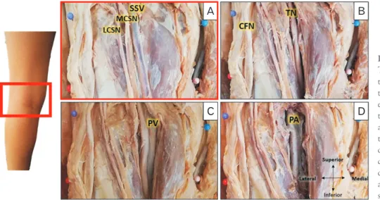
A vertical incision was made in the skin of the posterior knee extending from the mid-thigh to a point just distal to the knee joint. The skin was reflected laterally to expose the defined borders of the popliteal fossa. Soft tissue, including fascia and subcutaneous fat was removed around the distal femur and proximal tibia both medially and laterally. Four bony landmarks were palpated and a pin was placed at each of these landmarks, namely: the medial most point of the medial epicondyle of the femur (MFE), the medial most point of the medial condyle of the tibia (MTC), the lateral most point of the lateral epicondyle of the femur (LEF) and the lat- eral most point of the lateral condyle of the tibia (LTC). A pin was used to mark all four bony landmarks (Fig. 1). The neuro- vascular structures (the small saphenous vein [SSV], medial cutaneous sural nerve [MCSN], lateral cutaneous sural nerve [LCSN], tibial nerve [TN], common fibular nerve [CFN],
A B
C D
Inferior
Fig. 1. The dissected left popliteal fossa.
The superficial to deep dissection of the posterior cadaveric knee to expose the neurovascular structures. (A) The exposure of the LCSN, MCSN and the SSV. (B) The exposure of the CFN and TN. (C) The dissection to expose the PV. (D) The dissected PA. CFN, common fibular nerve; LCSN, lateral cutaneous sural nerve; MCSN, medial cutaneous sural nerve; PA, popliteal artery; PV, popliteal vein; SSV, small saphenous vein; TN, tibial nerve.
popliteal vein [PV] and popliteal artery [PA]) were exposed from superficial to deep, with a photograph taken at each level to capture the relevant structures in situ. Great care was taken to ensure that both distance and angles of each photo- graph were identical by using a tripod. Each photograph in- cluded a scale and the photographs were taken perpendicular to the longitudinal axis of the femur and parallel to the floor (Fig. 1). The following distances between the four bony land- marks were then measured with a sliding calliper, calibrated to 0.01 millimetres (Fig. 2):
• M1 distance between the MFE and LFE
• M2 distance between the MTC and LTC
• M3: distance between the MFE and MTC
• M4: distance between the LFE and LTC
The rectangle created by connecting the four bony landmarks was defined as the popliteal fossa (Fig. 2). The photographs were imported into ImageJ software (National Institute of Health, Bethesda, MD, USA) and digital mea- surements of the distance between the predefined neurovas- cular structures and the pinned bony landmarks were taken (Table 1). The scale was calibrated using ImageJ on each in- dividual image. All neurovascular structures were measured from the medial bony landmarks to their medial borders, except for the CFN and LCSN, which were measured from the lateral bony landmarks to their lateral borders (Figs. 3-5).
The point of measurement of the bony landmark was taken from the centre of the pinhead.
LFE LCSN
MCSN
MFE
MTC
LTC SSV
Fig. 2. The definition of the left popliteal fossa. The LFE and the MFE were identified and a pin was placed. Similarly, the LTC and the MTC were identified and marked with a pin. The four pinned bony landmark points were connected to demarcate the area of the popliteal fossa (M1–4). LCSN, ateral cutaneous sural nerve; LFE, lateral most point of the lateral epicondyle of the femur; LTC, lateral most point of the lateral condyle of the tibia; MCSN, medial cutaneous sural nerve; MFE, medial most point of the medial epicondyle of the femur;
MTC; medial most point of the medial condyle of the tibia; SSV, small saphenous vein.
Superior
LFE LCSN
MCSN MFE
LTC MTC
MP
CFN
SSV
MP
Fig. 3. The measurements of the MCSN and SSV were performed from the MFE, MTC and the MP between these landmarks. The measures for the LCSN and CFN were performed from the LFE, LTC and MP between these landmarks. Measures for LFE, LTC, MFE and MTC are shown. CFN, common fibular nerve; LCSN, lateral cutaneous sural nerve; LFE, lateral most point of the lateral epicondyle of the femur; LTC, lateral most point of the lateral condyle of the tibia; MCSN, medial cutaneous sural nerve; MFE, medial most point of the medial epicondyle of the femur; MP, midpoint; MTC;
medial most point of the medial condyle of the tibia; SSV, small saphenous vein.
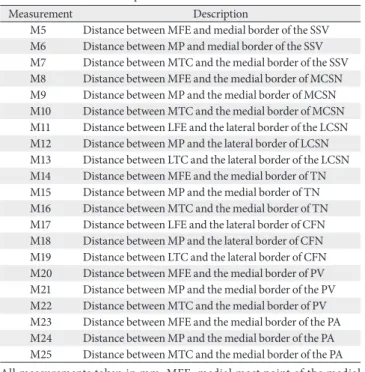
Table 1. Measurement descriptions
Measurement Description
M5 Distance between MFE and medial border of the SSV M6 Distance between MP and medial border of the SSV M7 Distance between MTC and the medial border of the SSV M8 Distance between MFE and the medial border of MCSN M9 Distance between MP and the medial border of MCSN M10 Distance between MTC and the medial border of MCSN M11 Distance between LFE and the lateral border of the LCSN M12 Distance between MP and the lateral border of LCSN M13 Distance between LTC and the lateral border of the LCSN M14 Distance between MFE and the medial border of TN M15 Distance between MP and the medial border of TN M16 Distance between MTC and the medial border of TN M17 Distance between LFE and the lateral border of CFN M18 Distance between MP and the lateral border of CFN M19 Distance between LTC and the lateral border of CFN M20 Distance between MFE and the medial border of PV M21 Distance between MP and the medial border of the PV M22 Distance between MTC and the medial border of PV M23 Distance between MFE and the medial border of the PA M24 Distance between MP and the medial border of the PA M25 Distance between MTC and the medial border of the PA All measurements taken in mm. MFE, medial most point of the medial epicondyle of the femur; SSV, small saphenous vein; MP, midpoint; MTC, medial most point of the medial condyle of the tibia; MFE, medial most point of the medial epicondyle of the femur; MCSN, medial cutaneous sural nerve;
LFE, lateral most point of the lateral epicondyle of the femur; LCSN, lateral cutaneous sural nerve; LTC, lateral most point of the lateral condyle of the tibia; TN, tibial nerve; CFN, common fibular nerve; PV, popliteal vein; PA, popliteal artery.
Statistical analysis
Descriptive statistics were used for the distance measure- ments. Mean length, standard deviation, 95% confidence in- tervals and minimum and maximum values were calculated.
Normal data distribution was assessed with the Shapiro- Wilks Test. Homogeneity of variance was verified with Levene’s test. A series of unpaired t-tests was used to com- pare sex and laterality differences. If significant differences were observed between male and female distance measures were analysed separately. Intra- and inter-rater reliability (intraclass correlation coefficients, ICC) were established by repeating the measures on two consecutive days in five cadaveric specimens. The algorithm of Landis and Koch [4]
was used to assess the rate of agreement. Values above 0.80 represented excellent agreement, values between 0.62–0.79 were considered good agreement, values between 0.41–0.61 indicated moderate agreement, and values below 0.4 sug- gested fair to poor agreement [4]. Pearson correlations coef- ficients were used to determine the relationships between the bony landmarks and the distance from the neurovascular structures. Pearson correlations coefficients were also used determined to the relationships between weight/height and the anatomic measures. For this part of the study an a-priori sample size calculation was performed. Using G*Power 3.1.9.2 (G*Power, Heinrich Heine University, Duesseldorf, Germa- ny) the following variables were used: H1 corr p_ab of 0.1, H1 corr p_ac of –0.46, alpha of 0.05, beta of 0.8 and a critical z of –1.644 the minimum sample size was calculated to be n=41.
Based All analyses were conducted using STATA SE (Version
12.0; StataCorp, College Station, TX, USA) for Windows.
Results
Intra- and inter-rater reliability (ICC) were scored be- tween three raters and ranged from 0.89–0.98 for interrater reliability and 0.95–0.97 for intra-rater reliability, which represents excellent agreement values [4]. All data were nor- mally distributed and unpaired t-tests did not demonstrate statistical significance between left and right knees (P=0.36).
There were significant differences between male and female knees (P=0.01) and therefore, the statistical analysis was re- ported separately for males and female.
The distances between MFE and LFE, MTC and LTC, MFE and MTC, and LFE and LTC are summarized in Table 2.
All distances were observed to be longer in males compared to females with the distance between MFE and LFE on aver- age 8% longer, between MTC and LTC and between MFE and MTC 7% longer, and between MFE and LTC only 3%
longer.
Measurements were taken from the medial bony land- marks to the medial lying structures namely the SSV, MCSN, TN, PV, and PA (Table 3). It is important to note that the values for M5–M10 and M14–M16 for the SSV, MCSN and TN are very similar between male and female. This shows a difference in proportion between male and female neurovas- cular layout for these three structures. The male bony land- mark values were consistently larger than the female (Table 2),
LFE
LTC
MP MP
TN
MTC MFE
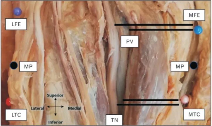
PV
Fig. 4. The PV and TN were measured from the MFE, MTC and the MP between these landmarks. LFE, lateral most point of the lateral epicondyle of the femur; LTC, lateral most point of the lateral condyle of the tibia; MFE, medial most point of the medial epicondyle of the femur; MP, midpoint; MTC; medial most point of the medial condyle of the tibia; PV, popliteal vein; TN, tibial nerve.
LFE
PA
MP MFE
MTC LTC
Fig. 5. The PA was measured from the MFE, MTC and the MP between these landmarks. LFE, lateral most point of the lateral epicondyle of the femur; LTC, lateral most point of the lateral condyle of the tibia; MFE, medial most point of the medial epicondyle of the femur; MP, midpoint; MTC; medial most point of the medial condyle of the tibia; PA, popliteal artery.
meaning that in males the SSV, MCSN and TN will lie more medially when compared as a proportion. For example, M5 for both male and female was very similar, around 37 mm, however, the male femoral epicondylar distance (M1) was 84.8 mm and the female 78.9 mm. The male epicondylar dis- tance being significantly larger than the female means that the SSV will lie more medially in the male popliteal fossa.
The LCSN and CFN were measured from the lateral bony landmarks. The CFN in male and female has a mean minimum
distance from the lateral landmarks of 0.0 mm (Table 4). This shows that the CFN ran over those landmarks in a few speci- mens. Of the sample of 45 knees, there was a 22.2% (n=10/45) occurrence of the CFNs coursing over at least one of the lat- eral bony landmarks. Neither the CFN nor LCSN displayed proportional differences between male and female as noted with the SSV, MCSN, and TN.
The medial or lateral deviations where calculated for each neurovascular structure. The deviation is between the femo-
Table 2. The definition of the popliteal fossa: the distances between the four bony landmarks
Measurement Male Female
N Min Max Mean±SD 95% CI N Min Max Mean±SD 95% CI
MFE-LFE 20 77.5 98.3 84.8±5.4 2.4 25 62.2 94.3 78.9±6.4 2.5
MTC-LTC 20 70.2 97.1 80.3±6.6 2.9 25 59.1 91.5 75.4±7.9 3.1
MFE-MTC 20 31.6 54.0 40.5±6.3 2.8 25 31.1 46.7 38.0±4.4 1.7
LFE-LTC 20 27.2 49.0 38.7±6.5 2.9 25 20.4 50.3 37.6±6.8 2.6
Measurements in mm. N, sample size; Min, minimum; Max, maximum; SD, standard deviation; CI, confidence interval; MFE, medial most point of the medial epicondyle of the femur; LFE, lateral most point of the lateral epicondyle of the femur; MTC, medial most point of the medial condyle of the tibia; LTC, lateral most point of the lateral condyle of the tibia.
Table 4. The measured distances of each neurovascular structure from the lateral bony landmarks in male and female cadavers
Measurement Male Female
N Min Max Mean±SD 95% CI N Min Max Mean±SD 95% CI
LCSN M11 12 15.1 43.1 24.9±7.3 4.1 19 0.0 49.6 18.4±10.4 4.7
M12 12 15.0 47.7 25.9±9.0 5.1 19 2.0 44.6 18.2±9.6 4.3
M13 12 16.7 50.9 27.3±10.8 6.1 19 6.3 40.3 19.9±9.0 4.1
CFN M17 20 0.0 37.0 13.4±8.2 3.6 25 0.0 46.4 8.4±9.1 3.6
M18 20 0.0 27.6 10.3±6.6 2.9 25 0.0 38.8 6.7±7.8 3.1
M19 20 0.0 18.1 7.7±4.8 2.1 25 0.0 26.0 5.3±6.0 2.4
Measurements in mm. N, sample size; Min, minimum; Max, maximum; SD, standard deviation; CI, confidence interval; LCSN, lateral cutaneous sural nerve;
CFN, common fibular nerve.
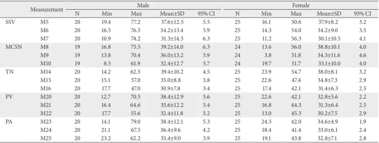
Table 3. The measured distances of each neurovascular structure from the medial bony landmarks in male and female cadavers
Measurement Male Female
N Min Max Mean±SD 95% CI N Min Max Mean±SD 95% CI
SSV M5 20 19.4 77.2 37.6±12.5 5.5 25 16.1 50.6 37.9±8.2 3.2
M6 20 16.5 76.5 34.2±13.4 5.9 25 14.3 54.0 34.2±9.0 3.5
M7 20 10.9 74.2 31.3±14.3 6.3 25 11.2 56.3 30.1±10.5 4.1
MCSN M8 19 16.8 75.5 39.2±14.0 6.3 24 13.6 56.0 38.8±10.1 4.0
M9 19 13.8 70.4 36.0±13.2 5.9 24 3.8 51.8 34.3±11.6 4.6
M10 19 8.5 61.9 32.4±12.7 5.7 24 19.7 51.7 33.1±10.0 4.0
TN M14 20 14.2 62.5 39.4±10.2 4.5 25 23.9 54.7 38.0±8.1 3.2
M15 20 15.1 57.0 35.0±8.8 3.8 25 22.6 47.4 34.8±7.3 2.9
M16 20 17.7 47.0 30.9±7.8 3.4 25 17.4 42.1 31.4±6.3 2.5
PV M20 20 12.7 70.5 38.4±12.9 5.6 25 22.6 42.1 32.8±5.6 2.2
M21 20 16.4 64.6 35.6±12.2 5.4 25 16.8 44.3 31.3±6.4 2.5
M22 20 17.7 55.6 32.4±11.8 5.2 25 13.0 45.3 30.2±7.5 2.9
PA M23 20 14.1 79.0 38.4±12.1 5.3 25 24.3 42.0 34.6±4.9 1.9
M24 20 21.1 67.5 36.4±9.6 4.2 25 18.4 41.4 33.0±6.1 2.4
M25 20 23.2 62.2 35.4±9.0 3.9 25 19.1 43.8 32.8±7.1 2.8
Measurements in mm. N, sample size; Min, minimum; Max, maximum; SD, standard deviation; CI, confidence interval; SSV, small saphenous vein; MCSN, medial cutaneous sural nerve; TN, tibial nerve; PV, popliteal vein; PA, popliteal artery.
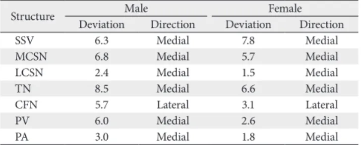
ral bony landmark proximally and the tibial bony landmark distally (Table 5). These results demonstrate the course of the neurovascular structure through the popliteal fossa and highlight the differences in neurovascular relations between the femoral and tibial plane. All structures deviated medially when passing through the popliteal fossa with exception to the CFN which deviated laterally (Table 5).
No correlation was found between weight and the femoral epicondylar distance (r=–0.004) or the tibial condylar dis- tance (r=–0.09). There was a moderate correlation between height and the femoral epicondylar distance (r=0.6) and between height and tibial intercondylar distance (r=0.5).
The distance between the two femoral epicondyles or tibial condyles showed no or very little correlation to the distance of the neurovascular structures from those bony landmarks (r=0.10–0.38).
Discussion
This study defined the popliteal fossa by reliable bony landmarks and provided a comprehensive map of the neuro- vascular structures which can be applied in direct posterior surgical access to the knee. The ‘inverted L-shape’ incision used for posterolateral tibial plateau fractures is traced along the medial edge of the biceps femoris muscle spanning over the articulate line [5]. The positioning of the CFN relative to this landmark is pertinent in protecting the nerve. The mean distance between the LFE and CFN in this study was 10.6 mm, 10.3 mm at the midpoint between LFE and LCT and 6.4 mm at the LTC. In 9 of the 45 knees, the CFN was traversing directly over either the LFE or LTC or both. There is very limited research available on measurements of the CFN from the bony landmarks used in this study. Thi et al. [6] demon-
strated a mean distance between the lateral femoral condyle and the CFN of 29.6 mm, which is substantially more medial when compared to a mean of 11 mm in this study. Unfortu- nately, Thi et al. [6] have failed to specify the exact anatomic landmark they have utilized for measurement, making com- parisons extremely difficult.
Median incisions must take the underlying popliteal neu- rovascular bundle layout into consideration. The popliteal neurovascular bundle consists of, superficial to deep, the TN, PV and PA [7]. The TN lies laterally in relation to the PA and PV [8]. The popliteal neurovascular bundle courses deep to and in between the two heads of the gastrocnemius muscle.
The TN lies superficially in the bundle; therefore, surgeons must practice care during incision and dissection to avoid iatrogenic damage to the nerve. The distance between the MFE and the TN is 33–45 mm in males and 34–42 mm in females. Sora et al. [9] measured the distance between the MFE and the TN to be 51.1–52.2 mm. The PV and PA is ly- ing deep to the TN and Sora et al. [9] reported the distances between the MFE and the medial borders of the PA and PV to be 42.3–49.7 mm and 46.0–53.5 respectively. The findings of this study documented distances of 33–45 mm and 31–34 mm respectively for males and 33–38 mm and 30–36 mm re- spectively for females. The ranges of mean distances between the MFE and the above-mentioned structures were lower than the ones defined by Sora et al. [9]. This explains the larger difference between Sora et al.’s results and this study’s female results due to the significant difference between male and female knees. In addition, Sora et al. [9] measured frozen cadaveric knees that had been sliced and scanned whereas this study measured embalmed cadaveric knees.
The TN, PV and PA all deviated medially within the popliteal fossa. Between the femoral epicondylar level and the tibial condyle level the TN, PV and PA deviated medi- ally by 8.5 mm, 6.0 mm and 3.0 mm respectively in males.
In females the TN, PV and PA deviated medially by 6.6 mm, 2.6 mm and 1.8 mm respectively. This implies that the more inferior aspect of the popliteal fossa will have a smaller me- dial surgical safe zone due to the course of these three struc- tures. Similarly, Sora et al. [9] also noticed medial deviation of these structures. However, Sora et al. [9] utilized different landmarks making direct comparisons quite difficult.
The very similar male and female distances between me- dial bony landmarks and the SSV, MCSN and TN indicate that these neurovascular structures lie more medially in the male popliteal fossa than in the female. The difference in
Table 5. The mean deviation and direction of deviation for each neurova- scular structure within the popliteal fossa in males and females
Structure Male Female
Deviation Direction Deviation Direction
SSV 6.3 Medial 7.8 Medial
MCSN 6.8 Medial 5.7 Medial
LCSN 2.4 Medial 1.5 Medial
TN 8.5 Medial 6.6 Medial
CFN 5.7 Lateral 3.1 Lateral
PV 6.0 Medial 2.6 Medial
PA 3.0 Medial 1.8 Medial
Measurements in mm. SSV, small saphenous vein; MCSN, medial cutaneous sural nerve; LCSN, lateral cutaneous sural nerve; TN, tibial nerve; CFN, common fibular nerve; PV, popliteal vein; PA, popliteal artery.
relative positioning between these neurovascular structures could be due to sexual dimorphism. The positioning of nerves in the popliteal fossa have been found to show signifi- cant differences between male and female after puberty due to the formation of muscle mass [10]. Along with the relative positioning differences displayed between male and female, the superficial structures; the SSV, LCSN and MCSN display a high level of variation. The LCSN and MCSN in particular are highly variable in pattern and presence [11, 12]. Incisions made in the median plane should therefore, factor in sex and sural nerve pattern variability.
The distances between bony landmark parameters in male and female knees were significantly larger by an aver- age of 7% to 8% with the exception for the distance between LFE and LTC (3%). This can possibly be explained by the dif- ference in Q-angle between males and females. Females tend to have a larger Q-angle, therefore placing the LEF and the LTC closer together than observed in males [13].
Hafez et al. [14] compared mediolateral dimensions at the femoral condyles and tibial plateaus and reported smaller di- mensions for both femoral epicondyle and tibial plateau dis- tances in an Arabic population. Similarly, Miyatake et al. [15]
measured mediolateral dimensions in Japanese patients and also reported smaller dimensions. Hafez et al. [14] compared their results to other population groups and were able to show that European groups had larger knees when compared to Asian groups highlighting possible population differences.
However, both studies also demonstrated that the measures in females are consistently smaller compared to males and their ratios (6%–9%) were comparable to the results of this study (6%–8%). Sexual dimorphism is the obvious reason for these findings as males are generally larger and more robust than females [16].
This study has several limitations. Due to the character- istically stiff cadaveric knees, superficial dissection is dif- ficult, and it cannot be entirely excluded that MCSN, LCSN and SSV were compromised which would have resulted in different measures. However, on careful inspection of all 45 specimens, macroscopic damage to these structures was not observed. Photographs were taken at every level of the knee dissection to measure the structures on ImageJ. Whilst great care was taken to ensure reliable and reproducible images, it is possible that measurement bias was introduced, which may have influenced measures. Inclusion and exclusion criteria of this study were strictly defined, but it cannot entirely exclude the possibility that the neurovascular structures within the
posterior knee had pathology that was not detected on mac- roscopic inspection. It is acknowledged that embalmment of human tissue results in significant stiffness when compared to fresh or fresh frozen tissue [17]. However, it is unlikely that these biomechanical changes influence tissue anatomy unless viscoelastic properties are investigated [17]. Theoreti- cally embalmment could also result in changes of anatomic morphology and increasing difficulty with dissection [18].
However, Kennel et al. [19] were able to demonstrate that embalming was unlikely to influence dissection, tissue han- dling and the anatomy.
This study defined the popliteal fossa by reliable bony landmarks and provided a comprehensive map of the neu- rovascular structures. This will assist surgeons in avoiding injuries to important neurovascular structures during surgi- cal procedures by using reliable and clearly identifiable land- marks and measure distances between these landmarks and the neurovascular structures in the popliteal fossa.
ORCID
Kelsi Greenwood: https://orcid.org/0000-0002-3923-7828 Reinette van Zyl: https://orcid.org/0000-0002-5059-1000 Natalie Keough: https://orcid.org/0000-0001-8249-8727 Erik Hohmann: https://orcid.org/0000-0003-1991-8682
Author Contributions
Conceptualization: KG, RvZ, EH. Data acquisition: KG.
Data analysis or interpretation: KG, NK, EH. Drafting of the manuscript: KG, RvZ. Critical revision of the manuscript:
NK, EH. Approval of the final version of the manuscript: all authors.
Conflicts of Interest
No potential conflict of interest relevant to this article was reported.
References
1. Faucett SC, Gannon J, Chahla J, Ferrari MB, LaPrade RF. Pos- terior surgical approach to the knee. Arthrosc Tech 2017;6:e391- 5.
2. Alpert JM, McCarty LP, Bach BR Jr. The direct posterior ap- proach to the knee: surgical and anatomic approach. J Knee Surg 2008;21:44-9.
3. Dagur G, Gandhi J, Smith N, Khan SA. Anatomical approach to clinical problems of popliteal fossa. Curr Rheumatol Rev 2017;13:126-38.
4. Landis JR, Koch GG. The measurement of observer agreement for categorical data. Biometrics 1977;33:159-74.
5. Chen H, Wu L. Surgical options for posterior tibial plateau fracture. Int J Clin Exp Med 2015;8:21421-7.
6. Thi C, Van Huy N, Nguyen NC, Thanh TH. Applied anatomy of common peroneal nerve: a cadaveric study. Int J Med Pharm 2018;6:6-10.
7. Hyland S, Sinkler MA, Varacallo M. Anatomy, bony pelvis and lower limb, popliteal region. Treasure Island: StatPearls Pub- lishing; 2020.
8. Kim TK, Savino RM, McFarland EG, Cosgarea AJ. Neurovas- cular complications of knee arthroscopy. Am J Sports Med 2002;30:619-29.
9. Sora MC, Dresenkamp J, Gabriel A, Matusz P, Wengert GJ, Bartl R. The relationship of neurovascular structures to the posterior medial aspect of the knee: an anatomic study us- ing plastinated cross-sections. Rom J Morphol Embryol 2015;56:1035-41.
10. Byun S, Gordon J, Morris S, Jacob T, Pather N. A computed to- mography and magnetic resonance imaging study of the varia- tions of the sciatic nerve branches of the pediatric knee: impli- cations for peripheral nerve blockade. Clin Anat 2019;32:836- 50.
11. Aktan Ikiz ZA, Uçerler H, Bilge O. The anatomic features of the sural nerve with an emphasis on its clinical importance.
Foot Ankle Int 2005;26:560-7.
12. Paraskevas GK, Natsis K, Tzika M, Ioannidis O. Fascial entrap- ment of the sural nerve and its clinical relevance. Anat Cell Biol 2014;47:144-7.
13. Nguyen AD, Boling MC, Levine B, Shultz SJ. Relationships between lower extremity alignment and the quadriceps angle.
Clin J Sport Med 2009;19:201-6.
14. Hafez MA, Sheikhedrees SM, Saweeres ES. Anthropometry of Arabian arthritic knees: comparison to other ethnic groups and implant dimensions. J Arthroplasty 2016;31:1109-16.
15. Miyatake N, Sugita T, Aizawa T, Sasaki A, Maeda I, Kamimura M, Fujisawa H, Takahashi A. Comparison of intraoperative anthropometric measurements of the proximal tibia and tibial component in total knee arthroplasty. J Orthop Sci 2016;21:635- 9.
16. Ubelaker DH, DeGaglia CM. Population variation in skeletal sexual dimorphism. Forensic Sci Int 2017;278:407.e1-7.
17. Hohmann E, Keough N, Glatt V, Tetsworth K, Putz R, Imhoff A. The mechanical properties of fresh versus fresh/frozen and preserved (thiel and formalin) long head of biceps tendons: a cadaveric investigation. Ann Anat 2019;221:186-91.
18. Eisma R, Lamb C, Soames RW. From formalin to thiel em- balming: what changes? One anatomy department’s experi- ences. Clin Anat 2013;26:564-71.
19. Kennel L, Martin DMA, Shaw H, Wilkinson T. Learning anat- omy through thiel- vs. formalin-embalmed cadavers: student perceptions of embalming methods and effect on functional anatomy knowledge. Anat Sci Educ 2018;11:166-74.