This is an Open Access article distributed under the terms of the Creative Commons Attribution Non-Commercial License (http://creativecommons.org/licenses/by-nc/4.0/) which permits unrestricted non-commercial use, distribution, and reproduction in any medium, provided the original work is properly cited.
Copyright © 2019. Anatomy & Cell Biology
Introduction
The cutaneous innervation of the dorsum of the foot shows various branching patterns. Dorsum skin is supplied by the terminal branches of tibial nerve and common peroneal nerve. Branches of the superficial peroneal nerve (SPN) sup- plies major portion of the dorsum of the foot and toes except the areas supplied by the deep peroneal nerve (DPN) and su- ral nerve (SN).
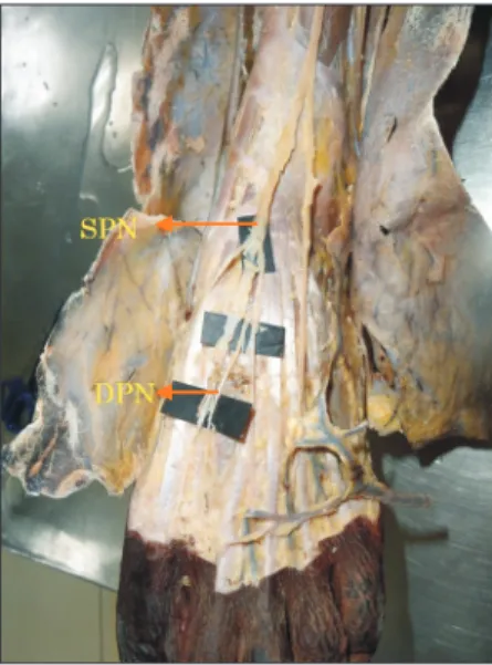
SPN which is a branch of the common peroneal nerve in the lower third of the leg pierces the deep fascia and becomes
cutaneous. It divides into medial and lateral dorsal cutaneous nerves, which in turn gives off dorsal digital nerves to supply the skin over the dorsum of the foot and the digits. SN sup- plies the lateral side of the fifth toe, saphenous nerve supplies the medial border of the foot up to the base of the great toe and the DPN supply the skin of the first interdigital cleft.
Dorsal branches of the medial and lateral plantar nerves sup- ply the nail beds [1].
SPN can be easily subjected to entrapment as it penetrates deep fascia of leg. It can also be affected in compartment syndrome of the lateral compartment of the leg. Clinicians working in emergency set up always come across patients who have injury to the dorsum of the foot that necessitate an- esthesia for repair. As the major part of the dorsum of the foot is innervated by SPN, regional block of this nerve guarantees quick anesthesia of this area [2]. According to review by Asp et al. [3] on SPN it was concluded that SPN has highly vari- able anatomy from its origin at the level of the knee to its ter-
Original Article
https://doi.org/10.5115/acb.2019.52.1.34 pISSN 2093-3665 eISSN 2093-3673
Corresponding author:
Vanishri S. Nayak
Department of Anatomy, Kasturba Medical College, Manipal Academy of Higher Education, Manipal, Karnataka 576104, India
Tel: +91-0820-2922327, Fax: +91-0820-2571927, E-mail: vanishri_nayak@
yahoo.co.in, vanishri.nayak@manipal.edu.
Anatomical variations in the cutaneous innervation on the dorsum of the foot
Vanishri S. Nayak
1, Nandini Bhat
1, Sunil S. Nayak
2, Suhani Sumalatha
11Department of Anatomy, Kasturba Medical College, Manipal Academy of Higher Education, Manipal, 2Department of Oral and Maxillofacial Surgery, Manipal College of Dental Sciences, Manipal Academy of Higher Education, Manipal, India
Abstract: Generally among the branches of common peroneal nerve, the superficial peroneal nerve provides cutaneous innervation to major part of the dorsum of the foot whereas the deep peroneal nerve innervates the skin over the first interdigital cleft region. The sural and saphenous nerves supplies the smaller lateral and medial margins of the dorsum respectively. The present study has been taken to classify the patterns of innervations of the nerves on the dorsum of the foot in South Indian population. A total of 40 formalin fixed lower limbs from 20 adult cadavers (15 males, 5 females) aged between 35 to 60 years were dissected and the branching patterns of nerves on the dorsum of the foot were noted and specimens were photographed. Gross anatomical variations were noted in the branching pattern of superficial peroneal, deep peroneal and sural nerve on the dorsum of foot. Results obtained in our study were classified into four groups. The cutaneous nerves are at risk of iatrogenic injuries during surgeries involving ankle, open reduction and internal fixation of fracture, arthroscopy etc.
Knowledge of such anatomical variations of the nerves provides information to clinicians to avoid injury to them in real clinical situations.
Key words: Peroneal nerve, Innervation of foot
Received July 2, 2018; 1st Revised August 29, 2018; 2nd Revised August 29, 2018; Accepted August 31, 2018
Cutaneous innervation on dorsum of foot
https://doi.org/10.5115/acb.2019.52.1.34
Anat Cell Biol 2019;52:34-37
35
www.acbjournal.org
minal branches to the dorsum of the foot. The key anatomical locations where the nerve is at risk includes many points in its course including over the first metatarso phalangeal joint which is supplied consistently by a medial dorsal cutaneous branch of the nerve [3]. The SPN is also at risk of injury in anterior ankle arthroscopy or fracture of ankle [4].
SPN and SN are used as donor nerve especially when mul- tiple and very long nerve grafts are necessary. They have been harvested with good results [5-7]. Knowledge of such varia- tions are essential for both orthopedic and trauma surgeons.
Therefore, this study is conducted to know the different pat- terns of nerve distribution on the dorsum of the foot and also to compare it with earlier studies.
Materials and Methods
Forty formalin fixed lower limbs (30 males and 10 females) of adult cadavers without specification to the side it belongs were dissected in the Department of Anatomy, Kasturba Medical College, Manipal Academy of Higher Education, Manipal. The specimens are dissected as per the instructions given in Cunningham’s manual of Anatomy [8]. The pattern of cutaneous innervation on the dorsum of the foot were care- fully dissected, noted, classified into four groups and photo- graphed. Group I is a classical pattern in which DPN supply- ing first interdigital cleft region, SN on lateral side of fifth toe, saphenous nerve along medial side of dorsum upto the base
of big toe and SPN supplying remaining part of the dorsum.
In group II, SPN supplies the entire dorsum except its medial and lateral sides. In group III, SN supplies lateral 2 1/2 toes, first interdigital cleft is supplied by DPN and remaining part is by SPN. In group IV, DPN communicates with the medial division of SPN and supplies first interdigital cleft, SN to the lateral side of the fifth toe and SPN to remaining part of the dorsum.
Results
Results obtained in our study were classified into four groups as follows.
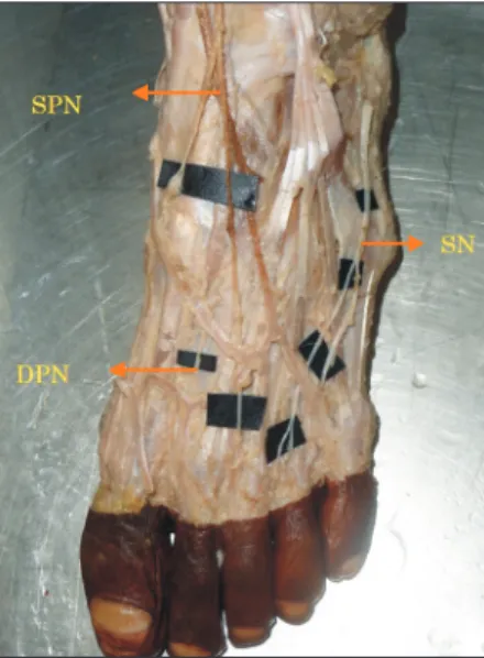
Group I (Fig. 1): DPN supplying the skin over first inter- digital cleft; SN, lateral side of the fifth toe; SPN, remaining part of the dorsum in 18 limbs (45% of specimens).
Group II (Fig. 2): SN, lateral side of fifth toe; SPN, remain- ing part of the dorsum in 12 limbs (30% of specimens).
Group III (Fig. 3): SN, lateral 2 1/2 toes; DPN, first inter- digital cleft; SPN, remaining part of the dorsum in 6 limbs (15% of specimens).
Group IV (Fig. 4): DPN communicates with the medial division of SPN and supplies first interdigital cleft. SN, lateral side of the fifth toe; SPN, remaining part of the dorsum in 4 limbs (10% of the specimens).
Fig. 1. Deep peroneal nerve (DPN) supplies the first interdigital cleft.
Rest of the dorsum of the foot is by the superficial peroneal nerve (SPN).
Fig. 2. In this group, first interdigital cleft is supplied by the branch of superficial peroneal nerve (SPN) instead of deep peroneal nerve (DPN).
Sural nerve, lateral side of fifth toe; SPN, remaining part of the dorsum of the foot.
Anat Cell Biol 2019;52:34-37 Vanishri S. Nayak, et al
36
www.acbjournal.org https://doi.org/10.5115/acb.2019.52.1.34
Discussion
In a study, 35.38% specimens the lateral side of the fifth toe was supplied by SN and rest was by SPN [9]. In our study, 30% of cases had this pattern. In the same study, 24.61% spec- imens lateral 2 1/2 digits were supplied by SN. In our study, 15% were supplied in this way. The innervation patterns found in other groups were not found in our study.
The most common distribution of the SN in the foot was to the lateral side of the fifth toe seen in 60% of specimens, in contrast we got 90%. In 26.7% cases, the SN supplied lateral 2 1/2 digits [10], whereas in our study it is seen in 15% of the cases.
In a study conducted by Drizenko et al. [11], the commu- nicating branches between SN and SPN were seen in more than 50% of cases. In our study, communication between SPN and DPN were seen only in 10% of cases.
Prakash et al. [12], found in 33.33% cases an additional branch of SPN called as accessory DPN which was seen sup- plying the ankle and dorsum of the foot. Wahee et al. [13]
found a variation pattern in 53.3% of cases which is similar to our group I, i.e., first interdigital cleft by DPN, lateral border of fifth digit by SN and remaining by SPN [13].
Four distinct patterns of innervation was observed by Gupta et al. [14]. In 55% of cases, our group I type of innerva- tion was seen (i.e., SN on lateral border of fifth digit, DPN in
the first interdigital cleft, rest of the dorsum by SPN). In 35%
cases, the pattern is similar to our group III (i.e., SN, lateral 2 1/2 toes; DPN, first interosseous space; SPN, remaining part of the dorsum). In the same study, the communicating branches were found between SPN and DPN in 25% cases, between SPN and SN in 15% of cases. A study conducted on communicating branches between SPN and SN of the foot 58% of the cases found to have communicating branches [11].
The rich sensory innervation of foot is manifest through the communicating branches linking the neural trunks. In our study the communicating branch was found between SPN and DPN in 10% of cases. There was no communicating branch found between SPN and SN. The knowledge of com- municating branches and their variations will help to decrease iatrogenic injury to the nerve.
Numerous variations do exist in the cutaneous innervation of the dorsum of the foot. Knowledge of such variations are useful for physicians while assessing sensory deficits and pain on the dorsum of the foot. The cutaneous nerves are at risk of iatrogenic injuries during surgeries involving ankle, open reduction and internal fixation of fracture, arthroscopy, local anesthetic block, nerve grafting, and surgical decompression of neurovascular bundle [15]. Therefore, a detailed knowledge of the cutaneous innervation pattern is helpful in diminishing the damage to these nerves during operative procedures.
Fig. 3. In this picture lateral 2 and 1/2 finger is supplied by sural nerve (SN) instead of superficial peroneal nerve (SPN). Deep peroneal nerve (DPN), first interdigital cleft; SN, lateral 2 1/2 toes. Rest of the dorsum of the foot is by the SPN.
Fig. 4. Deep peroneal nerve (DPN) communicates with the medial divi sion of superficial peroneal nerve (SPN) by communicating branch (CB) supplies first interdigital cleft. Sural nerve supplies lateral side of fifth toe. Rest of the dorsum of the foot is by the SPN.
Cutaneous innervation on dorsum of foot
https://doi.org/10.5115/acb.2019.52.1.34
Anat Cell Biol 2019;52:34-37
37
www.acbjournal.org
References
1. Standring S, Borley NR, Collins P, Crossman AR, Gatzoulis MA, Healy JC, Johnson D, Mahadevan V, Newell RL, Wigley CB.
Gray’s anatomy: the anatomical basis of clinical practice. 40th ed.
London: Churchill Livingstone; 2008. p.1427-9.
2. Tassone H, Silver MA. Superficial peroneal nerve block [In- terent]. Medscape; 2015 [cited 2018 Oct 1]. Available from:
http://emedicine.medscape.com/article/83218-overview.
3. Asp R, Marsland D, Elliot R. The superficial peroneal nerve: a review of its anatomy and surgical relevance. OA Anat 2014;2:6.
4. Blair JM, Botte MJ. Surgical anatomy of the superficial pe- roneal nerve in the ankle and foot. Clin Orthop Relat Res 1994;(305):229-38.
5. Buntic RF, Buncke HJ, Kind GM, Chin BT, Ruebeck D, Buncke GM. The harvest and clinical application of the superficial pero- neal sensory nerve for grafting motor and sensory nerve defects.
Plast Reconstr Surg 2002;109:145-51.
6. Slutsky DJ. A practical approach to nerve grafting in the upper extremity. Atlas Hand Clin 2005;10:e92.
7. Tallis R, Staniforth P, Fisher TR. Neurophysiological studies of autogenous sural nerve grafts. J Neurol Neurosurg Psychiatry 1978;41:677-83.
8. Romanes GJ. Cunningham’s manual of practical anatomy. 15th ed. Vol. 1. Oxford: Oxford University Press; 1986. p.16-8.
9. Madhavi C, Isaac B, Antoniswamy B, Holla SJ. Anatomical varia- tions of the cutaneous innervation patterns of the sural nerve on the dorsum of the foot. Clin Anat 2005;18:206-9.
10. Aktan Ikiz ZA, Uçerler H, Bilge O. The anatomic features of the sural nerve with an emphasis on its clinical importance. Foot Ankle Int 2005;26:560-7.
11. Drizenko A, Demondion X, Luyckx F, Mestdagh H, Cassagnaud X. The communicating branches between the sural and superfi- cial peroneal nerves in the foot: a review of 55 cases. Surg Radiol Anat 2004;26:447-52.
12. Prakash, Bhardwaj AK, Singh DK, Rajini T, Jayanthi V, Singh G.
Anatomic variations of superficial peroneal nerve: clinical impli- cations of a cadaver study. Ital J Anat Embryol 2010;115:223-8.
13. Wahee P, Aggarwal A, Harjeet, Sahni D. Variable patterns of cutaneous innervation on the dorsum of foot in fetuses. Surg Radiol Anat 2010;32:469-75.
14. Gupta C, Kiruba NL, Dsouza AS, Radhakrishnan P. A morpho- logical study to note the variable patterns of cutaneous innerva- tion on the dorsum of foot in south Indian human foetuses and its clinical implications. Adv Biomed Res 2013;2:15.
15. Halm JA, Schepers T. Damage to the superficial peroneal nerve in operative treatment of fibula fractures: straight to the bone?
Case report and review of the literature. J Foot Ankle Surg 2012;
51:684-6.