• 검색 결과가 없습니다.

The Current Status of Intervention for Intermediate Coronary Stenosis in the Korean Percutaneous Coronary Intervention (K-PCI) Registry

N/A
N/A
Protected

Academic year: 2021

Share "The Current Status of Intervention for Intermediate Coronary Stenosis in the Korean Percutaneous Coronary Intervention (K-PCI) Registry"

Copied!
11
0
0

로드 중.... (전체 텍스트 보기)

전체 글

(1)

ABSTRACT

Background and objectives: Intermediate coronary lesion that can be under- or over- estimated by visual estimation frequently results in stenting of functionally nonsignificant lesions or deferral of percutaneous coronary intervention (PCI) of significant lesions inappropriately. We evaluated current status of PCI for intermediate lesions from a standardized database in Korea.

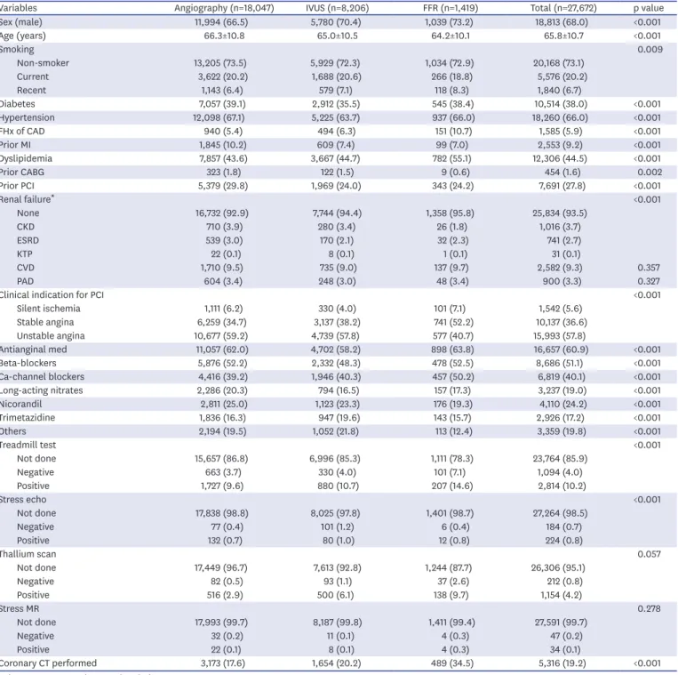
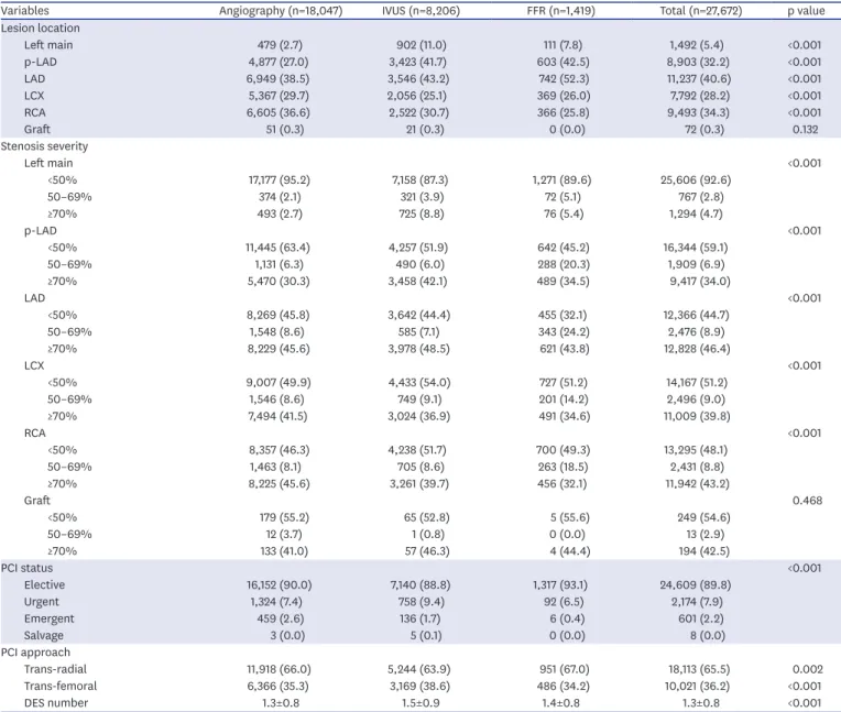
Methods: We analyzed the Korean percutaneous coronary intervention (K-PCI) registry data which collected a standardized PCI database of the participating hospitals throughout the country from January 1, 2014, through December 31, 2014. Intermediate lesion was defined as a luminal narrowing between 50% and 70% by visual estimation and then compared whether the invasive physiologic or imaging study was performed or not.

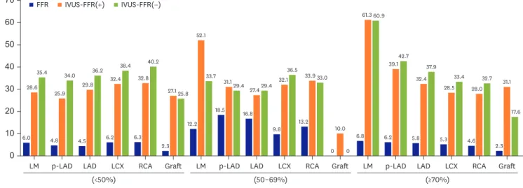
Results: Physiology-guided PCI for intermediate lesions was performed in 16.8% for left anterior descending artery (LAD), 9.8% for left circumflex artery (LCX), 13.2% for right coronary artery (RCA). PCI was more frequently performed using intravascular ultrasound (IVUS) than using fractional flow reserve (FFR) for coronary artery segments (27.7% vs.

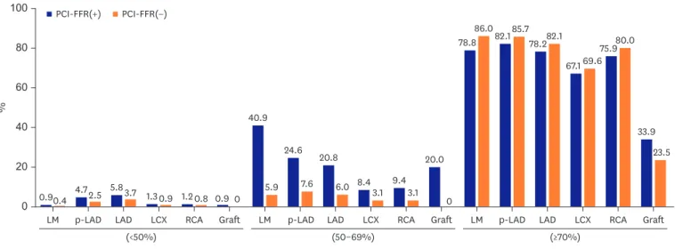
13.9% for LAD, 32.9% vs. 8.1% for LCX, and 33.8% vs. 10.8% for RCA). In accordance with or without FFR, PCI for intermediate lesions was more frequently performed in the hospitals with available FFR device than without FFR, especially in left main artery (LM), proximal LAD lesion (40.9% vs. 5.9% for LM, 24.6% vs 7.6% for proximal LAD).

Original Article

Jin-Ho Kim , MD 1 , Woonggil Choi , MD 1 , Ki-Chang Kim , MD 1 ,

Chang-Wook Nam , MD 2 , Bum-Kee Hong , MD 3 , June-Hong Kim , MD 4 , Doo Soo Jeon , MD 5 , Jang-Whan Bae , MD 6 , Sang-Hyun Kim , MD 7 , Keon-Woong Moon , MD 8 , Byung-Ryul Cho , MD 9 , Doo Il Kim , MD 10 , and Jae-Sik Jang , MD 11

1

Department of Cardiology, Konkuk University School of Medicine, Chungju, Korea

2

Division of Cardiology, Department of Internal Medicine, Keimyung University Dongsan Hospital, Daegu, Korea

3

Division of Cardiology, Yonsei University College of Medicine, Gangnam Severance Hospital, Seoul, Korea

4

Department of Cardiology, Pusan National University Yangsan Hospital, Busan, Korea

5

Department of Cardiology, The Catholic University of Korea, Incheon St. Mary's Hospital, Incheon, Korea

6

Department of Internal Medicine, Chungbuk National University College of Medicine, Cheongju, Korea

7

Division of Cardiology, Department of Internal Medicine, Seoul National University College of Medicine, Seoul Metropolitan Government-Seoul National University Boramae Medical center, Seoul, Korea

8

Division of Cardiology, The Catholic University of Korea, St.Vincent's Hospital, Suwon, Korea

9

Division of Cardiology, Department of Internal Medicine, Kangwon National University School of Medicine, Kangwon National University Hospital, Chuncheon, Korea

10

Division of Cardiology, University of Inje College of Medicine, Inje University Heaundae Paik Hospital, Busan, Korea

11

Division of Cardiology, University of Inje College of Medicine, Busan Paik Hospital, Busan, Korea

The Current Status of Intervention for Intermediate Coronary Stenosis in the Korean Percutaneous Coronary Intervention (K-PCI) Registry

Received: Feb 25, 2019 Revised: Apr 22, 2019 Accepted: May 16, 2019 Correspondence to Jae-Sik Jang, MD

Division of Cardiology, Busan Paik Hospital, University of Inje College of Medicine, 75, Bokji-ro, Busanjin-gu, Busan 47392, Korea.

E-mail: jsjang71@gmail.com

Copyright © 2019. The Korean Society of Cardiology

This is an Open Access article distributed under the terms of the Creative Commons Attribution Non-Commercial License (https://

creativecommons.org/licenses/by-nc/4.0) which permits unrestricted noncommercial use, distribution, and reproduction in any medium, provided the original work is properly cited.

ORCID iDs Jin-Ho Kim

https://orcid.org/0000-0003-3585-7859 Woonggil Choi

https://orcid.org/0000-0002-2235-8041 Ki-Chang Kim

https://orcid.org/0000-0003-0610-2502 Chang-Wook Nam

https://orcid.org/0000-0002-3370-5774 Bum-Kee Hong

https://orcid.org/0000-0002-6456-0184 June-Hong Kim

https://orcid.org/0000-0002-9973-2177 Doo Soo Jeon

https://orcid.org/0000-0001-6270-6621 Jang-Whan Bae

https://orcid.org/0000-0003-1362-9804

(2)

Sang-Hyun Kim

https://orcid.org/0000-0001-8026-1582 Keon-Woong Moon

https://orcid.org/0000-0003-2726-3209 Byung-Ryul Cho

https://orcid.org/0000-0003-2095-1121 Doo Il Kim

https://orcid.org/0000-0001-9194-6288 Jae-Sik Jang

https://orcid.org/0000-0001-7865-124X Conflict of Interest

The authors have no financial conflicts of interest.

Author Contributions

Conceptualization: Choi W, Kim KC;

Supervision: Nam CW, Hong BK, Kim JH, Jeon DS, Bae JW, Kim SH, Moon KW, Cho BR, Kim DI, Jang JS; Writing - original draft: Kim JH;

Writing - review & editing: Nam CW, Jang JS.

Conclusions: These data provide the current PCI practice pattern with the use of FFR and IVUS in intermediate lesion. More common use of FFR for intermediate lesion should be encouraged.

Keywords: Coronary artery disease; Percutaneous coronary intervention;

Fractional flow reserve, myocardial

INTRODUCTION

Revascularization for intermediate coronary stenosis (defined as 50% to 70% stenosis by quantitative coronary angiography) has been still under debate due to the limitations of coronary angiography itself. Coronary angiography, which evaluates lesions based on projection images, often under- and overestimates a lesion's severity because of contrast- dependent coronary dilation, high interobserver variability and poor ability to analyze plaque characteristics, especially in intermediate lesion.

1-3)

Current guidelines recommend the use of fractional flow reserve (FFR) and intravascular ultrasound (IVUS) to guide and optimize revascularization in intermediate coronary lesion.

1)4)5)

FFR measurement is generally used as index for the functional severity of coronary artery stenosis, and well known as modality to decide whether to proceed with percutaneous coronary intervention (PCI) or defer in stable coronary disease.

6)7)

On the other hand, IVUS has been widely used to optimize for PCI and assess accurate stenotic lesion and stent implantation.

8)

The use of FFR and IVUS has been associated with improved clinical and procedural

outcomes.

9)10)

In Korea, FFR measurement has been covered by the national insurance system under some limited cases like an intermediate lesion. However, it was not well known about contemporary patterns of FFR and IVUS among patients undergoing PCI in Korea. We aimed to investigate the current status of intervention using FFR and IVUS for intermediate stenotic lesions from Korean percutaneous coronary intervention (K-PCI) registry.

METHODS

We analyzed the K-PCI registry, as a retrospective multicenter registry, that collected a standardized database for PCI case analysis of the participating 92 hospitals throughout the country from January 1, 2014, through December 31, 2014. Two reports of the K-PCI registry have already been published in the Korean Circulation Journal.

11)12)

We excluded 17,295 patients with acute myocardial infarction from the original registry data (44,967 patients), and analyzed 27,672 patients who underwent PCI under angiography-only, FFR-, or IVUS- guidance. Demographic characteristics, medical history, procedural data and PCI related complications in hospital were collected retrospectively using case report forms (CRFs). All variables corresponded with written definitions in the CRF dictionary to standardize the data, as detailed in the previous article (K-PCI registry 2014 CRF, Supplementary Table 1 in the online-only data Supplement).

11)

The local Institutional Review Board (IRB) at each participating center approved this

study. The informed consent document was waived at all center, considering retrospective

enrollment of the study, without clinical follow-up. Data were collected using a web-based

reporting system.

(3)

We classified enrolled patients to 3 groups according to intervention approach, such as angiography-only, IVUS and FFR. When both IVUS and FFR were used, those cases were considered to be FFR groups. The centers that were equipped with FFR device accounted for about 77.1% among the K-PCI registry. We divided enrolled patients according to the severity of diameter stenosis (<50%, 50–69%, and ≥70%), the use of devices (IVUS and FFR).

Intermediate coronary stenosis was defined as 50% to 70% narrowing of coronary artery in this registry, considering stenotic severity range (50–70%) for reimbursements by National Health Insurance (NHI) about FFR measurement. The reimbursement in our country only would cover as follows: 1) to evaluate the need of PCI in case which intermediate coronary stenosis of 50–70% by quantitative coronary angiography (QCA) was confirmed in patients who have multivessel disease or more than two lesions in the same artery, 2) to evaluate the need of additional PCI for the side branch after the stent insertion at the main branch. We investigated the rates of coronary intervention and the use of FFR device according to the target vessels, lesion severity, and the presence or absence of FFR device. In-hospital clinical outcomes before discharge were also analyzed.

Continuous variables are presented as mean±standard deviation and compared using Student's t-tests or analysis of variance (ANOVA). Categorical variables are summarized as counts and percentages and compared using the chi-square test or Fisher's exact test, as appropriate. All statistical analyses were conducted with R version 3.3.2 (R foundation for Statistical Computing, Vienna, Austria). A 2-sided p-value of <0.05 was considered statistically significant.

RESULTS

The mean age was 65.8±10.7 years and 68.0% were male in this study, as shown in Table 1.

Hypertension was the most common concomitant medial history (66.0%), followed by dyslipidemia (44.5%) and diabetes mellitus (38.0%). Unstable angina (57.8%) was more common than stable angina (36.6%) as clinical indication for PCI. FFR was used in 1,419 cases (5.1%) and IVUS was used in 8,206 cases (29.6%) among all coronary lesions in K-PCI registry.

Most of procedure were conducted in elective cases (89.8%). Left anterior descending artery (LAD, 40.6%) was the most commonly detected coronary vessel, followed by right coronary artery (RCA, 34.3%) and left circumflex artery (LCX, 28.2%). Mean number of stents implanted was 1.3±0.8, and trans-radial approach (65.5%) was performed more frequently than trans-femoral approach (36.2%) (Table 2).

The rates of PCI for intermediate lesions in left main artery (LM) (32.3%) and LAD (18.1%) were relatively higher than RCA (8.2%) and LCX (7.5%), as shown in Figure 1 (detailed in Supplementary Table 1). On the other hand, the incidence of deferring PCI for significant lesion with stenosis >70% was highest in LCX (32.5%), followed by RCA (23.4%), LM (20.2%), LAD (21.1%), except graft. Especially, the rates of PCI for proximal LAD was relatively higher than other coronary vessel of all stenotic severity.

The rate of FFR and IVUS measurement in intermediate lesion performed PCI was

respectively 11.5% and 32.6%. The rates of using FFR in intermediate lesion was low in

the hospitals with FFR device (16.8% for LAD, 13.2% for RCA, 12.2% for LM, and 9.8% for

LCX) (Figure 2, detailed in Supplementary Table 2). The rates of using IVUS in intermediate

(4)

Table 1. Baseline patient and clinical characteristics

Variables Angiography (n=18,047) IVUS (n=8,206) FFR (n=1,419) Total (n=27,672) p value

Sex (male) 11,994 (66.5) 5,780 (70.4) 1,039 (73.2) 18,813 (68.0) <0.001

Age (years) 66.3±10.8 65.0±10.5 64.2±10.1 65.8±10.7 <0.001

Smoking 0.009

Non-smoker 13,205 (73.5) 5,929 (72.3) 1,034 (72.9) 20,168 (73.1)

Current 3,622 (20.2) 1,688 (20.6) 266 (18.8) 5,576 (20.2)

Recent 1,143 (6.4) 579 (7.1) 118 (8.3) 1,840 (6.7)

Diabetes 7,057 (39.1) 2,912 (35.5) 545 (38.4) 10,514 (38.0) <0.001

Hypertension 12,098 (67.1) 5,225 (63.7) 937 (66.0) 18,260 (66.0) <0.001

FHx of CAD 940 (5.4) 494 (6.3) 151 (10.7) 1,585 (5.9) <0.001

Prior MI 1,845 (10.2) 609 (7.4) 99 (7.0) 2,553 (9.2) <0.001

Dyslipidemia 7,857 (43.6) 3,667 (44.7) 782 (55.1) 12,306 (44.5) <0.001

Prior CABG 323 (1.8) 122 (1.5) 9 (0.6) 454 (1.6) 0.002

Prior PCI 5,379 (29.8) 1,969 (24.0) 343 (24.2) 7,691 (27.8) <0.001

Renal failure * <0.001

None 16,732 (92.9) 7,744 (94.4) 1,358 (95.8) 25,834 (93.5)

CKD 710 (3.9) 280 (3.4) 26 (1.8) 1,016 (3.7)

ESRD 539 (3.0) 170 (2.1) 32 (2.3) 741 (2.7)

KTP 22 (0.1) 8 (0.1) 1 (0.1) 31 (0.1)

CVD 1,710 (9.5) 735 (9.0) 137 (9.7) 2,582 (9.3) 0.357

PAD 604 (3.4) 248 (3.0) 48 (3.4) 900 (3.3) 0.327

Clinical indication for PCI <0.001

Silent ischemia 1,111 (6.2) 330 (4.0) 101 (7.1) 1,542 (5.6)

Stable angina 6,259 (34.7) 3,137 (38.2) 741 (52.2) 10,137 (36.6)

Unstable angina 10,677 (59.2) 4,739 (57.8) 577 (40.7) 15,993 (57.8)

Antianginal med 11,057 (62.0) 4,702 (58.2) 898 (63.8) 16,657 (60.9) <0.001

Beta-blockers 5,876 (52.2) 2,332 (48.3) 478 (52.5) 8,686 (51.1) <0.001

Ca-channel blockers 4,416 (39.2) 1,946 (40.3) 457 (50.2) 6,819 (40.1) <0.001

Long-acting nitrates 2,286 (20.3) 794 (16.5) 157 (17.3) 3,237 (19.0) <0.001

Nicorandil 2,811 (25.0) 1,123 (23.3) 176 (19.3) 4,110 (24.2) <0.001

Trimetazidine 1,836 (16.3) 947 (19.6) 143 (15.7) 2,926 (17.2) <0.001

Others 2,194 (19.5) 1,052 (21.8) 113 (12.4) 3,359 (19.8) <0.001

Treadmill test <0.001

Not done 15,657 (86.8) 6,996 (85.3) 1,111 (78.3) 23,764 (85.9)

Negative 663 (3.7) 330 (4.0) 101 (7.1) 1,094 (4.0)

Positive 1,727 (9.6) 880 (10.7) 207 (14.6) 2,814 (10.2)

Stress echo <0.001

Not done 17,838 (98.8) 8,025 (97.8) 1,401 (98.7) 27,264 (98.5)

Negative 77 (0.4) 101 (1.2) 6 (0.4) 184 (0.7)

Positive 132 (0.7) 80 (1.0) 12 (0.8) 224 (0.8)

Thallium scan 0.057

Not done 17,449 (96.7) 7,613 (92.8) 1,244 (87.7) 26,306 (95.1)

Negative 82 (0.5) 93 (1.1) 37 (2.6) 212 (0.8)

Positive 516 (2.9) 500 (6.1) 138 (9.7) 1,154 (4.2)

Stress MR 0.278

Not done 17,993 (99.7) 8,187 (99.8) 1,411 (99.4) 27,591 (99.7)

Negative 32 (0.2) 11 (0.1) 4 (0.3) 47 (0.2)

Positive 22 (0.1) 8 (0.1) 4 (0.3) 34 (0.1)

Coronary CT performed 3,173 (17.6) 1,654 (20.2) 489 (34.5) 5,316 (19.2) <0.001

Values are presented as number (%), or mean±SD.

CABG = coronary artery bypass graft; CAD = coronary artery disease; CKD = chronic kidney disease; CT = computed tomography; CVD = cerebrovascular disease; ESRD

= end-stage renal disease; FFR = fractional flow reserve; FHx = family history; GFR = glomerular filtration rate; IVUS = intravascular ultrasound; KTP = kidney transplant patients; MI = myocardial infarction; MR = magnetic resonance; PAD = peripheral arterial disease; PCI = percutaneous coronary intervention; SD = standard deviation.

* GFR ≤60mL/min/1.73m

2

.

(5)

lesions, even in FFR-present group, was higher than the rate of using FFR in all coronary vessels (52.1% vs. 12.2% for LM, 27.4% vs. 16.8% for LAD, 32.1% vs. 9.8% for LCX, and 33.9% vs. 13.2% for RCA). And also, the rate of PCI in intermediate lesion was higher in the FFR-present group than in the FFR-absent group, especially in LM (40.9% vs. 5.9%) and LAD (20.8% vs. 6.0%) (Figure 3, detailed in Supplementary Table 3).

There was no significant difference in composite in-hospital clinical outcomes among three groups, as shown in Table 3. However, non-fatal myocardial infarction (MI) was more frequently occurred in the FFR group than other groups (0.3% vs. 1.0% vs. 1.6%, p<0.001).

And also, the rates of death and transfusion were significantly different between 3 groups.

Table 2. Baseline angiographic and procedural characteristics

Variables Angiography (n=18,047) IVUS (n=8,206) FFR (n=1,419) Total (n=27,672) p value

Lesion location

Left main 479 (2.7) 902 (11.0) 111 (7.8) 1,492 (5.4) <0.001

p-LAD 4,877 (27.0) 3,423 (41.7) 603 (42.5) 8,903 (32.2) <0.001

LAD 6,949 (38.5) 3,546 (43.2) 742 (52.3) 11,237 (40.6) <0.001

LCX 5,367 (29.7) 2,056 (25.1) 369 (26.0) 7,792 (28.2) <0.001

RCA 6,605 (36.6) 2,522 (30.7) 366 (25.8) 9,493 (34.3) <0.001

Graft 51 (0.3) 21 (0.3) 0 (0.0) 72 (0.3) 0.132

Stenosis severity

Left main <0.001

<50% 17,177 (95.2) 7,158 (87.3) 1,271 (89.6) 25,606 (92.6)

50–69% 374 (2.1) 321 (3.9) 72 (5.1) 767 (2.8)

≥70% 493 (2.7) 725 (8.8) 76 (5.4) 1,294 (4.7)

p-LAD <0.001

<50% 11,445 (63.4) 4,257 (51.9) 642 (45.2) 16,344 (59.1)

50–69% 1,131 (6.3) 490 (6.0) 288 (20.3) 1,909 (6.9)

≥70% 5,470 (30.3) 3,458 (42.1) 489 (34.5) 9,417 (34.0)

LAD <0.001

<50% 8,269 (45.8) 3,642 (44.4) 455 (32.1) 12,366 (44.7)

50–69% 1,548 (8.6) 585 (7.1) 343 (24.2) 2,476 (8.9)

≥70% 8,229 (45.6) 3,978 (48.5) 621 (43.8) 12,828 (46.4)

LCX <0.001

<50% 9,007 (49.9) 4,433 (54.0) 727 (51.2) 14,167 (51.2)

50–69% 1,546 (8.6) 749 (9.1) 201 (14.2) 2,496 (9.0)

≥70% 7,494 (41.5) 3,024 (36.9) 491 (34.6) 11,009 (39.8)

RCA <0.001

<50% 8,357 (46.3) 4,238 (51.7) 700 (49.3) 13,295 (48.1)

50–69% 1,463 (8.1) 705 (8.6) 263 (18.5) 2,431 (8.8)

≥70% 8,225 (45.6) 3,261 (39.7) 456 (32.1) 11,942 (43.2)

Graft 0.468

<50% 179 (55.2) 65 (52.8) 5 (55.6) 249 (54.6)

50–69% 12 (3.7) 1 (0.8) 0 (0.0) 13 (2.9)

≥70% 133 (41.0) 57 (46.3) 4 (44.4) 194 (42.5)

PCI status <0.001

Elective 16,152 (90.0) 7,140 (88.8) 1,317 (93.1) 24,609 (89.8)

Urgent 1,324 (7.4) 758 (9.4) 92 (6.5) 2,174 (7.9)

Emergent 459 (2.6) 136 (1.7) 6 (0.4) 601 (2.2)

Salvage 3 (0.0) 5 (0.1) 0 (0.0) 8 (0.0)

PCI approach

Trans-radial 11,918 (66.0) 5,244 (63.9) 951 (67.0) 18,113 (65.5) 0.002

Trans-femoral 6,366 (35.3) 3,169 (38.6) 486 (34.2) 10,021 (36.2) <0.001

DES number 1.3±0.8 1.5±0.9 1.4±0.8 1.3±0.8 <0.001

Values are presented as number (%), or mean±SD.

DES = drug-eluting stent; FFR = fractional flow reserve; IVUS = intravascular ultrasound; LAD = left anterior descending artery; LCX = left circumflex artery; p-LAD =

proximal left anterior descending artery; PCI = percutaneous coronary intervention; RCA = right coronary artery; SD = standard deviation.

(6)

p-LAD

LM LAD LCX RCA Graft LM p-LAD LAD LCX RCA Graft LM p-LAD LAD LCX RCA Graft 0

40 60 80 100

%

20 99.2

0.8

95.7 4.3

94.6 5.4

98.8 1.2

98.9 1.1

99.2 0.8

67.7 32.3

78.5 21.5

81.9 18.1

92.5 7.5

91.8 8.2

84.6 15.4

20.2 79.8

17.3 82.7

21.1 78.9

32.5 67.5 76.6

23.4 67.0 33.0 No PCI PCI

(<50%) (50–69%) (≥70%)

Figure 1. Comparison of PCI rates for coronary vessels according to stenotic severity.

LAD = left anterior descending artery; LCX = left circumflex artery; LM = left main artery; p-LAD = proximal left anterior descending artery; PCI = percutaneous coronary intervention; RCA = right coronary artery.

p-LAD

LM LAD LCX RCA Graft LM p-LAD LAD LCX RCA Graft LM p-LAD LAD LCX RCA Graft

0 30

10 40 50 70

%

60

20

FFR IVUS-FFR(+) IVUS-FFR(−)

(<50%) (50–69%) (≥70%)

6.0 28.6

35.4

4.8 25.9

34.0

4.5 29.8

36.2

6.2 32.4

38.4

6.3 32.8

40.2

2.3 27.1 25.8

12.2 52.1

33.7

18.5 31.1 29.4

16.8 27.4 29.4

9.8 32.1

36.5

13.2 33.9 33.0

0 10.0

0 6.8

61.3 60.9

6.2 39.1 42.7

5.8 32.4

37.9

5.3 28.5

33.4

4.6 28.0

32.7

2.3 31.1

17.6

Figure 2. Use of adjunctive PCI tools according to the availability of FFR.

FFR = fractional flow reserve; FFR(+) = Hospitals that were equipped with FFR device; FFR(−) = Hospitals that were not equipped with FFR device; IVUS = intravascular ultrasound; LAD = left anterior descending artery; LCX = left circumflex artery; LM = left main artery; p-LAD = proximal left anterior descending artery; PCI = percutaneous coronary intervention; RCA = right coronary artery.

Table 3. In-hospital outcomes

Variables Angiography (n=18,047) IVUS (n=8,206) FFR (n=1,419) Total (n=27,563) p value

Death 84 (0.5) 18 (0.2) 1 (0.1) 103 (0.4) 0.002

Cardiac death 32 (0.2) 10 (0.1) 0 (0.0) 42 (0.2) 0.181

Nonfatal MI 58 (0.3) 79 (1.0) 22 (1.6) 159 (0.6) <0.001

Stent thrombosis 28 (0.2) 9 (0.1) 0 (0.0) 37 (0.1) 0.237

Stroke 24 (0.1) 7 (0.1) 1 (0.1) 32 (0.1) 0.503

Urgent PCI 23 (0.1) 8 (0.1) 0 (0.0) 31 (0.1) 0.345

Transfusion 271 (1.5) 92 (1.1) 14 (1.0) 377 (1.4) 0.022

Composite outcome 439 (2.4) 194 (2.4) 36 (2.5) 669 (2.4) 0.904

Values are presented as number (%).

FFR = fractional flow reserve; IVUS = intravascular ultrasound; MI = myocardial infarction; PCI = percutaneous coronary intervention.

(7)

DISCUSSION

The major findings of our study are as follows: 1) the rate of FFR measurement among non- acute MI patients who performed PCI was relatively low (5.1%); 2) PCI was more frequently performed using IVUS than using FFR in intermediate lesion (32.6% vs 11.5%); 3) regardless of whether FFR is used or not, PCI for intermediate lesion was more frequently performed in the hospitals with available FFR device than without it; and 4) there were no significant differences in in-hospital clinical outcomes such as cardiac death, stent thrombosis, and stroke among angiographic-guided vs IVUS-guided vs. FFR-guided PCI groups.

The rate of FFR measurement in intermediate lesions among PCI cases in our K-PCI registry, even though it did not have deferred-PCI cases, was quite low (11.5%). Despite a significant increase in the use of FFR and IVUS according to the national database

13)

and data from the registry,

14)

FFR was only performed in 3.7% of patients (2.5% in ACS patients, 7.2% in non-ACS patients) according to the original report of the 2014 K-PCI registry.

11)

Prolonged procedure time due to catheter reinsertion, the use of drug like adenosine, high cost of catheter, and poor reimbursement could be considered as factors that can be considered as causes for the low rates of FFR use.

15)

The low rates of FFR use was similarly observed in other country, 6.1% in US (among 61,874 attempted PCI for intermediate lesion between April 2009 and September 2010) and 8.2% in Japan.

15)16)

There might have been more obstacles in using the FFR for intermediate coronary lesions due to strict national insurance coverage than in Japan, where application for national insurance coverage is relatively easy.

Interestingly, we also found that PCI for intermediate lesion was more frequently performed in all coronary lesions in the hospitals equipped with FFR device with or without FFR guidance, compared with the hospitals without FFR device. As the most important factor of this result, the difference in cardiovascular center size (high- vs. low-volume centers) could be considered. Previous study using K-PCI registry reported that the low-volume centers had fewer patients with stable coronary disease, and performed less elective PCI and more frequently urgent/emergency PCI for patients with ACS than high-volume centers.

11)

The differences in centers volume, clinical indication for PCI, and composition of patients may p-LAD

LM LAD LCX RCA Graft LM p-LAD LAD LCX RCA Graft LM p-LAD LAD LCX RCA Graft

0 40 60 80 100

%

20

0.90.4 4.72.5 5.83.7 1.30.9 1.20.8 0.9 0 40.9

5.9 24.6

7.6 20.8

6.0 8.4 3.1 9.4 3.1

20.0 78.8

86.0 82.1 85.7 78.2 82.1

67.1 69.6 75.9 80.0

33.9 23.5

0 PCI-FFR(+) PCI-FFR(−)

(<50%) (50–69%) (≥70%)

Figure 3. Comparison of PCI rates according to availability of FFR.

FFR = fractional flow reserve; FFR(+) = Hospitals that were equipped with FFR device; FFR(−) = Hospitals that were not equipped with FFR device; IVUS =

intravascular ultrasound; LAD = left anterior descending artery; LCX = left circumflex artery; LM = left main artery; p-LAD = proximal left anterior descending

artery; PCI = percutaneous coronary intervention; RCA = right coronary artery.

(8)

have caused the difference in the rates of PCI for intermediate lesions. However, this analysis is not powered to draw definitive conclusion, and additional research would be needed to elucidate the factors responsible for differences between high- vs. low-volume centers.

The rate of IVUS use was higher than that of FFR in coronary intervention for intermediate lesion (32.6% vs. 11.5%) in K-PCI registry, similar to data from National Cardiovascular Data Registry in US (20.3% vs. 6.1%).

15)

Optimization of PCI using IVUS has been well known to improve clinical outcomes compared to angiography-only guided PCI.

10)17)18)

However, IVUS guided PCI may not help to decide whether to proceed PCI, but for optimization of PCI.

IVUS-derived minimal lumen area (MLA), which can be used as an indicator of functionally significant stenosis, had a relatively low positive predictive value, and use of IVUS-derived MLA to guide PCI may result in unnecessary or inappropriate PCI.

19)20)

And, mean MLA cut- off value was different by ethnicity between Asians and Westerners, maybe due to lower body mass index and smaller myocardial masses in Asian people.

21)22)

Unlike angiography- or IVUS-guided anatomical approach, physiologic approach using FFR measurement in intermediate lesions could be used as an accurate indicator to guide and proceed PCI. FFR-guided PCI improved clinical outcome when compared with angiography- guided PCI or medical therapy alone, during long-term follow-up.

9)23-25)

Furthermore, use of FFR could reduce unnecessary or inappropriate PCI without any disadvantages, and ensure economic efficiency by reducing medical cost related to PCI.

16)26)27)

Previous study related to PCI for intermediate coronary lesion reported that clinical outcomes were not different between FFR-guided vs. IVUS-guided PCI and FFR-guided PCI reduced the need for revascularization (FFR vs. IVUS; relative risk, 0.02).

28)

In our study, we could observe relatively higher use of FFR in LAD compared with other vessels (16.8% for LAD, 13.2% for RCA, and 9.8% for LCX). Physicians might have concerned about a higher association of LAD stenosis with heart failure and poor prognosis as previously reported.

22)25)

Based on clinical importance and lots of clinical data for FFR-guided physiologic approach PCI, current guidelines recommended FFR measurements before PCI in patients with stable coronary artery disease.

5)29)

Composite in-hospital clinical outcome in our K-PCI registry was not significantly different among angiographic-guided vs IVUS-guided vs. FFR-guided PCI groups. Even though the rate of clinical outcomes such as death, nonfatal MI, and transfusion had significant difference;

we think this might be explained by extremely large disparity in the number of cases between

the 3 groups (angiographic-guided group: 18,047, IVUS-guided group: 8,206, and FFR-

guided group: 1,419). Originally, this study is retrospective, cross-sectional study, and was

not designed for randomized comparison of FFR-vs. image-guided PCI of intermediate

lesions. Furthermore, data on specific causes and timing for death and transfusion at each

92 hospitals could not be uniformly verified in this registry. Nevertheless, our study is the

first analysis about real-world pattern of PCI with the use of FFR and IVUS for intermediate

coronary lesion from the nationwide standardized database by K-PCI registry. Although

results of our study did not show improvement in the clinical outcomes or reduction of

complication, we think our study might suggest what to supplement and what to do for

improvement of clinical outcomes, by presenting current status of PCI and obstacles to use

FFR device. Meanwhile, nonfatal MI was higher in IVUS-guided and FFR-guided group than

angiographic-guided group. Possible mechanism for this difference could be explained by

transient increase of cardiac marker that was caused by manipulation of adjunctive device, or

prolonged procedure time.

(9)

There are several limitations to be addressed in our study that should be considered when interpreting the results. First, even though the K-PCI registry is the largest PCI registry in Korea, these results represent only those patients treated in 92 high volume hospitals, not all patients who underwent PCI in Korea. Also, the rate of FFR or imaging studies might be underreported due to the absence of data for the patients who underwent only diagnostic angiography with those studies. Second, this registry had only in-hospital clinical outcomes before hospital discharge. It was limited to evaluate long-term clinical effect according to use of IVUS or FFR in intermediate lesion. Further long-term follow-up study would be needed to evaluate the effect of adjunctive device such as FFR and IVUS. Third, because this study used a multicenter retrospective registry, procedural features may have influenced by uncontrolled variable factors in terms of diversity of operator in different region or different hospitals.

Fourth, because this K-PCI registry is a database for PCI case which was performed on 92 hospitals, the FFR-guided deferred cases are not included in this study. Finally, there are no data regarding exact timing of IVUS examination in the IVUS-guided group.

In conclusion, these data may provide the current PCI practice pattern with the use of FFR and IVUS in intermediate lesion. To reduce unnecessary PCI and help to decide whether to proceed PCI, more common use of FFR for intermediate lesion should be encouraged.

ACKNOWLEDGMENTS

This study was co-sponsored by the Korean Society of Cardiology and the Korean Society of Interventional Cardiology.

SUPPLEMENTARY MATERIALS

Supplementary Table 1

Comparison of PCI rate for coronary vessel according to stenotic severity Click here to view

Supplementary Table 2

Use of adjunctive PCI tools according to the availability of FFR Click here to view

Supplementary Table 3

Comparison of PCI rates according to availability of FFR Click here to view

REFERENCES

1. Park SJ, Kang SJ, Ahn JM, et al. Visual-functional mismatch between coronary angiography and fractional flow reserve. JACC Cardiovasc Interv 2012;5:1029-36.

PUBMED | CROSSREF

(10)

2. Trask N, Califf RM, Conley MJ, et al. Accuracy and interobserver variability of coronary cineangiography:

a comparison with postmortem evaluation. J Am Coll Cardiol 1984;3:1145-54.

PUBMED | CROSSREF

3. Katritsis D, Webb-Peploe M. Limitations of coronary angiography: an underestimated problem? Clin Cardiol 1991;14:20-4.

PUBMED | CROSSREF

4. Patel MR, Calhoon JH, Dehmer GJ, et al. ACC/AATS/AHA/ASE/ASNC/SCAI/SCCT/STS 2017 appropriate use criteria for coronary revascularization in patients with stable ischemic heart disease: a report of the American College of Cardiology Appropriate Use Criteria Task Force, American Association for Thoracic Surgery, American Heart Association, American Society of Echocardiography, American Society of Nuclear Cardiology, Society for Cardiovascular Angiography and Interventions, Society of Cardiovascular Computed Tomography, and Society of Thoracic Surgeons. J Am Coll Cardiol 2017;69:2212-41.

PUBMED | CROSSREF

5. Authors/Task Force members, Windecker S, Kolh P, et al. 2014 ESC/EACTS Guidelines on myocardial revascularization: The Task Force on Myocardial Revascularization of the European Society of Cardiology (ESC) and the European Association for Cardio-Thoracic Surgery (EACTS)Developed with the special contribution of the European Association of Percutaneous Cardiovascular Interventions (EAPCI). Eur Heart J 2014;35:2541-619.

PUBMED | CROSSREF

6. Pijls NH, van Schaardenburgh P, Manoharan G, et al. Percutaneous coronary intervention of functionally nonsignificant stenosis: 5-year follow-up of the DEFER Study. J Am Coll Cardiol 2007;49:2105-11.

PUBMED | CROSSREF

7. Safi M, Mehrabi MA, Alipour Parsa S, Khaheshi I, Naderian M. The role of FFR in clinical decision making in patients with moderate coronary lesions: a pilot study. Acta Biomed 2018;89:378-81.

PUBMED | CROSSREF

8. Parise H, Maehara A, Stone GW, Leon MB, Mintz GS. Meta-analysis of randomized studies comparing intravascular ultrasound versus angiographic guidance of percutaneous coronary intervention in pre- drug-eluting stent era. Am J Cardiol 2011;107:374-82.

PUBMED | CROSSREF

9. Tonino PA, De Bruyne B, Pijls NH, et al. Fractional flow reserve versus angiography for guiding percutaneous coronary intervention. N Engl J Med 2009;360:213-24.

PUBMED | CROSSREF

10. Jang JS, Song YJ, Kang W, et al. Intravascular ultrasound-guided implantation of drug-eluting stents to improve outcome: a meta-analysis. JACC Cardiovasc Interv 2014;7:233-43.

PUBMED | CROSSREF

11. Jang JS, Han KR, Moon KW, et al. The current status of percutaneous coronary intervention in Korea:

based on year 2014 cohort of Korean percutaneous coronary intervention (K-PCI) registry. Korean Circ J 2017;47:328-40.

PUBMED | CROSSREF

12. Gwon HC, Jeon DW, Kang HJ, et al. The practice pattern of percutaneous coronary intervention in Korea:

based on year 2014 cohort of Korean percutaneous coronary intervention (K-PCI) registry. Korean Circ J 2017;47:320-7.

PUBMED | CROSSREF

13. Pothineni NV, Shah NN, Rochlani Y, et al. U.S. trends in inpatient utilization of fractional flow reserve and percutaneous coronary intervention. J Am Coll Cardiol 2016;67:732-3.

PUBMED | CROSSREF

14. Ahn JM, Park DW, Shin ES, et al. Fractional flow reserve and cardiac events in coronary artery disease:

data from a prospective IRIS-FFR registry (Interventional Cardiology Research Incooperation Society Fractional Flow Reserve). Circulation 2017;135:2241-51.

PUBMED | CROSSREF

15. Dattilo PB, Prasad A, Honeycutt E, Wang TY, Messenger JC. Contemporary patterns of fractional flow reserve and intravascular ultrasound use among patients undergoing percutaneous coronary intervention in the United States: insights from the National Cardiovascular Data Registry. J Am Coll Cardiol 2012;60:2337-9.

PUBMED | CROSSREF

16. Tanaka N, Kohsaka S, Murata T, et al. Treatment strategy modification and its implication on the medical cost of fractional flow reserve-guided percutaneous coronary intervention in Japan. J Cardiol 2019;73:38-44.

PUBMED | CROSSREF

17. Hong SJ, Kim BK, Shin DH, et al. Effect of intravascular ultrasound-guided vs angiography-guided everolimus-eluting stent implantation: the IVUS-XPL randomized clinical trial. JAMA 2015;314:2155-63.

PUBMED | CROSSREF

(11)

18. Witzenbichler B, Maehara A, Weisz G, et al. Relationship between intravascular ultrasound guidance and clinical outcomes after drug-eluting stents: the assessment of dual antiplatelet therapy with drug-eluting stents (ADAPT-DES) study. Circulation 2014;129:463-70.

PUBMED | CROSSREF

19. Inohara T, Kohsaka S, Miyata H, et al. Appropriateness ratings of percutaneous coronary intervention in Japan and its association with the trend of noninvasive testing. JACC Cardiovasc Interv 2014;7:1000-9.

PUBMED | CROSSREF

20. Cho YK, Nam CW, Han JK, et al. Usefulness of combined intravascular ultrasound parameters to predict functional significance of coronary artery stenosis and determinants of mismatch. EuroIntervention 2015;11:163-70.

PUBMED | CROSSREF

21. Han JK, Koo BK, Park KW, et al. Optimal intravascular ultrasound criteria for defining the functional significance of intermediate coronary stenosis: an international multicenter study. Cardiology 2014;127:256-62.

PUBMED | CROSSREF

22. Jang JS, Shin HC, Bae JS, et al. Diagnostic performance of intravascular ultrasound-derived minimal Lumen area to predict functionally significant non-left main coronary artery disease: a meta-analysis.

Korean Circ J 2016;46:622-31.

PUBMED | CROSSREF

23. De Bruyne B, Fearon WF, Pijls NH, et al. Fractional flow reserve-guided PCI for stable coronary artery disease. N Engl J Med 2014;371:1208-17.

PUBMED | CROSSREF

24. Xaplanteris P, Fournier S, Pijls NHJ, et al. Five-year outcomes with PCI guided by fractional flow reserve. N Engl J Med 2018;379:250-9.

PUBMED | CROSSREF

25. Chen ZW, Yu ZQ, Yang HB, et al. Rapid predictors for the occurrence of reduced left ventricular ejection fraction between LAD and non-LAD related ST-elevation myocardial infarction. BMC Cardiovasc Disord 2016;16:3.

PUBMED | CROSSREF

26. Fearon WF, Bornschein B, Tonino PA, et al. Economic evaluation of fractional flow reserve-guided percutaneous coronary intervention in patients with multivessel disease. Circulation 2010;122:2545-50.

PUBMED | CROSSREF

27. Fearon WF, Nishi T, De Bruyne B, et al. Clinical outcomes and cost-effectiveness of fractional flow reserve-guided percutaneous coronary intervention in patients with stable coronary artery disease:

three-year follow-up of the FAME 2 trial (Fractional Flow Reserve Versus Angiography for Multivessel Evaluation). Circulation 2018;137:480-7.

PUBMED | CROSSREF

28. Nam CW, Yoon HJ, Cho YK, et al. Outcomes of percutaneous coronary intervention in intermediate coronary artery disease: fractional flow reserve-guided versus intravascular ultrasound-guided. JACC Cardiovasc Interv 2010;3:812-7.

PUBMED | CROSSREF

29. Levine GN, Bates ER, Blankenship JC, et al. 2011 ACCF/AHA/SCAI guideline for percutaneous coronary intervention. A report of the American College of Cardiology Foundation/American Heart Association Task Force on Practice Guidelines and the Society for Cardiovascular Angiography and Interventions. J Am Coll Cardiol 2011;58:e44-122.

PUBMED | CROSSREF

수치

Table 1. Baseline patient and clinical characteristics
Table 2. Baseline angiographic and procedural characteristics
Figure 2. Use of adjunctive PCI tools according to the availability of FFR.
Figure 3. Comparison of PCI rates according to availability of FFR.

참조

관련 문서

Since the API gateway is the entry point for microservices, it should be equipped with the necessary infrastructure services (in addition to its main service of request

PWM mode implements current limiting using an internal comparator that trips at 2 A for both bucks (typical). If the output is shorted to ground the device enters a timed

1st association group – “Lifeline” reports the device status and al- lows for assigning single device only (main controller by default)4. 2nd association group –

The students of Gwangju Secondary School had to compete with the students of Gwangju Intermediate School, who were Japanese nationals for access to a

The staff of outstanding hospitals (doctors, nurses, administrative personnel) provide proper medical services that meet the

The sample for this research are current patients in hospitals or alternative medical centre and were diagnosed to be not in need for special equipments

The result of this study on the intervention of function words in children with language development delay shows that intervention of function words by grammatically

Desirably today's Korean education is to serve with an ultimate object for forming human beings as well-rounded persons who are equipped with