http://dx.doi.org/10.14316/pmp.2014.25.2.72
Quasi-breath-hold (QBH) Biofeedback in Gated 3D Thoracic MRI: Feasibility Study
Taeho Kim*
†‡, Robert Pooley
§, Danny Lee
∥, Paul Keall
∥, Rena Lee
‡, Siyong Kim*
¶*Department of Radiation Oncology, University of Virginia, Virginia, USA,
†Department of Radiation Oncology, Mayo Clinic, Florida, USA,
‡Department of Radiation Oncology, Ewha Womans University,
Seoul, Korea,
§Department of Radiology, Mayo Clinic, Florida, USA,
∥Radiation Physics Laboratory, Sydney Medical School, University of Sydney, Sydney, Australia,
¶Department of Radiation Oncology,
Virginia Commonwealth University, Virginia, USA
The aim of the study is to test a hypothesis that quasi-breath-hold (QBH) biofeedback improves the residual respiratory motion management in gated 3D thoracic MR imaging, reducing respiratory motion artifacts with insignificant acquisition time alteration. To test the hypothesis five healthy human subjects underwent two gated MR imaging studies based on a T2 weighted SPACE MR pulse sequence using a respiratory navigator of a 3T Siemens MRI: one under free breathing and the other under QBH biofeedback breathing. The QBH biofeedback system utilized the external marker position on the abdomen obtained with an RPM system (Real-time Position Management, Varian) to audio-visually guide a human subject for 2s breath-hold at 90% exhalation position in each respiratory cycle. The improvement in the upper liver breath-hold motion reproducibility within the gating window using the QBH biofeedback system has been assessed for a group of volunteers. We assessed the residual respiratory motion management within the gating window and respiratory motion artifacts in 3D thoracic MRI both with/without QBH biofeedback. In addition, the RMSE (root mean square error) of abdominal displacement has been investigated. The QBH biofeedback reduced the residual upper liver motion within the gating window during MR acquisitions (∼6 minutes) compared to that for free breathing, resulting in the reduction of respiratory motion artifacts in lung and liver of gated 3D thoracic MR images. The abdominal motion reduction in the gated window was consistent with the residual motion reduction of the diaphragm with QBH biofeedback.
Consequently, average RMSE (root mean square error) of abdominal displacement obtained from the RPM has been also reduced from 2.0 mm of free breathing to 0.7 mm of QBH biofeedback breathing over the entire cycle (67% reduction, p-value=0.02) and from 1.7 mm of free breathing to 0.7 mm of QBH biofeedback breathing in the gated window (58% reduction, p-value=0.14). The average baseline drift obtained using a linear fit was reduced from 5.5 mm/min with free breathing to 0.6 mm/min (89% reduction, p-value=0.017) with QBH biofeedback. The study demonstrated that the QBH biofeedback improved the upper liver breath-hold motion reproducibility during the gated 3D thoracic MR imaging. This system can provide clinically applicable motion management of the internal anatomy for gated medical imaging as well as gated radiotherapy.
Key Words: Gated 3D thoracic MRI, Quasi-breath-hold biofeedback, Respiratory motion management
This study was supported by the Industrial Strategic Technology Development Program (10035527) funded by the Ministry of Knowle- dge Economy (MKE, Korea).
Received 8 May 2014, Revised 26 May 2014, Accepted 2 June 2014 Correspondence: Siyong Kim ([email protected])
Tel: 1-804-628-0981, Fax: 1-804-827-1861
Co-correspondence: Rena Lee ([email protected]) Tel: 82-2-2650-5337, Fax: 82-2-2654-0363
cc This is an Open-Access article distributed under the terms of the Creative Commons Attribution Non-Commercial License (http://creativecommons.org/licenses/by-nc/3.0) which permits unrestricted non-commercial use, distribution, and reproduction in any medium, provided the original work is properly cited.
Introduction
One of the most critical steps in the radiotherapy process is
anatomic image acquisition. Obviously, any error induced dur-
ing imaging such as motion artifacts in four-dimensional com-
puted tomography (4DCT)
1,2)and Positron Emission Tomography
(PET)
3)images is systematic and remains the same through the
whole treatment process, resulting in adverse impact on clin-
ical outcome.
4-6)For instance, incorrect target and healthy sur-
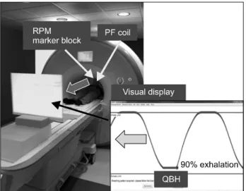
Fig. 1. QBH biofeedback system in 3 Tesla Siemens MRI con- sisting of the RPM system and audio-visual devices. The screen of the QBH biofeedback system shows a guiding wave (blue curve) and a marker position (red ball) in real time. 2s breath- hold at 90% exhalation position in respiratory cycle is shown.
rounding tissue delineation can be implemented in treatment planning due to these artifacts, leading to the irradiation of healthy surrounding tissues in addition to the tumor, which re- sults in the increased risk of radiation-related toxicity.
4-6)Various respiratory motion-guidance systems using an ex- ternal surrogate
7-12)were developed in medical imaging and treatment procedures to improve respiratory motion manage- ment in clinical practice. In multiple breath-holds based Magnetic Resonance Imaging (MRI), an audio breath-hold prompt was used to improve coronary imaging
8)and an MR-compatible active breathing control (ABC) system was ap- plied to lung MRI.
13)In addition, a respiratory biofeedback with an adaptive gating windowing was implemented in volun- tary breathing guidance for thoracic aorta MR imaging.
Breath-hold practice, however, often suffers from limited pul- monary function of the patient.
14)On the other hand, the respi- ratory gating technique is frequently subject to non-negligible residual motion during acquisition
15)and lengthened practice.
Recently, Park et al.
16)proposed a hybrid technique combin- ing free-breathing-based gating and multiple breath-holds called Quasi-breath-hold (QBH) biofeedback, and demonstrated its feasibility that the QBH biofeedback reduced phase-shift, re- sidual motion, complexity, and patient’s discomfort. However, the previous study has not been fully investigated in the feasi- bility of the QBH biofeedback in medical imaging. The cur- rent study is the first to investigate the feasibility of QBH bio- feedback to demonstrate the residual motion management of the internal anatomy with gated 3D thoracic MRI. The specific aim of this study is to verify that QBH biofeedback would re- duce respiratory motion artifacts in gated 3D thoracic MR imaging by reducing the residual respiratory motion in acquisi- tion.
Materials and Methods
1. QBH biofeedback
The QBH biofeedback system was proposed to provide vol- untary multiple breath-holds guidance in medical imaging and treatment practices.
16)QBH is a respiratory biofeedback techni- que which utilizes a hybrid technique combining free-breath- ing-gating and breath-hold. In the current study, the QBH bio- feedback system utilized real-time respiratory motion signals
obtained from the real-time position management (RPM) sys- tem (Varian Medical Systems, Palo Alto, USA) consisting of an infrared camera and a marker block on the abdomen as shown in Fig. 1. The RPM system was combined with MR compatible audiovisual equipment including a projector, a semi-transparent screen and headphones. In addition, the QBH biofeedback system utilized an in-house developed software in- terfaced to the RPM system,
17)including a multiple breath- holds maneuver to audio-visually guide a human subject for 2s breath-hold at 90% exhalation position in each respiratory cy- cle during gated 3D thoracic MR imaging. A patient-specific visual guiding wave, utilized for the QBH guidance, has been formulated from the patients’ own breathing within the MRI magnet.
2. MRI studies with QBH biofeedback and residual motion assessment
The improvement in the upper liver breath-hold motion re- producibility within the gating window using the QBH bio- feedback system has been assessed for a group of volunteers.
Five healthy human subjects underwent two gated MR imaging
studies: one under free breathing and the other under QBH bi-
ofeedback breathing (mean age: 43, range: 38∼51). In the
QBH biofeedback breathing session, the QBH biofeedback sys-
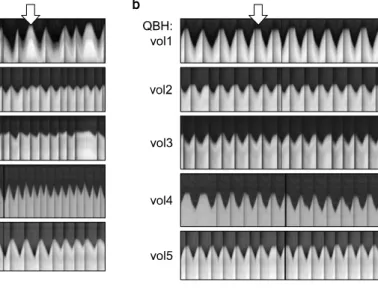
Fig. 2. Upper liver breath-hold mo- tion obtained using the RF naviga- tor pre-pulse with (a) FB and (b) QBH biofeedback breathing. The upper liver breath-hold motion reproducibility has been consider- ably improved at 90% exhalation using QBH biofeedback (arrow in (b)). Dark vertical lines indicate signal acquisition for MR images with a pause of the navigator.
tem has been combined with a gated 3D thoracic MRI acquis- ition with an upper dome scout navigator. The respiratory gat- ed T2-weighted SPACE MR pulse sequence with an MR res- piratory navigator of a 3T Siemens MRI has been employed for 3D thoracic imaging (range of acceptance gated position:
±4 mm and triggering position: 90% exhalation position).
Typical MR imaging parameters were TR/TE=∼4400/89 ms, FOV=380×380 mm
2, Pixel size: 1.19×1.19×4 mm
3and image matrix=320×320×52.
The residual upper liver dome motion using the navigator signal has been qualitatively evaluated. In this study, the navi- gator utilized an additional MR pre-pulse to measure the posi- tion of the liver dome before obtaining image signal with imaging pulses thus, navigator signals were not obtained dur- ing image signal acquisition, resulting in discontinued in- formation on liver dome displacement with time. Therefore, visual evaluation of liver dome displacement was made only during the navigator signal available. In the respiratory motion artifact analysis from the obtained gated 3D thoracic MR im- ages, the region of interest (ROI) was selected on the liver since the left side of the chest showed considerable cardiac motion artifacts including signals from blood vessels and the heart. Qualitative analysis of respiratory motion artifacts with/without QBH biofeedback has been assessed. The residual respiratory motion management within the gating window has been evaluated using the synchronized respiratory signal ob- tained from the RPM system. The RMSE (root mean square
error) of abdominal displacement in the gated window has been investigated in the phase domain and the RMSE in peri- od was also computed from each waveform. The slope of the linear fit on the collected entire data has been investigated as a baseline drift.
Quantitative statistical comparison of RMSE in displacement and period from the different breathing conditions was per- formed using the paired Student’s t-test (Microsoft Excel 2010, TTEST).
Results
Five healthy human subjects participated across ten MRI sessions including five free breathing and five QBH biofeed- back sessions. The upper liver dome motion data from navi- gator signals, the abdominal motion data from the RPM sys- tem, and gated 3D thoracic MRI have been obtained simul- taneously. However, continuous navigator signals were not ac- quired during image signal acquisition due to a current techni- cal limitation.
1. Upper liver breath-hold motion management in the gating window
Fig. 2 shows 1D RF navigator signal of each subject that is
basically the upper liver dome position according to time un-
der (a) free breathing (FB) and (b) QBH. As can be easily
seen, the liver dome, when compared to FB, remained at the
Fig. 3. Gated T2 weighted 3D thoracic MR images obtained with (a) FB and (b) QBH biofeedback breathing. Respiratory-related motion artifacts are shown distinctly in liver and lungs with free breathing [arrows in (a)]. The artifacts have been reduced by controlling residual motions within the gating window during acquisitions using QBH biofeedback [arrows in (b)].
designated position longer under QBH in vol 1, 2, 4 and 5, re- sulting in reduction of the residual motion within the gating window during MR acquisitions (arrows in Fig. 2). In addi- tion, QBH biofeedback made the liver dome motion more re- producible through the entire session in both period and dis- placement aspect. In Fig. 2, each dark vertical line indicates a pause of the navigator during which imaging signal was obtained. More irregular pattern of the dark vertical lines with FB indicates unsuccessful signal acquisition in certain respira- tory cycle. In contrast, the dark vertical lines under the QBH biofeedback are more regular and reproducible, implying suc- cessful signal acquisition in each respiratory cycle.
2. Respiratory motion artifact analysis in gated T2 weighted 3D thoracic MR images
Fig. 3 shows coronal images corresponding to the navigator sequence in Fig. 2 [(a) FB and (b) QBH]. Anatomical features such as the liver, heart and lung, were clearly observable in addition to the diaphragm in all sets of Fig. 3. The MR signal of the organs with a higher proton density is more prominent than that of the lungs. Corresponding to the reduced residual
motion in the gating window in Fig. 2(a), the QBH biofeed- back reduced respiratory motion artifacts in lungs and liver of 3D thoracic MR images (especially in vol1, vol3 and vol5), while the scan time with QBH biofeedback increased from 355±78 s with FB to 377±35 s due to repeated breath-holds.
However, non-negligible cardiac motion artifacts such as blur- ring artifacts due to the signal from the blood vessels and the heart still can be seen in Fig. 3.
3. Residual motion and RMSE analysis in abdominal displacement
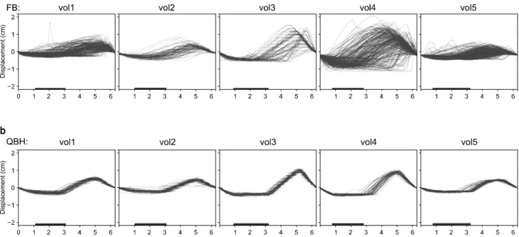
In Fig. 4, five sets of abdominal motion data obtained from
the RPM with/without QBH biofeedback are shown. The ab-
dominal motion with free breathing shows large variations in
displacement, period and baseline. However, the variations
have been significantly reduced by using the QBH biofeedback
system. The results are significant reductions of RMSE in dis-
placement due to QBH biofeedback from 2.0 mm of free
breathing to 0.7 mm over the entire cycle (67% reduction,
p-value: 0.02) and from 1.7 mm of free breathing to 0.7 mm
in the gated window (58% reduction, p-value: 0.14). The aver-
Fig. 4. Example of RMSE analysis in displacement with (a) FB and (b) QBH biofeedback. Average RMSE of abdominal displacement has been reduced from 1.7 mm of free breathing to 0.7 mm of QBH biofeedback within the gating window (bar on the bottom of each figure), providing more consistent breathing patterns.
age baseline drift obtained using a linear fit was reduced from 5.5 mm/min with free breathing to 0.6 mm/min (89% reduc- tion) with QBH biofeedback (p-value=0.017). In addition, the average RMSE of the respiratory cycle length was reduced from 1.8 sec with free breathing to 0.3 sec (i.e., 83% reduc- tion) with the implementation of QBH biofeedback, resulting in more consistent breathing patterns.
Discussion
In this study, the feasibility of QBH biofeedback to improve the residual motion management of the internal anatomy with gated 3D thoracic MRI was investigated for the first time. The need of MR imaging in radiotherapy is continuously increasing because of its non-ionizing radiation property and superior soft tissue contrast compared to CT imaging. However, current 3D thoracic MR imaging with a long scan time often suffers from respiratory motion artifacts such as blurring, causing non-diag- nostic quality of MR images and needs to be improved.
The most advanced technique in routine clinical practice is gated 3D thoracic MR imaging. One of the state of the art gated techniques is a real-time MR navigator that utilizes pre- RF-pulse to monitor real-time internal organ motion (usually
diaphragm motion) during MR imaging.
18)However, repeated 1.5 sec long signal acquisition time after respiratory triggering and a 4 mm wide gating window can cause substantial motion artifact due to residual motion within the gated window. In ad- dition, unpredictable variations in the cycle-to-cycle free-breath- ing (FB) may lead to the scan being on incorrect locations.
Under the 1.5 sec of signal acquisition window in 3D thoracic MR imaging, 2 sec QBH is considered appropriate in that it secures necessary information acquirement as well keeps rea- sonable imaging efficiency. Regarding the relationship between the signal acquisition window and the total scan time, if signal acquisition window decreases, the residual motion can be re- duced but the total scan time substantially lengthens.
In the image acquisition process, we set the imaging trigger point at 90% exhalation position with a 4 mm wide gating window in each respiratory cycle not to lose scan time effi- ciency too much (∼6 minutes) in free breathing condition.
Because the exhalation position is very reproducible over the
respiratory cycles,
14)a success rate of the acquisition triggering
is very high. With the acquisition trigger point near at the in-
halation position, the scan time can substantially increase due
to the likely low success rate of the acquisition triggering with
poor position reproducibility, and the image acquisition cannot
be completed without significant increase in the gating win- dow, resulting in more residual motions and a non-practical imaging protocol in practice. In fact, several respiratory cycles did not have dark vertical lines in the navigator signals under the FB condition [Fig. 2(a)], indicating unsuccessful signal ac- quisition sometimes with the current gating scheme (at 90%
exhalation position with a 4 mm wide gating window). How- ever, the QBH biofeedback increased the reproducibility of the respiratory motion in the entire respiratory cycles and the sta- bility of breath-hold position in the gating window, resulting in increase in the success rate of the acquisition triggering without substantial residual motion.
In this study, we demonstrated that the proposed system is feasible with current thoracic medical imaging in terms of mo- tion artifact reduction without significant scan time alternation.
This system can be applicable to regions can be affected by respiratory motion, such as the lung, pancreas, liver, kidney and esophagus in medical imaging and radiotherapy.
Conclusions
The study demonstrated that the QBH biofeedback improved the upper liver breath-hold motion reproducibility during the gated 3D thoracic MR imaging. This system can provide clin- ically applicable motion management of the internal anatomy for gated medical imaging and gated radiotherapy.
References
1. Yamamoto T, Langner U, Loo Jr BW, Shen J, Keall PJ:
Retrospective analysis of artifacts in four-dimensional CT Images of 50 abdominal and thoracic radiotherapy patients.
International Journal of Radiation Oncology*Biology*Physics 72(4):1250-1258 (2008)
2. Langner UW, Keall PJ: Quantification of artifact reduction with real-time cine four-dimensional computed tomography ac- quisition methods. International Journal of Radiation Oncology*
Biology*Physics 76(4):1242-1250 (2010)
3. Yang J, Yamamoto T, Cho B, Seo Y, Keall PJ: The im- pact of audio-visual biofeedback on 4D PET images: results of a phantom study. Medical Physics 39(2):1046-1057 (2012) 4. Hugo GD, Campbell J, Zhang T, Di Yan DS. Cumulative
lung dose for several motion management strategies as a func-
tion of pre-treatment patient parameters. Int J Radiat Oncol Biol Phys 74:593-601 (2009)
5. Theuws J, Kwa SLS, Wagenaar AC, et al: Prediction of overall pulmonary function loss in relation to the 3-D dose dis- tribution for patients with breast cancer and malignant lympho- ma. Radiotherapy and Oncology 49(3):233-243 (1998) 6. Marks LB, Bentzen SM, Deasy JO, et al: Radiation
dose-volume effects in the lung. International Journal of Radiation Oncology*Biology*Physics 76(3):S70-S76 (2010) 7. Lim S, Park SH, Ahn SD, et al: Guiding curve based on
the normal breathing as monitored by thermocouple for regular breathing. Medical Physics 34(11):4514-4518 (2007)
8. Wang Y, Christy PS, Korosec FR, et al: Coronary MRI with a respiratory feedback monitor: the 2D imaging case.
Magnetic Resonance in Medicine 33(1):116-121 (1995) 9. Venkat RB, Sawant A, Suh Y, George R, Keall PJ:
Development and preliminary evaluation of a prototype audio- visual biofeedback device incorporating a patient-specific guid- ing waveform. Physics in Medicine and Biology 53(11):N197 (2008)
10. George R, Chung TD, Vedam SS, et al: Audio-visual bio- feedback for respiratory-gated radiotherapy : Impact of audio in- struction and audio-visual biofeedback on respiratory-gated radiotherapy. Int J Radiat Oncol Biol Phys 65(3):924-933 (2006) 11. Locklin JK, Yanof J, Luk A, Varro Z, Patriciu A, Wood
BJ: Respiratory biofeedback during CT-guided procedures.
Journal of Vascular and Interventional Radiology 18(6):749-755 (2007)
12. Kini VR, Vedam SS, Keall PJ, Patil S, Chen C, Mohan R: Patient training in respiratory-gated radiotherapy. Medical Dosimetry 28(1):7-11 (2003)
13. Arnold JFT, Mörchel P, Glaser E, Pracht ED, Jakob PM. Lung MRI using an MR-compatible active breathing control (MR-ABC). Magnetic Resonance in Medicine 58(6):1092-1098 (2007)
14. Keall P, Mageras G, Balter J, et al: The management of respiratory motion in radiation oncology report of AAPM Task Group 76. Med Phys 33:3874-3900 (2006)
15. Berbeco RI, Nishioka S, Shirato H, Chen GT, Jiang SB: Residual motion of lung tumours in gated radiotherapy with external respiratory surrogates. Physics in Medicine and Biology 50(16):3655 (2005)
16. Park YK, Kim S, Kim H, Kim IIH, Lee K, Ye SJ: Quasi- breath-hold technique using personalized audio-visual biofeed- back for respiratory motion management in radiotherapy.
Medical Physics 38:3114 (2011)
17. Kim T, Pollock S, Lee D, O’Brien R, Keall P: Audiovisual biofeedback improves diaphragm motion reproducibility in MRI.
Medical Physics 39:6921 (2012)
18. McRobbie DW, Moore EA, Graves MJ, Prince MR: MRI
from Picture to Proton Cambridge University Press 2006.
게이트 흉부자기 공명 영상법과 함께 사용할 수 있는 의사호흡정지(QBH) 바이오 피드백
*Department of Radiation Oncology, University of Virginia, Virginia, USA,
†Department of Radiation Oncology, Mayo Clinic, Florida, USA,
‡이화여자대학교 방사선종양학과,
§Department of Radiology, Mayo Clinic, Florida, USA,
∥
Radiation Physics Laboratory, Sydney Medical School, University of Sydney, Sydney, Australia,
¶