∙ Received: March 8, 2010. Accepted: March 31, 2010.
∙ Corresponding author: Seok Hwan Yoon
Department of Nuclear Medicine, Seoul National University Hospital, 28 Yungun-dong, Jongno-gu, Seoul, 110-744, Korea
Tel: +82-2-2072-2535, Fax: +82-2-766-9083 E-mail: [email protected]
Original Article 동적 영상 획득 방식을 이용한 정적 영상의 움직임 보정
서울대학교병원 핵의학과
윤석환⋅승종민⋅김계환⋅김재일⋅이형진⋅김진의⋅김현주
The Application of Dynamic Acquisition with Motion Correction for Static Image
Seok Hwan Yoon, Jong Min Seung, Kye Hwan Kim, Jae Il Kim, Hyung Jin Lee, Jin Eui Kim and Hyun Joo Kim Department of Nuclear Medicine, Seoul National University Hospital
Purpose: The static image of nuclear medicine study should be acquired without a motion, however, it is difficult to acquire static image without movement for the serious patients, advanced aged patients. These movements cause decreases in reliability for quantitative and qualitative analysis, therefore re-examination was inevitable in the some cases. Consequently, in order to improve the problem of motion artifacts, the authors substituted the dynamic acquisition technique for the static acquisition, using motion correction. Materials and Methods: A capillary tube and IEC body phantom were used. First, the static image was acquired for 60 seconds while the dynamic images were acquired with a protocol, 2 sec/frame×30 frames, under the same parameter and the frames were summed up into one image afterwards. Also, minimal motion and excessive motion were applied during the another dynamic acquisition and the coordinate correction was applied towards X and Y axis on the frames where the motion artifact occurred. But the severe blurred images were deleted. Finally, the resolution and counts were compared between the static image and the summed dynamic images which before and after applying motion correction, and the signal of frequency was analysed after frequency spatial domain was transformed into 2D FFT. Supplementary examination, the blind test was performed by the nuclear medicine department staff. Results: First, the resolution in the static image and summed dynamic image without motion were 8.32 mm, 8.37 mm on X-axis and 8.30 mm, 8.42 mm on Y-axis, respectively. The counts were 484 kcounts, 485 kcounts each, so there was nearly no difference. Secondly, the resolution in the image with minimal motion applying motion correction was 8.66 mm on X-axis, 8.85 mm on Y-axis and had 469 kcounts while the image without motion correction was 21.81 mm, 24.02 mm and 469 kcounts in order. So, this shows the image with minimal motion applying motion correction has similar resolution with the static image. Lastly, the resolution in the images with excessive motion applying motion correction were 9.09 mm on X-axis, 8.83 mm on Y-axis and had 469 kcounts while the image without motion correction was 47.35 mm, 40.46 mm and 255 kcounts in order.
Although there was difference in counts because of deletion of blurred frames, we could get similar resolution.
And when the image was transformed into frequency, the high frequency was decreased by the movement.
However, the frequency was improved again after motion correction. In the blind test, there was no difference between the image applying motion correction and the static image without motion. Conclusion: There was no significant difference between the static image and the summed dynamic image. This technique can be applied to patients who may have difficulty remaining still during the imaging process, so that the quality of image can be improved as well as the reliance for analysis of quantity. Moreover, the re-examination rate will be considerably decreased. However, there is a limit of motion correction, more time will be required to successfully image the patients applying motion correction. Also, the decrease of total counts due to deletion of the severe blurred images should be calculated and the proper number of frames should be acquired. (Korean J Nucl Med Technol 2010;14(1):46-53)
Key Words : Dynamic acquisition, Motion correction, 2D FFT, Resolution, Frequency
Fig. 1. Capillary tube.
Fig. 2. IEC body phantom.
Table 1. Summary of image acquisition and processing parameters
Subject Scan Type Movement Processing
Static acquisition Static None None
Dynamic acquisition Dynamic None Summation
Minimal motion Dynamic Minimal Summation & Motion correction
Excessive motion Dynamic Excessive Summation & Motion correction, Frame exception
서 론
핵의학 영상은 시간 정보에 따라 정적(static), 동적(dynamic), 게이트(gate), 검사 등으로 분류할 수 있다. 정적 영상은 핵의 학 영상 중 가장 일반적인 영상이며, 일정 시간 동안 얻어진 계수를 특정 좌표에 표시해 줌으로써 방사성 추적자 분포를 영상화하는 기술이다. 더불어 정적 영상을 통해서 특정 목적 장기의 섭취율과 배설율 등 정량적 분석을 통해 진단 및 치 료를 결정할 수 있다. 이러한 정적 영상을 얻는 시간에 대해 서 특별히 정해진 바는 없으나 일정 시간 동안 움직임 없이 영상을 획득하여야 한다.1) 하지만 일정 시간 동안 움직임 없 이 한 자세를 유지하는 것이 환자에게 어려운 일이며 특히 소아 환자, 고령 환자, 중환자에게 있어서 움직임이 빈번하 게 발생한다.
이러한 움직임은 정량적 분석의 결과에도 영향을 주어 검 사의 신뢰도가 감소될 수밖에 없으며 해상력과 같은 정성적 분석에도 영향을 끼치게 된다. 또한 정적 영상 획득 방식에 서는 움직임에 의해 발생되는 문제점을 해결할 수 있는 방 법이 없어 재검사가 불가피하며, 재검사로 인해 환자들과 검 사자에게 불필요한 시간 소요와 어려움이 있다. 이러한 문제 점을 해결하기 위해 영상을 획득하기에 앞서 움직임의 예상 시 시간적으로 정지된 정적 영상 획득 방식 대신 시간 정보 가 부가된 동적 영상 획득 방식을 이용하여 영상을 획득한 다. 그리고 움직임이 발생한 시간의 frame 보정과 각 frame 을 더하여 정적 영상의 움직임에 대한 문제점을 해결하고자 하였다.2)
실험재료 및 방법
실험재료와 실험기기로는 capillary tube와 IEC body phan- tom을 이용하여 e-cam (Siemens Medical System, Erlangen, Germany) 감마카메라로 실험하였다.
실험방법은 capillary tube와 IEC body phantom을 이용하 여 움직임이 없을 때 정적 영상 획득 방식과 동적 영상 획득 방식의 영상을 비교 분석하기 위해 capillary tube 4개를 상
하좌우로 위치시키고 동일한 조건으로 영상을 획득하기 위 해 정적 영상 획득 방식에서는 time set method (60초)를 적 용하였으며 동적 영상 획득 방식에서는 30 frame×2 sec (60 초)를 적용하였다. 모든 실험은 256×256 matrix, zoom 1.0으 로 설정하였다.
동일한 조건으로 동적 영상 획득 방식을 적용하여 임의의 상하좌우 최소한의 움직임(0~3 cm)과 과도한 움직임(3~10 cm)을 주었다. 움직인 영상에 대해 움직임 보정(motion cor- rection)을 적용하여 motion correction 적용 전후의 영상을 비교 분석하였다. 여기서 최소한의 움직임과 과도한 움직임
Fig. 4. The images is acquired using capillary tube without motion. (A) Static image (B) Summed dynamic image (C) X-profile gaussian curve of static image (D) X-profile gaussian curve of summed dynamic image (E) Y-profile gaussian curve of static image (F) Y-profile gaussian curve of summed dynamic image.
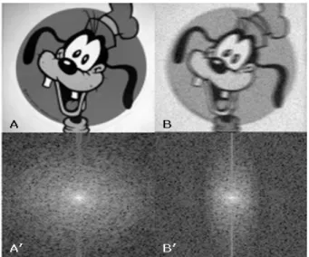
Fig. 3. Spatial domain (A, B) is transformed into frequency domain (A', B') by 2D FFT. The high frequency of the right image (B') is decreased by blur compared to the left image (A').
의 차이는 움직임의 속도와 움직임의 범위 차이이므로 움직 임의 속도와 범위가 클수록 흐림 현상(blur)이 많이 발생하 여 움직임의 차이에 대한 motion correction에 대해서도 알 아보고자 하였다.
영상의 처리는 Syngo software version 7.7 (Siemens Medical System, Erlangen, Germany)을 이용하였다. 동적 영상 획득 방식으로 얻은 각각의 frame을 확인한 후 움직임이 발생한 frame에 대해서 움직이지 않은 frame을 기준으로 X, Y축 좌 표 보정을 실시하며 과도한 움직임에 대해 흐림 현상이 심 하게 발생한 frame에 대해 좌표 보정이 어려운 경우 frame을 제외하였다. 그리고 보정된 각각의 frame을 하나의 영상으 로 더하였다(Table 1).
실험결과 대한 분석방법은 X, Y축으로 11개의 profile을 그려 움직임이 없을 때 정적 영상 획득 방식으로 얻은 영상 과 동적 영상 획득 방식으로 얻은 각각의 frame을 더한 영상
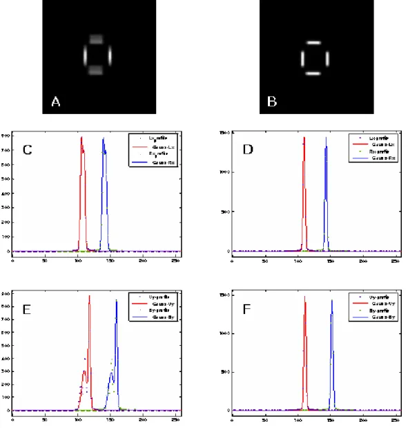
Fig. 5. The images is acquired using capillary tube with minimal motion. (A) Before motion correction (B) After motion correction (C) X-profile gaussian curve of static image before motion correction (D) Y-profile gaussian curve after motion correction (E) X-profile gaussian curve before motion correction (F) Y-profile gaussian curve after motion correction.
에 대해 resolution을 비교 분석하였다. 그리고 움직임이 있 는 영상에 대해 motion correction 전후의 resolution을 비교 분석하였다.
2D Fast Fourier Transform (FFT)을 이용하여 2차원 공간 영역의 영상을 2차원 주파수 영역으로 변환하여 움직임에 의한 주파수 변화와 motion correction 전후의 영상의 주파 수를 비교 분석해 보았다. 2D FFT란 2차원 고속 퓨리에 변 환을 뜻하는데, 퓨리에 변환은 공간적 위치에서의 신호 크기 를 가지고 있는 영상 신호를 신호의 성분 즉, 주기 함수의 변 동성을 분석하기 위한 영상 처리 기법이다. 저주파수 영역은 주기가 길어 크기가 느리게 변하는 신호를 나타내며 영상의
대략적인 모양을 나타낸다. 고주파수 영역은 주기가 짧아 크 기가 빠르게 변하는 신호와 영상의 경계선 부위를 나타내며, 영상의 자세한 변화를 나타낸다.4) 흐림 현상이 발생한 영상 에 대해서는 경계선 부분이 모호해져 고주파수 영역이 감소됨 을 알 수 있어 영상의 움직임 변화를 분석할 수 있다(Fig. 3).
움직임이 없을 때 정적 영상 획득 방식으로 얻은 영상과 동적 영상 획득 방식으로 얻은 영상에 대해 total counts를 비교 분석 하였으며, 최소한의 움직임과 과도한 움직임의 영 상에 대해 motion correction 전후의 total counts를 비교 분 석 하였다.
서울대학교병원 핵의학과 방사선사를 대상으로 움직임이
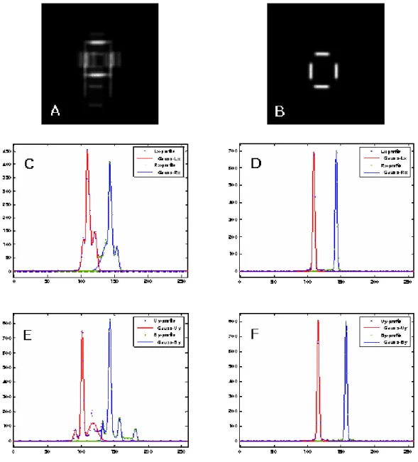
Fig. 6. The images is acquired using capillary tube with excessive motion. (A) Before motion correction (B) After motion correction (C) X-profile gaussian curve before motion correction (D) Y-profile gaussian curve after motion correction (E) X-profile gaussian curve before motion correction (F) Y-profile gaussian curve after motion correction.
Table 2. Resolution data
(unit: mm)
Subject F W H M
X-profile (left) X-profile (right) Y-profile (upper) Y-profile (bottom)
Static acquisition 8.32 8.23 8.37 8.56
Dynamic acquisition 8.30 8.23 8.42 8.54
Motion (minimal) 21.81 19.27 24.02 25.48
Motion correction 8.66 8.52 8.85 9.07
Motion (excessive) 27.67 47.35 40.46 37.80
Motion correction 9.09 9.16 8.83 8.73
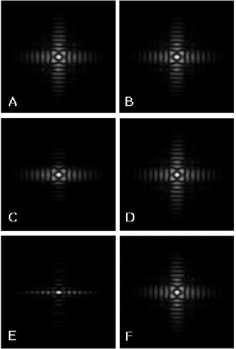
Fig. 7. The images are transformed into frequency domain by 2D FFT. (A) Static image without motion (B) Summed dynamic image without motion (C) Before motion correction (minimal motion) (D) After motion correction (minimal motion) (E) Before motion correction (excessive motion) (F) After motion correction (excessive motion).
없을 때 정적 영상 획득 방식으로 얻은 영상과 동적 영상 획 득 방식으로 얻은 영상, 움직임이 있는 영상의 motion cor- rection 전후의 영상에 대해 설문지를 통한 영상을 비교 분 석하였다(Appendix 1).
결 과
1. Resolution 평가에 의한 비교 분석
Capillary tube를 이용한 실험에서 움직임이 없을 때 정적 영상 획득 방식으로 얻은 영상과 동적 영상 획득 방식으로 얻은 각각의 frame을 더한 영상에 대해 resolution은 크게 차 이가 나지 않았다. 그러나 최소한의 움직임과 과도한의 움직 임을 주었을 경우 Full Width Half Maximum (FWHM) 값이 상당히 증가하여 resolution이 감소되었음을 알 수 있다. 움 직인 영상의 motion correction을 적용한 영상에 대해서는 FWHM 값이 감소하여 resolution이 움직임이 없을 때의 영 상과 비슷한 결과 값을 보였다(Fig. 4, Fig. 5, Fig. 6, Table 2).
2. Frequency 평가에 의한 비교 분석
2D FFT를 이용하여 공간 영역의 영상을 주파수 영역으 로 변환하게 되면 영상의 가운데 영역은 저주파수 영역을 나타내고 가장자리와 경계 부분은 고주파수 영역으로 나타 나게 된다. 움직임이 없는 경우 정적 영상 획득 방식을 이용 하여 얻은 영상과 동적 영상 획득 방식을 이용하여 얻은 영 상에서 동일한 주파수 영역을 나타내었다. 움직임이 발생한 영상에 대해 변환하였을 경우 움직임에 의해 경계선이 모호 해져 고주파수 영역이 감소됨을 알 수 있다. 움직임이 많을 경우 고주파수 영역은 급격하게 감소되었으며 motion cor- rection 적용 후 고주파수 영역이 보정되었음을 알 수 있다 (Fig. 7).
3. Total counts 평가에 의한 비교 분석
움직임이 없을 때의 정적 영상 획득 방식으로 얻은 영상 과 동적 영상 획득 방식으로 얻은 각각의 frame을 더한 영상 의 total counts는 각각 484, 485 kcounts로 거의 차이가 없었 다. 또한 최소한의 움직임을 준 영상에 대해 motion correc- tion 전후의 total counts는 각각 474, 471 kcounts로 거의 차 이가 없었다. 그러나 과도한 움직임을 준 경우, 최소한의 움
직임을 주었을 경우보다 흐림 현상이 많이 발생하여 X, Y축 의 좌표 보정이 어려운 경우 frame을 제외하고 각 frame을 더하였기 때문에 frame의 삭제된 수만큼 total counts에서 각 각 471, 227 kcounts로 차이를 보였다.
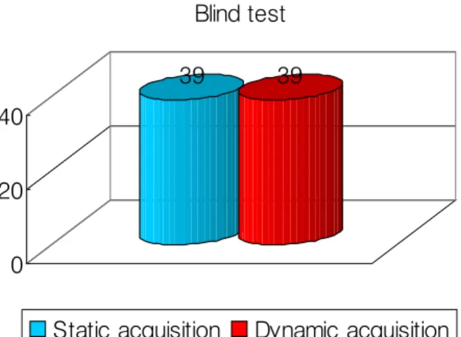
4. Blind test 평가에 의한 비교 분석
IEC body phantom을 이용하여 움직임이 없는 경우 정적 영상 획득 방식으로 얻은 영상과 동적 영상 획득 방식으로 얻은 각각의 frame을 더한 영상, 최소한의 움직임과 과도한 움직임에 의해 motion correction 전후의 영상을 육안적 평 가로 비교한 결과는 움직임이 없는 경우의 정적 영상 획득 방식의 영상과 동적 영상 획득 방식으로 획득한 각각의 frame을 더한 영상에 대해서는 거의 차이가 없었으며 최소
39 39
0 20 40
Blind test
Static acquisition Dynamic acquisition
Fig. 8. Blind test for static image and summed dynamic image.
75
131
31
118
0 20 40 60 80 100 120 140
Blind test
Minimal motion Motion correction (minimal) Excessive motion Motion correction (excessive)
Fig. 9. Blind test for the images with minimal motion and excessive motion before and after motion correction.
한의 움직임과 과도한의 움직임에 의한 motion correction 전후의 영상 비교에서는 motion correction 후의 영상의 질 이 월등히 우수하다고 평가되었다(Fig. 8, Fig. 9).
결론 및 고찰
핵의학과 검사 중 정적 검사는 가장 일반적인 검사이며 정적 영상을 통해 특정 목적 장기의 섭취율과 배설율 등 정 량적 분석을 통해 진단 및 치료를 결정하게 된다. 정적 영상 을 획득하는 시간에 대해서 특별히 정해진 바는 없으나 일 정 시간 동안 움직임 없이 영상을 획득하여야 한다. 움직임 에 의해 발생되는 문제점은 정성적 분석과 정량적 분석의 신뢰도가 감소하며 재검사 또한 불가피한 실정이었다. 이에 본 연구는 기존의 정적 영상 획득 시 동적 영상 획득 방식으 로 바꾸어 motion correction을 통해 움직임 발생 시 발생되
는 문제점을 해결하고자 하는 데 목적이 있다. Phantom 실 험에서 정적 영상 획득 방식을 이용한 영상과 동적 영상 획 득 방식을 이용하여 각각의 frame을 더한 영상의 resolution, frequency, total counts, blind test 비교 분석에서 결과 값의 차이가 없음을 알 수 있었다. 또한 임의적인 최소한의 움직 임과 과도한의 움직임을 주어 실험 한 경우, motion correc- tion 전후의 영상 비교에서는 motion correction 후 영상의 resolution, frequency, blind test의 결과 값이 움직임이 없을 때의 영상 결과와 비슷한 결과를 보였다. 하지만 과도한 움 직임을 준 경우 frame당 흐림 현상이 많이 발생하여 좌표 보 정으로 인한 motion correction이 어려워 frame을 제외하는 방법을 이용하였기 때문에 과도한 움직임에 대한 motion correction 비교 분석에서는 frame을 제외한 만큼 total counts의 차이를 보였다. 그러므로 움직임이 예상되는 환자 검사 시 초 당 카운트 수(cps)를 계산하여 충분한 frame 수를 확보함으로써 검사에 필요한 최소한의 counts를 획득하여야 할 것이다.3,5) 이 방법을 적용함으로써 기존의 정적 영상 획 득 시 발생되었던 움직임에 의해 감소되었던 영상의 해상력 과 정량적 분석의 신뢰도가 증가할 것이며 재검사 발생률도 현저히 줄어들 것으로 기대된다. 그러나 움직임 보정을 하기 위해서는 움직임의 좌표 보정이 X, Y축으로의 국한된 보정 의 한계와 정확한 움직임 보정을 하기 위해서는 움직임 보 정 시간이 오래 소요될 수도 있다. 또한 기존의 정적 영상 획득 시 움직임을 보정할 수 있는 방법이 거의 없었다. 그러 므로 정확한 움직임 보정을 하기 위해서는 프로그램 개발 또한 필요하다고 생각된다. 그리고 핵의학 정적 검사에 움직 임 보정 방법을 모두 적용시킬 수 없으므로 임상 적용에 대 한 광범위한 연구가 현실적으로 필요하며 향후 지속적인 연 구가 기대되는 바이다.
요 약
정적 영상 획득 시 기존의 정적 영상 획득 방식 대신 동적 영상 획득 방식을 이용하여 움직임에 의한 motion correc- tion을 적용함으로써 정적 영상 획득 시 발생되는 움직임에 의한 문제점을 해결하고자 하였다.
실험은 capillary tube와 IEC body phantom을 이용하여 움직임이 없을 때 정적 영상 획득 방식으로 얻은 영상과 동 적 영상 획득 방식으로 얻은 각각의 frame을 더한 영상에 대 해 resolution, frequency, total counts, blind test를 비교 분석 하였으며 임의로 최소한의 움직임과 과도한 움직임을 주어
motion correction 전후의 영상에 대해서도 resolution, fre- quency, total counts, blind test를 비교 분석 하였다. 기존의 정적 영상 획득 방식으로 얻은 영상과 동적 영상 획득 방식 으로 얻은 각각의 frame을 더한 영상의 resolution, fre- quency, total counts, blind test의 결과 값의 차이가 없었다.
또한 최소한의 움직임과 과도한 움직임을 준 영상에 대해 motion correction 적용 전후의 비교 결과 값은 motion cor- rection 후 resolution, frequency, blind test의 결과 값이 움직 임이 없을 때의 정적 영상과 거의 차이가 없었다. 하지만 과 도한 움직임에 대한 보정 시 frame당 흐림 현상이 많이 발생 하였으므로 좌표 보정이 어려워 frame을 제외하는 방법을 적용하였기 때문에 과도한 motion correction 후 삭제한 frame 수만큼 total counts에서 차이를 보였다. 정적 영상 획 득 시 움직임이 예상되는 환자에게 기존의 정적 영상 획득 방식이 아닌 동적 영상 획득 방식을 이용하여 움직임 발생 시 좌표 보정과 흐림 현상이 심한 frame 제외 방법을 이용하 여 정적 영상에서 움직임에 의해 발생되었던 영상의 질 저 하와 정량적 분석의 신뢰도 감소, 재검사에 대한 문제점을 해결할 수 있을 것이라고 생각되며 motion correction에 제 공되는 다양한 프로그램 개발과 임상 적용에 대한 광범위한 연구가 현실적으로 필요하며 향후 지속적인 연구가 기대되 는 바이다.
Appendix 1. Image quality questionnaire
Question 1.
Looking at image.
How clear and sharp edges do circles have on each image ? Please check appropriate score
1 2 3 4 5
5 = Defined sharp & clear 1 = Defined poorly
REFERENCES
1. 고창순. 제2판 핵의학. 고려의학 1997;125-137.
2. Alliance Medical, Home Farm Drive, Upton, Banbury, Oxfordshire OX15 6HU, UK. An analysis of motion correction for 99mTc- DMSA renal imaging in paediatrics. Radiography May 2007;
13:109-121.
3. J. S. Fleming. A technique for motion correction in dynamic scintigraphy. J Nucl Med. September 1984;9:397-402.
4. http://www.cs.unm.edu/~brayer/vision/fourier.html. Introduction to fourier transforms for image processing.
5. David A. Turner, Ernest W. Fordham, Amjd Ali, Paul D. Rubin, Robert C. Motion Corrected Hepatic Scintigraphy: An Objective Clinical Evaluation. J Nucl Med. 1978;19:142-148.