Review https://doi.org/10.14478/ace.2017.1041
다양한 소수성 물질이 개질된 키토산 나노입자의 약물전달체로서 응용성 고찰
정경원⋅나재운†⋅박준규*,†
순천대학교 공과대학 고분자공학과, *(주)시지바이오
(2017년 5월 8일 접수, 2017년 5월 23일 심사, 2017년 5월 26일 채택)
Application of Various Hydrophobic Moiety-modified Chitosan Nanoparticle as a Drug Delivery Carrier
Gyeong-Won Jeong, Jae-Woon Nah†, and Jun-Kyu Park*,†
Department of Polymer Science and Engineering, Sunchon National University, Jeonnam 57922, Republic of Korea
*CGbio Co.Ltd, Jeonnam Jangseong-gun 57248, Republic of Korea (Received May 8, 2017; Revised May 23, 2017; Accepted May 26, 2017)
초 록
천연고분자 키토산은 생체적합하고 생분해성의 특성뿐만 아니라 항암, 항균, 콜레스테롤 저하 등의 다양한 생체활성 을 갖고 있어 의료용 분야에서 많이 응용되고 있다. 현재 키토산을 약물전달시스템에 응용한 다양한 약물이 담지 된 키토산 나노입자를 개발하여 질병을 치료할 수 있는 연구가 활발히 진행 중에 있다. 키토산에 존재하는 free 아민 (-NH2) 그룹은 다양한 소수성기를 물리적⋅화학적 개질을 통해 결합이 가능하며 소수성기가 도입된 키토산은 물에 분산시 자기회합에 의한 shell-core 나노입자를 형성하고 core 부분에 다양한 난용성 약물을 담지하여 물에 대한 용해 도를 증가시킬 수 있으며, 단백질, 항암제, 백신 등의 다양한 약물을 담지하여 기존 약물의 부작용을 최소화하여 치료 효과를 극대화할 수 있다. 또한, 키토산에 도입된 소수성기에 따라 입자의 크기 및 방출 속도를 제어할 수 있어 다양한 의료용 분야에 응용이 가능하다. 본 총설에서는 다양한 소수성기가 도입된 키토산 나노입자의 제조 및 특성과 특성에 따른 약물전달시스템의 응용성에 관하여 논의 하고자 한다.
Abstract
Natural polymer chitosan has been widely applied to medical fields due to its biochemical activities such as anticancer, anti- bacterial and lowering cholesterol in addition to biocompatibility and biodegradability. Currently, researches are being actively conducted to develop various drug-encapsulated chitosan nanoparticles for curing different diseases by applying chitosan to a drug delivery system. The free amine (-NH2) group present in chitosan can bind to various hydrophobic groups by physical and chemical modification and the chitosan with hydrophobic groups can form shell-core nanoparticles by self-assembly when dispersed in water. In addition, an insoluble drug can increase the solubility against water when it was encapsulated in the core of chitosan nanoparticles. Also, the therapy effect can be maximized by minimizing side effects of drugs such as proteins, anticancer drugs and vaccines when they were encapsulated in the core of chitosan nanoparticles. Moreover, it is possible to control the particle size and release rate according to the hydrophobic group introduced to chitosan, so that it can be applied to a wide range of medical fields. The purpose of this review is to discuss the preparation and property of chitosan nano- particles modified with various hydrophobic groups, and the application to drug delivery systems according to their property.
Keywords: chitosan, hydrophobic group, self-assembly, shell-core, drug-encapsulated chitosan nanoparticle
1. 서 론
1)
약물전달시스템(DDS, drug delivery system)은 기존의 약물의 부작 용을 최소화하고 적은 양의 약물을 효과적으로 전달하여 약효를 극대
† Corresponding Author: J.-W. Nah, Sunchon National University, Department of Polymer Science and Engineering, Jeonnam 57922, Republic of Korea / J.-K. Park, CGbio Co.Ltd, Jeonnam Jangseong-gun 515-898, Republic of Korea Tel: +82-61-750-3566, +82-61-392-9840
e-mail: jwnah@sunchon.ac.kr, pjk23@cgbio.co.kr
pISSN: 1225-0112 eISSN: 2288-4505 @ 2017 The Korean Society of Industrial and Engineering Chemistry. All rights reserved.
화하기 위한 제형으로 정의할 수 있다[1,2]. 최근 DDS 분야에 나노기 술을 이용한 나노 입자가 개발되어 나노 약물전달체, 나노진단시약, 나노의료용 물질, 그리고 나오바이오물질 등이 다양하게 활용되고 있 다[3]. 현재 이러한 나노 기술을 접목한 약물전달시스템 응용 범위는 지속성 약물방출시스템, 제어방출시스템 그리고 표적 지향적 약물전 달시스템이 폭넓게 응용되어 지고 있다[4]. 지속성 약물방출 시스템은 생체 이용률이 낮거나 약물이 너무 서서히 흡수되거나 지나치게 빨리 체외로 소실되는 경우 약물의 방출속도를 늦춤으로써 이러한 문제점 을 줄이고자 설계된 제형이고, 제어방출시스템은 표적 부위의 농도를 제어함으로써 실제의 치료효과를 조절하는 것을 목적으로 하는 것으
Figure 1. Drug delivery system using EPR effect of drug-loaded chitosan nanoparticle (Ref.[30]).
로 약물전달시간을 연장할 수 있을 뿐만 아니라 약물방출 속도의 재 현 및 예측이 가능한 제형이다. 또한, 표적 지향적 약물전달시스템은 약물의 불필요한 분포를 억제하여 비 표적 부위를 보호하고 표적부위 로만 약물을 전달하는 방법으로 암 치료 시 정상세포에는 영향을 주 지 않고 암세포에만 특이적으로 반응하도록 설계한 제형이다[5-8]. 현 재 의료용 분야인 암을 치료하는 연구에서 정상세포에 대한 부작용을 최소화하고 암의 치료 효율을 극대화하기 위해서 표적 지향적 약물전 달시스템을 많이 응용하고 있다[9].
현재 DDS분야에서 다양한 기능과 성능을 가진 생체고분자 및 합성 고분자를 이용한 새로운 약물 전달체를 개발하는 연구가 활발하게 진 행되고 있다[10]. DDS용 재료로서 요구되는 고분자의 특성은 생체적 합성, 생분해성, 화학적⋅생물학적 무독성 등이 필수 조건이다[11,12].
하지만 DDS 재료로서 합성고분자는 체내에서 분해되지 않고 잔존하 게 되어 부작용을 유발할 수 있는 단점을 갖고 있다[13,14]. 이러한 문 제를 해결하기 위해 최근에는 생물학적 특성이 우수하고 생체 적합한 천연 고분자 키토산을 이용하여 다양한 기법의 약물전달 시스템이 활 발히 연구되고 있다[15,16].
키토산은 게나 새우 등의 갑각류, 오징어 연골 연체류 등에 분포되 어 있는 천연 고분자인 키틴을 농축 알칼리로 처리하여 얻어지는 물 질로서 β-(1,4)-glycosidic 결합으로 연결된 D-glucosamine과 N-acetyl D-glucosamine 두 단위체로 구성된 생분해성 천연 다당류이다[17]. 키 토산은 항암효과, 콜레스테롤 감소, 면역 활성 및 항균성 등 우수한 생리활성을 갖는 것으로 잘 알려져 있다[18,19]. 키토산을 이용한 응 용 분야로는 화장품, 수처리, 의약품, 식품, 바이오산업 등이 있으며 최근에는 의료용 분야로의 연구가 확대되고 있다[20]. 또한 생체 적합 성이 우수하고, 독성이 낮으며 강한 양전하를 띄고 있어 약물 전달체 나 유전자 전달체로 많이 응용되어지고 있다[21,22]. 최근에 연구 논 문에 따르면 키토산에 다양한 소수성기를 도입하여 shell-core를 형성 하는 micelle 나노입자를 제조하고 core 내부에 난용성 약물들을 담지 하여 물에 대한 용해도를 향상시키는 연구들이 보고된바 있으며[23], 이를 응용하여 소수성 물질들이 도입된 키토산 나노입자에 다양한 항 암제를 담지하여 암세포를 치료하는 연구들도 많이 진행 중에 있다 [24]. 이러한 키토산 나노입자는 도입된 소수성기에 따라 약물의 방출 속도를 제어할 수 있으며, 약물의 방출 속도에 따라 치료효과도 현저 하게 달라질 수 있다[25]. 그러므로 본 총설에서는 다양한 소수성기가 도입된 키토산 나노입자의 제조 및 특성과 특성에 따른 약물전달시스 템의 응용성에 관하여 논의하고자 한다.
2. 약물전달시스템에 응용하기 위한 키토산의 특성
키토산은 N-acetylglucosamine과 glucosamine이 β-1,4-glycosidic 결합으로 이루어진 반복단위로 구성되어 있으며 자연상에 존재하는 게나 새우 껍질인 키틴질로부터 탈아세틸화하여 얻어지는 풍부한 물 질이다[26]. 이러한 키토산은 점막에 접착능력이 우수하며, 생체적합 하고, 생분해성이며, 낮은 독성과 낮은 면역을 갖는다는 장점을 가지 고 있다[27,28]. 이러한 키토산의 물리화학적 특성과 생물학적 특성은 키토산의 분자량 및 탈아세틸화도에 따라 달라지기 때문에 다양한 분 야에서 그 응용성이 확대되고 있다[29]. 또한, 키토산에 free한 아민 (-NH2) 그룹이 존재하여 카르복실(-COOH) 그룹을 갖는 물질을 쉽게 물리적⋅화학적 개질이 가능하다며 소수성기가 도입된 키토산 나노 입자의 경우 암 치료에 적용 시 암 조직 주변의 느슨한 혈관의 높은 투과성 EPR (Enhanced permeability and retantion)효과에 의해 종양에
나노입자의 축적(acculmulation)을 증가시키는 수동적인 표적(passive targeting)이 가능하여 치료효율을 극대화할 수 있다(Figure 1)[30]. 또 한, 키토산의 pKa 값이 약 6.5의 특성을 갖고 있기 때문에 다양한 소 수성 물질을 도입하여 제조된 키토산 나노입자에 담지된 약물을 pH 변화에 따른 방출을 제어할 수 있다는 장점을 갖고 있다[31,32]. 이러 한 특성을 갖는 키토산을 약물 전달체로 응용했을 시체내의 부작용 을 줄일 수 있고, 다양한 소수성기를 도입하여 약물의 방출을 효율적 으로 제어하여 치료 효율을 증가시킬 수 있는 매우 우수한 물질이다.
3. 다양한 소수성 물질이 도입된 키토산 나노입자의 제조와 특성
3.1. 소수성기 포화 지방산(Saturated fatty acid)이 도입된 키토산 나노입자
지방산은 크게 포화 지방산과 불포화 지방산으로 분류되며 포화지 방산은 탄소사슬이 단일결합으로 구성되어 있고 불포화 지방산은 탄 소사슬에 중간에 단일결합과 이중결합이 교대로 존재한다. 이러한 불 포화 지방산의 경우 이중결합에 존재하는 파이 전자는 화학적 결합에 있어서 매우 불안정한 결합으로 외부 인자로부터 쉽게 공격받기 쉽기 때문에 불포화 지방산 보다 포화 지방산을 많이 이용하고 있다[33].
포화지방산은 탄화수소 사슬로 이루어져 말단에 카르복실(-COOH) 그룹이 존재하고 물에 대한 용해도가 매우 낮은 소수성 물질이다. 이 러한 지방산은 연결된 탄소(carbon)의 개수에 따라 Hexanoic acid (C6), Lauric acid (C12), Palmitic acid (C16), Stearic acid (C18) 등 다양 하게 명명되고 있다(Table 1)[34]. 이러한 지방산들은 말단에 카르복 실 그룹이 존재하여 키토산 아민에 쉽게 도입할 수 있는데, 지방산들 의 카르복실 그룹을 N-(3-Dimethylaminopropyl)-N’-ethylcarbodiimide hydrochloride (EDC)와 N-hydroxysuccinimide (NHS) 등의 가교제를 이용하여 활성화 시켜 키토산 아민 그룹과 아마이드 결합을 형성하여 화학적인 결합이 가능하다(Figure 2(A))[35]. 이때 지방산이 도입된 키 토산을 물에 분산 시켰을 시 친수성-소수성 결합에 의한 자기 회합 (self-assembly)이 일어나 shell-core 나노입자를 형성하게 된다(Figure 3)[36]. 이때 키토산 나노입자는 탄소수가 많은 지방산이 결합될수록 소수성-소수성 간의 상호작용이 증가하여 Core 부분에 소수성 밀도가 증가하게 되고, 이는 지방산의 탄소수에 따라 입자의 크기를 조절할 수 있을 뿐만 아니라, Core 내부에 난용성 약물 및 항암제를 담지하여 물에 대해 용해도를 향상 시킬 수 있다[37,38]. 또한, 탄소수가 다양한 지방산을 적절히 도입한다면 약물 방출 시간을 느리게 혹은 빠르게 조절할 수 있어 다양한 질병 치료에 응용이 가능할 것으로 사료된다.
Name Lipid number Chemical structure Reference
Hexanoic acid C6:0 [33]
Lauric acid C12:0
[34]
Palmitic acid C16:0
Stearic acid C18:0 [35]
Table 1. Type of Saturated Fatty Acid as Hydrophobic Moiety
(a)
(b)
Figure 2. Synthesis scheme of hydrophobic moiety-grafted chitosan. (a) Fatty acid-grafted chitosan, (b) Deoxycholic acid-grafted chitosan.
Figure 3. Structure of shell-core nanoparticle composed with hydrophilic chain and hydrophobic chain (Ref.[52]).
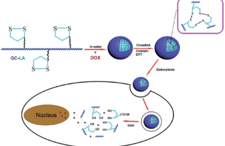
Figure 4. Illustration of the preparation of drug-loaded lipoic acid-grafted chtiosan micelle and drug-loaded core-crosslinked lipoic acid-grafted chtiosan micelle, and schematic representation of the response to the endogenous high GSH in tumor cells (Ref.[53]).
3.2. 담즙산(Bile acid)이 도입된 키토산 나노입자의 제조 및 특성 담즙산은 생체물질로서 간에서 만들어지며, 콜레스테롤 및 지방을 유화시켜 소화를 촉진시키는 물질 중에 하나이다. 또한 수용액에서 자 가적으로 소수성 중심을 갖는 미셀(Micelle)을 형성하고 소수성 약물을 효율적으로 담지 할 수 있다는 장점을 갖고 있다[39,40]. 이러한 담즙산 은 구조상으로 하이드록시기(-OH)의 위치, 입체적 배위, 및 곁사슬의 구조에 따라 리소콜릭산(Lithocholic acid), 데옥시콜릭산(Deoxycholic acid), 콜릭산(Cholic acid) 등으로 나눌 수 있고, 이것들은 약리작용이 우수하다는 장점을 갖고 있다. 이 중에서 데옥시콜릭산의 경우 소수 성이 가장 강한 것으로 보고되어져 있고 수많은 연구자들이 연구한바 있다[41]. 이러한 특성을 갖는 답즙산은 소수성 기능기로 카르복실 (-COOH)를 포함하고 있으며, 키토산에 존재하는 아민 그룹과 위에서 언급한 바와 같이 다양한 가교제를 이용하여 쉽게 화학적 결합을 할 수 있으며(Figure 2(B)), 물에 분산 시 자기회합에 의한 shell-core 나노 입자를 형성할 수 있다(Figure 3)[42,43]. 또한, 담즙산은 입체적으로 매우 큰 분자이기 때문에 키토산에 도입된 양이 증가할수록 입자의 크기가 증가되는 연구결과가 보고된 바 있으며, 이는 지방산과 차이 를 보이는데 이러한 입자크기의 차이를 보이는 이유는 소수성 물질의 분자 입체의 크기에 의한 것으로 보고하고 있다[44]. 이러한 키토산에 도입된 소수성 물질의 종류에 따라 입자 크기를 조절할 수 있으며, 이 는 입자의 크기에 따라 다양한 분야에 약물전달시스템을 응용할 수 있을 것으로 사료된다.
3.3. 리포익산(α-Lipoic acid)이 도입된 키토산 나노입자 제조 및 특성
리포익산은 천연적으로 만들어지는 화합물로서 건강에 매우 유익 한 물질이다[45]. 현재 리포익산은 하이드록실 라디칼 및 산화 라디칼 을 억제하는 항산화 효과가 매우 우수한 물질로 잘 알려져 있다[46].
이러한 특성을 갖는 리포익산은 생체 내에서 매우 유용한 물질이며, 이는 현재 약물전달 시스템에 응용하는 연구가 활발히 진행되고 있다. 리포익산은 말단에 disulfide bond (-S-S-)가 존재하는 lipoic ring 그룹 을 포함하고 있어 강한 소수성기를 나타낸다. 또한 말단에 카르복실 (-COOH) 그룹이 존재하기 때문에 다양한 가교제를 이용하여 키토산 의 아민(-NH2) 그룹과 화학적 결합을 할 수 있다는 장점을 갖고 있다
[47,48]. 현재 리포익산이 도입된 키토산 나노입자는 리포익산의 lipo- ic ring 그룹에 존재하는 disulfide bond가 특이적으로 암세포의 cyto- sol 내에 존재하는 glutathione (GSH)와 특이적으로 반응하여 -S-S- 결 합을 빠른 시간 내에 분해할 수 있다는 장점을 갖고 있어 암치료 연구 에 많이 응용되고 있다(Figure 4). 또한 이러한 리포익산이 도입된 키 토산 나노입자에 Core 부분에 항암제를 담지하여 암세포에 전달하였 을 시 전달된 키토산 나노입자는 암세포 내의 GSH에 의해 lipoic ring 그룹에 존재하는 -S-S- 결합이 분해되어 단시간에 담지된 항암제를 과 다 방출할 수 있어 짧은 시간 내에 암의 치료를 극대화할 수 있다는 장점을 갖고 있다[49,50]. 또한, 지방산과 답즙산과 같은 소수성기가 도입된 키토산 나노입자의 경우 자기회합을 할 수 있는 최소농도 값 인 critical aggregation concentration (CAC)가 존재하고 혈액 내에서 CAC 값이 높아짐에 따라 입자의 안정성이 떨어져 약물 전달 효율성 에 문제점이 있다고 보고하고 있다[51]. 그러나 리포익산이 도입된 키 토산 나노입자의 경우 lipoic ring 그룹의 -S-S- 결합을 crosslinking함 으로써 혈액 내의 입자의 안정성 및 암세포에 약물 전달 효율성을 증 가시켜 암 치료를 극대화할 수 있을 것으로 사료된다.
4. 결 론
생체적합하고 생분해성의 다양한 특성을 갖는 천연고분자 키토산 은 free 아민 그룹에 다양한 소수성기를 물리적⋅화학적 개질을 통하 여 결합이 가능하며 다양한 소수성기를 도입하여 제조된 키토산은 자 기회합에 의한 shell-core 나노입자를 형성하고, 제조된 키토산 나노입 자 내부 core 부분에 난용성 약물을 담지하여 물에 대한 용해도를 향 상시킬 수 있을 뿐만 아니라 도입된 소수성기에 따라 입자크기 및 담 지된 약물의 방출 속도를 조절할 수 있어 다양한 의료용 분야에 약물 전달시스템을 응용할 수 있을 것으로 사료된다. 또한 단백질, 항암제, 백신 등의 다양한 약물이 담지된 나노입자 제조가 가능함에 따라 기 존의 약물의 부작용을 최소화하여 약물을 효과를 극대화할 수 있는 특정 질병에 적합한 약물전달체의 개발이 가능할 것으로 사료된다.
감 사
This research was supported by The Leading Human Resource Training Program of Regional Neo industry through the National Research Foundation of Korea (NRF) funded by the Ministry of Science, ICT and future Planning (grant number) (NRF-2016H1D5A1910499) and grants from the Korean Health Technology R&D Project (HI16C0094), Ministry of Health & Welfare, Republic of Korea.
References
1. M. Jaiswal, R. Dudhe, and P. K. Sharma, Nanoemulsion: an ad- vanced mode of drug delivery system, 3 Biotech, 5, 123-127 (2015).
2. I. Khan, M. Khan, M. N. Umar, and D. H. Oh, Nanobiotechnology and its applications in drug delivery system: a review, IET Nanobiotechnol., 9, 396-400 (2015).
3. R. Pandey and G. K. Khuller, Nanotechnology based drug delivery system(s) for the management of tuberculosis, Indian J. Exp. Biol., 44, 357-366 (2006).
4. J. D. Kingsley, H. Dou, J. Morehead, B. Rabinow, H. E.
Gendelman, and C. J. Destache, Nanotechnology: A focus on nanoparticles as a drug delivery system, J. Neuroimmune Pharmacol., 1, 340-350 (2006).
5. B. Dineshkumar, K. Krishnakumar, A. R. Bhatt, D. Paul, J.
Cherian, A. John, and S. Suresh, Single-walled and multi-walled carbon nanotubes based drug delivery system: Cancer therapy: A review, Indian J. Cancer, 52, 262-264 (2015).
6. M. J. Tobin, G. Jenouri, I. Danta, C. Kim, H. Watson, and M. A.
Sackner, Response to bronchodilator drug administration by a new reservoir aerosol delivery system and a review of other auxiliary delivery systems, Am. Rev. Respir. Dis., 126, 670-675 (1982).
7. K. Hori, M. Suzuki, S. Tanda, S. Saito, and Q. Zhang, Functional-characterization of developing tumor vascular system and drug delivery (review), Int. J. Oncol., 2, 289-296 (1993).
8. A. Semalty, M. Semalty, R. Singh, S. K. Saraf, and S. Saraf, Iontophoretic drug delivery system: A review, Technol. Health Care, 15, 237-245 (2007).
9. C. Bharti, U. Nagaich, A. K. Pal, and N. Gulati, Mesoporous silica nanoparticles in target drug delivery system: A review, Int. J.
Pharm. Investig., 5, 124-133 (2015).
10. B. Krishnamoorthy, V. Karanam, V. R. Chellan, K. Siram, T. S.
Natarajan, and M. Gregory, Polymersomes as an effective drug de- livery system for glioma-a review, J. Drug Target., 22, 469-477 (2014).
11. Y. Hao, L. Wang, B. Zhang, D. Li, D. Meng, J. Shi, H. Zhang, Z. Zhang, and Y. Zhang, Manganese dioxide nanosheets-based re- dox/pH-responsive drug delivery system for cancer theranostic ap- plication, Int. J. Nanomed., 11, 1759-1778 (2016).
12. A. Vyas, A. Kumar Sonker, and B. Gidwani, Carrier-based drug delivery system for treatment of acne, ScientificWorldJournal, 2014, 276260-276273 (2014).
13. K. Songsurang, K. Siraleartmukul, and N. Muangsin, Mucoadhesive drug carrier based on functional-modified cellulose as poorly wa- ter-soluble drug delivery system, J. Microencapsul, 32, 450-459 (2015).
14. X. L. Bi, X. Liu, Q. Zu, and L. Q. Di, Application of oral mi- cro-carrier drug delivery system in studies on traditional Chinese medicine, Zhongguo Zhong Yao Za Zhi, 38, 3638-3644 (2013).
15. M. P. Patel, R. R. Patel, and J. K. Patel, Chitosan mediated tar- geted drug delivery system: a review, J. Pharm. Pharm. Sci., 13, 536-557 (2010).
16. K. Nagpal, S. K. Singh, and D. N. Mishra, Chitosan nanoparticles:
a promising system in novel drug delivery, Chem. Pharm. Bull., 58, 1423-1430 (2010).
17. L. Hu, X. Meng, R. Xing, S. Liu, X. Chen, Y. Qin, H. Yu, and P. Li, Design, synthesis and antimicrobial activity of 6-N-sub- stituted chitosan derivatives, Bioorg. Med. Chem. Lett., 26, 4548-4551 (2016).
18. J. Y. Je, Chitosan-phytochemical conjugates: Preparation, anti- oxidant, and NO inhibition in LPS-stimulated macrophages, J.
Chitin Chitosan, 20, 245-250 (2015).
19. A. Zimoch-Korzycka and L. Bobak, A. Jarmoluk, Antimicrobial and antioxidant activity of chitosan/hydroxypropyl methylcellulose film-forming hydrosols hydrolyzed by cellulase, Int. J. Mol. Sci., 17(9), 1436-1445 (2016).
20. L. Fan, S. Zou, H. Ge, Y. Xiao, H. Wen, H. Feng, M. Liu, and M. Nie, Preparation and characterization of hydroxypropyl chitosan modified with collagen peptide, Int. J. Biol. Macromol., 93,
636-643 (2016).
21. T. H. Kim, J. K. Park, C. Y. Choi, M. K. Jang, and J. W. Nah, Synthesis of low molecular water soluble chitosan conjugated bio- tin for utilizing target drug delivery system, J. Chitin Chitosan, 17, 37-42 (2012).
22. G. W. Jeong, S. C. Park, C. Y. Choi, J. P. Nam, T. H. Kim, S.
K. Choi, J. K. Park, and J. W. Nah, Anticancer effect of gene/pep- tide co-delivery system using transferrin-grafted LMWSC, Int. J.
Pharm., 488, 165-173 (2015).
23. C. Zhang, Y. Ding, Q. Ping, and L. L. Yu, Novel chitosan-derived nanomaterials and their micelle-forming properties, J. Agric. Food Chem., 54, 8409-8416 (2006).
24. M. Huo, Y. Zhang, J. Zhou, A. Zou, D. Yu, Y. Wu, J. Li, and H. Li, Synthesis and characterization of low-toxic amphiphilic chi- tosan derivatives and their application as micelle carrier for anti- tumor drug, Int. J. Pharm., 394, 162-173 (2010).
25. T. Yan, D. Li, J. Li, F. Cheng, J. Cheng, Y. Huang, and J. He, Effective co-delivery of doxorubicin and curcumin using a glycyr- rhetinic acid-modified chitosan-cystamine-poly(epsilon- capro- lactone) copolymer micelle for combination cancer chemotherapy, Colloids Surf. B, 145, 526-538 (2016).
26. C. Zhang, Y. Ding, L. L. Yu, and Q. Ping, Polymeric micelle sys- tems of hydroxycamptothecin based on amphiphilic N-alkyl-N-tri- methyl chitosan derivatives, Colloids Surf. B, 55, 192-199 (2007).
27. H. R. Lin and P. C. Chang, Novel pluronic-chitosan micelle as an ocular delivery system, J. Biomed. Mater. Res. B, 101, 689-699 (2013).
28. G. Qu, X. Zhu, C. Zhang, and Q. Ping, Modified chitosan de- rivative micelle system for natural anti-tumor product gambogic acid delivery, Drug Deliv., 16, 363-370 (2009).
29. J. Singh and P. K. Dutta, Preparation, circular dichroism induced helical conformation and optical property of chitosan acid salt complexes for biomedical applications, Int. J. Biol. Macromol., 45, 384-392 (2009).
30. M. Malekigorji, A. D. M. Curtis, and C. Hoskins, The use of iron oxide nanoparticles for pancreatic cancer therapy, J. Nanomed.
Res., 1(1), 1-12 (2014).
31. C. Liu, Y. Wu, L. Zhao, and X. Huang, Preparation of acetylsali- cylic acid-acylated chitosan as a novel polymeric drug for drug controlled release, Int. J. Biol. Macromol., 78, 189-194 (2015).
32. P. I. Siafaka, A. Titopoulou, E. N. Koukaras, M. Kostoglou, E.
Koutris, E. Karavas, D. N. Bikiaris, Chitosan derivatives as effec- tive nanocarriers for ocular release of timolol drug, Int. J. Pharm., 495, 249-264 (2015).
33. V. M. Heinze and A. B. Actis, Dietary conjugated linoleic acid and long-chain n-3 fatty acids in mammary and prostate cancer protection: a review, Int. J. Food Sci. Nutr., 63, 66-78 (2012).
34. A. B. Thomson, Unidirectional flux rate of cholesterol and fatty acids into the intestine of rats with drug-induced diabetes mellitus:
effect of variations in the effective resistance of the unstirred water layer and the bile acid micelle, J. Lipid Res., 21, 687-698 (1980).
35. Y. T. Xie, Y. Z. Du, H. Yuan, and F. Q. Hu, Brain-targeting study of stearic acid-grafted chitosan micelle drug-delivery system, Int.
J. Nanomed., 7, 3235-3244 (2012).
36. H. A. Tajmir-Riahi, S. Nafisi, S. Sanyakamdhorn, D. Agudelo, P.
Chanphai, Applications of chitosan nanoparticles in drug delivery, Methods Mol. Biol., 1141, 165-184 (2014).
37. L. Meng, W. Huang, D. Wang, X. Huang, X. Zhu, and D. Yan,
Chitosan-based nanocarriers with pH and light dual response for anticancer drug delivery, Biomacromolecules, 14, 2601-2610 (2013).
38. Z. Chen, L. Zhang, Y. Song, J. He, L. Wu, C. Zhao, Y. Xiao, W.
Li, B. Cai, H. Cheng, and W. Li, Hierarchical targeted hepatocyte mitochondrial multifunctional chitosan nanoparticles for anticancer drug delivery, Biomaterials, 52, 240-250 (2015).
39. J. Y. Lee, C. Crake, B. Teo, D. Carugo, M. de Saint Victor, A.
Seth, and E. Stride, Ultrasound-enhanced siRNA delivery using magnetic nanoparticle-loaded chitosan-deoxycholic acid nano- droplets, Adv. Healthc. Mater., 6, 1-9 (2017).
40. M. Wu, K. Guo, H. Dong, R. Zeng, M. Tu, and J. Zhao, In vitro drug release and biological evaluation of biomimetic polymeric mi- celles self-assembled from amphiphilic deoxycholic acid-phosphor- ylcholine-chitosan conjugate, Mater. Sci. Eng. C, 45, 162-169 (2014).
41. S. Y. Chae, S. Son, M. Lee, M. K. Jang, and J. W. Nah, Deoxycholic acid-conjugated chitosan oligosaccharide nanoparticles for efficient gene carrier, J. Control. Release, 109, 330-344 (2005).
42. H. Zhou, W. Yu, X. Guo, X. Liu, N. Li, Y. Zhang, and X. Ma, Synthesis and characterization of amphiphilic glycidol-chitosan-de- oxycholic acid nanoparticles as a drug carrier for doxorubicin, Biomacromolecules, 11, 3480-3486 (2010).
43. K. Kim, S. Kwon, J. H. Park, H. Chung, S. Y. Jeong, I. C. Kwon, and I. S. Kim, Physicochemical characterizations of self-assembled nanoparticles of glycol chitosan-deoxycholic acid conjugates, Biomacromolecules, 6, 1154-1158 (2005).
44. Y. H. Kim, S. H. Gihm, C. R. Park, K. Y. Lee, T. W. Kim, I.
C. Kwon, H. Chung, and S. Y. Jeong, Structural characteristics of size-controlled self-aggregates of deoxycholic acid-modified chito- san and their application as a DNA delivery carrier, Bioconjug.
Chem., 12, 932-938 (2001).
45. V. A. Shchelkonogov, G. M. Sorokoumova, O. A. Baranova, A.
V. Chekanov, A. V. Klochkova, K. D. Kazarinov, E. Y. Solovieva, A. I. Fedin, and V. I. Shvets, Liposomal form of lipoic acid: prep- aration and determination of antiplatelet and antioxidant activity, Biomed. Khim., 62, 577-583 (2016).
46. F. A. Moura, K. Q. de Andrade, J. C. dos Santos, and M. O.
Goulart, Lipoic acid: Its antioxidant and anti-inflammatory role and clinical applications, Curr. Top. Med. Chem., 15, 458-483 (2015).
47. G. Liu, K. Li, and H. Wang, Polymeric micelles based on PEGylated chitosan-g-lipoic acid as carrier for efficient intra- cellular drug delivery, J. Biomater. Appl., 31, 1039-1048 (2017).
48. S. D. Yang, W. J. Zhu, Q. L. Zhu, W. L. Chen, Z. X. Ren, F.
Li, Z. Q. Yuan, J. Z. Li, Y. Liu, X. F. Zhou, C. Liu, and X. N.
Zhang, Binary-copolymer system base on low-density lip- oprotein-coupled N-succinyl chitosan lipoic acid micelles for co-delivery MDR1 siRNA and paclitaxel, enhances antitumor ef- fects via reducing drug, J. Biomed. Mater. Res. Part B Appl.
Biomater., 105, 1114-1125 (2016).
49. S. C. How, Y. F. Chen, P. L. Hsieh, S. S. Wang, and J. S. Jan, Cell-targeted, dual reduction- and pH-responsive saccharide/lipoic acid-modified poly(L-lysine) and poly(acrylic acid) polyionic com- plex nanogels for drug delivery, Colloids Surf. B, 153, 244-252 (2017).
50. R. Wei, L. Cheng, M. Zheng, R. Cheng, F. Meng, C. Deng, and Z. Zhong, Reduction-responsive disassemblable core-cross-linked micelles based on poly(ethylene glycol)-b-poly(N-2-hydroxypropyl methacrylamide)-lipoic acid conjugates for triggered intracellular anticancer drug release, Biomacromolecules, 13, 2429-2438 (2012).
51. O. E. Philippova, E. V. Volkov, N. L. Sitnikova, A. R. Khokhlov, J. Desbrieres, and M. Rinaudo, Two types of hydrophobic ag- gregates in aqueous solutions of chitosan and its hydrophobic de- rivative, Biomacromolecules, 2, 483-490 (2001).
52. L. Zhu, C. Tu, B. Zhu, Y. Su, Y. Pang, D. Yan, J. Wu, and X.
Zhu, Construction and application of pH-triggered cleavable hyper- branched polyacylhydrazone for drug delivery, Polym. Chem., 2, 1761-1768 (2011).
53. Y. Zhou, J. Yu, X. Feng, W. Li, Y. Wang, H. Jin, H. Huang, Y.
Liud, and D. Fanac, Reduction-responsive core-crosslinked mi- celles based on a glycol chitosan-lipoic acid conjugate for trig- gered release of doxorubicin, RSC Adv., 6, 31391-31400 (2016).
![Figure 1. Drug delivery system using EPR effect of drug-loaded chitosan nanoparticle (Ref.[30]).](https://thumb-ap.123doks.com/thumbv2/123dokinfo/5143627.339846/2.892.469.830.117.252/figure-drug-delivery-using-effect-loaded-chitosan-nanoparticle.webp)