Renal Failure in a Female Muskrat
H M Arif Ullah
1,2†, A. K. Elfadl
3†, SunYoung Park
1,2, Myung-Jin Chung
1,2, Ji-Yoon Son
1,2, Hyun-Ho Yun
1,2, Jae-Min Park
1,2, Jae-Hyuk Yim
1,2, Seung-Jun Jung
1,2, Jin-Kyu Park
1and Kyu-Shik Jeong
1,2*
1College of Veterinary Medicine, Kyungpook National University, Daegu 41566, Korea
2Stem Cell Therapeutic Research Institute, Kyungpook National University, Daegu 41566, Korea
3Faculty of Veterinary Medicine, University of Khartoum, 13314, Khartoum, Sudan Received June 17, 2020 /Revised June 27, 2020 /Accepted June 29, 2020
Renal failure syndrome in wild mammals is infrequently reported. Muskrat (Ondatra zibethicus) is a medium-sized rodent known to carry many diseases but rarely exhibiting renal failure. A six-month old female muskrat was submitted to our laboratory for pathological diagnosis, and necropsy revealed severe renal damage with sand-like lithiasis in the ureter, renal calculi, and hydronephrosis. All major organs, including the cerebrum, also showed systemic hemorrhage and calcification which may have been due to uremia induced by renal failure. Histopathologically, necrosis and microcalcification were detected in the renal cortex and the medulla, especially in the proximal convoluted tubules and col- lecting ducts of the kidney. Significant hyalinization of the glomeruli was also observed, and this sug- gested chronic nephritis. These findings would support mycotoxic effects, particularly on the kidney.
Moreover, infiltration of neutrophils and mononuclear cells was observed in the lung and of plasma cells in the spleen. The definitive cause of the toxic effects in this case of muskrat renal failure could be attributed to contaminated food.
Key words : Calcification, muskrat, nephropathy, tubular necrosis, uremia
†Authors contributed equally.
*Corresponding author
*Tel : +82-53-950-5975, Fax : +82-53-950-5955
*E-mail : [email protected]
This is an Open-Access article distributed under the terms of the Creative Commons Attribution Non-Commercial License (http://creativecommons.org/licenses/by-nc/3.0) which permits unrestricted non-commercial use, distribution, and reproduction in any medium, provided the original work is properly cited.
ISSN (Online) 2287-3406 Journal of Life Science 2020 Vol. 30. No. 7. 630~633 DOI : https://doi.org/10.5352/JLS.2020.30.7.630
Introduction
The kidney is an important organ which performs a num- ber of crucial functions in the body including filtering the blood; remove the waste materials from food and toxic sub- stances; control the fluid balance in the body [6, 12]. Renal failure is the end stage of chronic kidney disease charac- terized by severe decline in renal functions [2].
Muskrat (Ondatra zibethicus) is the only semi-aquatic ro- dent that belongs to the tribe Ondatrini and it is found in different parts of the world [16, 17]. Muskrats inhabit lakes, ponds, streams, rivers and marshes. Their population is esti- mated to be 40 animals per hectare [5]. They have body char- acteristics that enable them to survive in aquatic environ- ments such as lips that close behind incisors, hind feet that are partially webbed, besides their ability to stay under wa- ter up to 20 minutes. They feed on aquatic vegetation (cattails
and horsetails) and occasionally mussels, turtles, mice, birds, frogs and fish [4, 10]. They are skilled architects that they use vegetations to build their houses above water level, while the entrance via underwater tunnels [5]. However, re- nal failure syndrome rarely reported in muskrat. Here, we describe the histopathological observations of major body organs in muskrat.
Materials and Methods
Female muskrat died at the age of 6 months was brought to our laboratory at department of Pathology, College of Veterinary Medicine, Kyungpook National University for necropsy. Animal was died suddenly without prior clinical signs. Muskrat was fed on Alfalfa, hay and commercial rab- bit diet. Animal was housed in appropriate conditions with water temperature of 15~16℃ and a shade temperature of 20℃. For microscopic examination, the tissue specimens were fixed in 10% neutral buffered formalin and embedded in paraffin. The tissue samples were sections at 5-µm slices and then sections were routinely processed with a graded ethanol series and toluene. Finally, the sections stained with hematoxylin and eosin (H&E) [8, 11].
- Note -
Journal of Life Science 2020, Vol. 30. No. 7 631
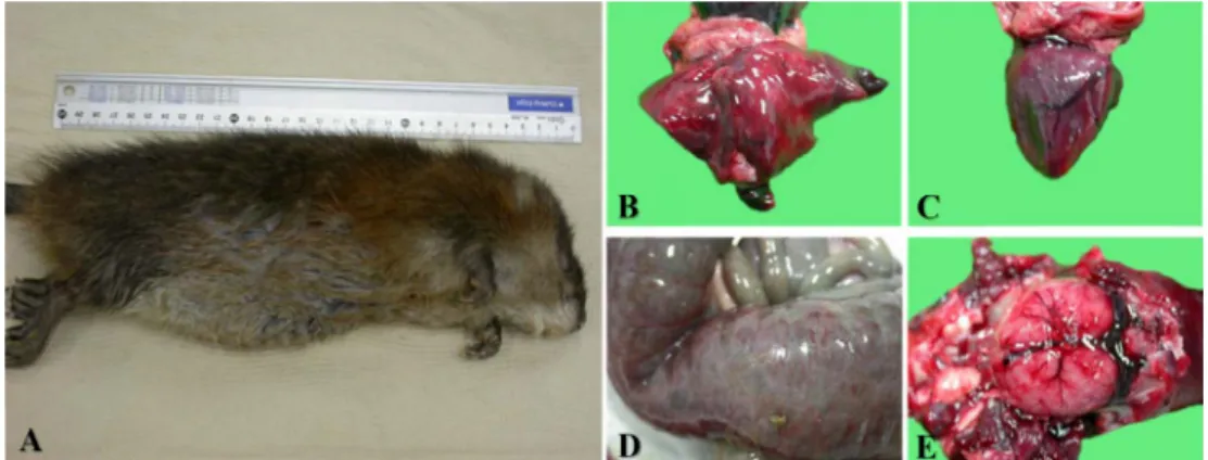
Fig. 1. Gross pictures representing pathological changes in various body organs. (A) Picture of a female musk rat. (B) Severe hemor- rhage in all lobes of the lung. (C) Diffuse hemorrhage and a little edema of the heart. (D) Serosal hemorrhage in the small intestine. They were also filled with brown to black contents with mucosal ulceration. (E) Severe hemorrhage and congestion of the meninges.
Fig. 2. Gross pictures representing pathological changes in kidneys, ureter and urinary bladder. (A) Moderate congestion of both kidneys. The left kidney also showed hydronephrosis and renal calculi on cut section. (B) The ureter contained sand-like green to yellowish lithiasis. (C) Hemorrhage on the serosal and mucosal membrane of urinary bladder with round and yellowish urolithiasis 2-2.5 cm in a diameter. RT, Right Kidney. LT, Left Kidney.
Results and Discussion
At necropsy, muskrat showed sand like Urolithiasis in the ureter, and kidneys; hydronephrosis was also evident (Fig.
1, Fig. 2). Systemic hemorrhage and calcification of all organs were observed.
Detailed histopathology of female muskrat showed severe congestion and hemorrhage of the lung accompanied with edema that occupying alveoli. Focal infiltration of neu- trophils and diffuse infiltration of mononuclear cells were observed in interstitial tissue and inside alveoli (Fig. 3A).
Severe hemorrhage and depletion of lymphoid follicles were evident in the spleen. Diffuse infiltration of plasma cells was also observed especially in sub-capsular region in the spleen (Fig. 3B). Both kidneys showed diffuse microcalcification in the cortex and medulla with tubular degeneration and in-
flammation remarkably, Calcification was mostly evident in proximal convoluted tubules and collecting ducts. Peri-glo- merular and inter-tubular hyalinization were remarkable and it was suggestive for chronic nephritis (Fig. 3C). The left ureter revealed desquamation of the lining epithelium with calcium salts that were detected in the lumen (Fig. 3D).
In the current case we are describing the pathology of renal failure in a muskrat for the first time, to the author’s best knowledge. Various nephrotoxic drugs are identified in- cluding cancer therapeutics, drugs of abuse, antibiotics, and radiocontrast agents [13]. Several Pollutants also found in environment and affect the kidney such as cadmium, mer- cury, arsenic, lead, trichloroethylene, bromate, brominated- flame retardants, diglycolic acid, and ethylene glycol [18].
Aristolochic acid and mycotoxins such as ochratoxin, fumo- nisin B1 and citrinin are natural toxins that target kidneys
632 생명과학회지 2020, Vol. 30. No. 7
Fig. 3. Histopathological findings of lung, spleen, kidney and ureter in died muskrat. (A) Severe hemorrhage and congestion with interstitial pneumoniae accompany- ing inflammatory cells infiltrates in the lung. Focal infiltrate of neutrophils (arrow), infiltration of mono- nuclear cells in interstitial tissue (arrow heads). (B) Severe hemorrhage and depletion of lymphocytes in the white pulp with infiltration of a large number of plasma cells (arrow heads). (C) Both kidneys showed diffuse microcalcification (arrows) in the cor- tex and medulla with tubular degeneration. Hyalini- zation of glomeruli (black arrow head) and interstitial tissue (red arrow heads) were suggestive for chronic inflammation. (D) The left ureter revealed desquama- tion of the lining epithelium with necrosis and micro- calcification. Hematoxylin and eosin (H&E) staining.
All scale bar = 100 μm.
[1, 9].
In our case, the histopathological observations were sup- portive of a chronic toxicity and mainly on convoluted tu- bules in kidney. Moreover, the calculi, the systemic hemor- rhages and calcification in several organs were compatible with uremia. The cause of renal failure in our case did not seem to be toxification by aflatoxins and fumonisin, since specific lesions in liver that are caused by theses toxins were not detected [3, 7, 14, 15]. Therefore, there is a high possi- bility that the renal failure in this case may have been pro- duced by other nephrotoxins in the foodstuffs. However, we couldn’t determine the exact toxins and fungi for this case.
This report presents evidences that support the mycotoxic hypothesis of diet-induced nephropathy [8]. Here, we have described a rare case report of renal failure with calcification in a muskrat displaying nephrotoxicity most likely by foodstuffs.
Acknowledgement
This research was funded by the Republic of Korea gov- ernment (Ministry of Science and ICT); grant number (NRF- 2017R1E1A1A01072781).
The Conflict of Interest Statement
The authors declare that they have no conflicts of interest with the contents of this article.
References
1. Adebo, O., Njobeh, P., Gbashi, S., Nwinyi, O. and Mavu- mengwana, V. 2017. Review on microbial degradation of aflatoxins. Crit. Rev. Food Sci. Nutr. 57, 3208-3217.
2. Atkins, R. C. 2005. The epidemiology of chronic kidney disease. Kidney. Int. 67, S14-S18.
3. Barnett, L. M. and Cummings, B. S. 2018. Nephrotoxicity and renal pathophysiology: a contemporary perspective.
Toxicol. Sci. 164, 379-390.
4. Gethöffer, F. and Siebert, U. 2019. Current knowledge of the Neozoa, Nutria and Muskrat in Europe and their environmental impacts. J. Wild. Bio.10.22120/jwb.2019.
109875. 1074
5. Heske, E. J. 1999. Mammalogy: adaptation, diversity, and ecology. J. Mammal. 80, 699.
6. Hosohata, K. 2016. Role of oxidative stress in drug-induced kidney injury. Int. J. Mol. Sci. 17, 1826.
7. Iranshahy, M., Etemad, L., Shakeri, A., Badibostan, H. and Karimi, G. 2020. Protective activity of melatonin against my- cotoxins-induced toxicity: a review. Toxicol. Environ. Chem.
435-450.
8. Jeong, W. I., Do, S. H., Jeong, D. H., Chung, J. Y., Yang, H.
J., Yuan, D. W., Hong, I. H., Park, J. K., Goo, M. J. and Jeong, K. S. 2006. Canine renal failure syndrome in three dogs. J. Vet. Sci. 7, 299-301.
9. Kabak, B., Dobson, A. D. and Var, I. 2006. Strategies to pre- vent mycotoxin contamination of food and animal feed: a review. Crit. Rev. Food Sci. Nutr. 46, 593-619.
10. Keddy, P. A. 2010. Wetland ecology: principles and conservation.
Cambridge University Press.
11. Kong, J. Y., Kim, H. S., Yeon, S. C., Park, J. K., Jeong, K.
S. and Hong, I. H. 2019. Preputial gland adenoma in a wild nutria (Myocastor coypus): a case report. J. Vet. Sci. 21, e1.
12. Long, M., Li, Q. M., Fang, Q., Pan, L. H., Zha, X. Q. and Luo, J. P. 2019. Renoprotective effect of Laminaria japonica polysaccharide in adenine-induced chronic renal failure.
Journal of Life Science 2020, Vol. 30. No. 7 633
초록:암컷 사향쥐( Ondatra zibethicus )의 신부전
울라아리프1,2†․아메드 엘파들3†․박선영1,2․정명진1,2․손지윤1,2․윤현호1,2․박재민1,2․임재혁1,2․ 정승준1,2․박진규1․정규식1,2*
(1경북대학교 수의과대학, 2경북대학교 줄기세포치료 연구소, 3하르툼대학교 수의과대학)
야생 포유류의 신부전 증후군은 거의 보고가 없다. Muskrat (Ondatra zibethicus)는 중간 크기의 설치류이며 많 은 질병을 가지고 있으나, 신부전 증후군은 보고된 바가 없으며, 본 케이스는 병리학적 진단을 위해 6개월령 암컷 사향쥐의 다른 임상증후군 없는 상태로 부검을 실시하였다. 요관, 신장결석과 수진증을 관찰하였고, 결석에 의한 심각한 신장 손상과, 뇌 손상을 포함한 전신 출혈과 석회화가 관찰되었고, 이는 신장 손상으로 인한 요로결석과 장기 손상에 기인한 것이다. 괴사 및 미세석회화는 신장 피질 및 수질에서, 특히 근위 곡 세뇨관 및 신장의 수집관 에서 검출되었다. 사구체의 초자양변성이 크게 관찰되었으며 이는 만성 신염을 나타내며. 이러한 소견은 특히 신 장에 대한 진균성 독성 효과를 나타내는 것으로 사료된다. 또한, 폐에서 호중구 및 단핵 세포의 침윤이 관찰되었 고, 비장에서도 만성 염증세포인 형질세포의 침윤이 관찰되었다. 본 소견에서는 사향쥐 신부전에 따른 사인은 오 염된 사료섭취로 의심되는 것으로 판단된다.
Molecules 24, 1491.
13. Naughton, C. A. 2008. Drug-induced nephrotoxicity. Am.
Fam. Physician 78, 743-750.
14. Otim, M. O., Mukiibi, M. G., Christensen, H. and Bisgaard, M. 2005. Aflatoxicosis, infectious bursal disease and im- mune response to Newcastle disease vaccination in rural chickens. Avian Pathol. 34, 319-323.
15. Şehu, A., Cakir, S., Cengiz, Ö. and Eşsiz, D. 2005. MYCOTOX®
and aflatoxicosis in quails. Br. Poult. Sci. 46, 520-524.
16. Skyrienė, G. and Paulauskas, A. 2012. Distribution of in- vasive muskrats (Ondatra zibethicus) and impact on ecosys- tem. Ekologija 58, 357-367.
17. Willner, G. R., Feldhamer, G. A., Zueker, E. E. and Chapman, J. A. 1980. Ondatra zibethicus. Mamm. Species 141, 1-8.
18. Xu, X., Nie, S., Ding, H. and Hou, F. F. 2018. Environmental pollution and kidney diseases. Nat. Rev. Nephrol. 14, 313.