CASE REPORT
만성C형간염을 동반한 만성 신부전 환자에서 인터페론 치료후 발생한 간질성 폐렴
강은정, 김동균, 전성란, 최현숙, 정승원, 장재영, 이준성, 어수택1
순천향대학교 의과대학 내과학교실 소화기연구소, 호흡기알레르기내과1
Interstitial Pneumonitis in a Patient with Chronic Hepatitis C and Chronic Renal Failure on Interferon Therapy
Eun Jung Kang, Dong Kyun Kim, Seong Ran Jeon, Hyun Sook Choi, Soung Won Jeong, Jae Young Jang, Joon Seong Lee and Soo Taek Uh1
Institute for Digestive Research, Department of Internal Medicine, 1Department of Allergy and Respiratory Medicine, Soonchunhyang University College of Medicine, Seoul, Korea
After 4-months of alpha interferon (IFN-α), a 64-year old woman with chronic hepatitis C developed a cough and dyspnea and showed diffuse infiltrative opacities on her chest X-ray. Her symptoms persisted after stopping the IFN-α therapy. Pulmonary function testing revealed a reduced forced vital capacity. High-resolution computed tomography of the lung showed peripheral and peribronchovascular ground glass attenuation and consolidation associated with reticulation. Bronchoalveolar lavage was performed for further evaluation and showed a lymphocyte level of 8.2%, an uncommon finding in IFN-α-induced interstitial pneumonitis. We performed a lung biopsy to diagnose her disease and it suggested interstitial pneumonitis. This was considered to be due to the immunomodulatory effects of INF-α. Although rare, any sign of significant pulmonary involvement should be evaluated. (Korean J Gastroenterol 2011;58:47-52)
Key Words: Chronic hepatitis C, Interferon, Chronic renal failure, Interstitial pneumonitis
Received December 4, 2009. Revised August 4, 2010. Accepted August 5, 2010.
CC This is an open access article distributed under the terms of the Creative Commons Attribution Non-Commercial License (http://creativecommons.org/licenses/
by-nc/3.0) which permits unrestricted non-commercial use, distribution, and reproduction in any medium, provided the original work is properly cited.
교신저자: 장재영, 140-743, 서울시 용산구 대사관길 22, 순천향대학교 의과대학 내과학교실 소화기연구소
Correspondence to: Jae Young Jang, Institute for Digestive Research, Department of Internal Medicine, Soonchunhyang University College of Medicine, Seoul Hospital, 657, Hannam-dong, Yongsan-gu, Seoul 140-743, Korea. Tel: +82-2-709-9863, Fax: +82-2-709-9696, E-mail: [email protected]
Financial support: None. Conflict of interest: None.
INTRODUCTION
Hepatitis C is caused by a small, single-stranded RNA virus which replicates in the liver at a high rate. The most common routes of HCV transmission are intravenous drug use, blood transfusion (before the advent of screening for the virus in the blood supply), and sexual exposure.1
Chronic infection (defined as detectable HCV RNA for more than 6 months) develops in 55-85% of patients with hepatitis C2 and is a major cause of chronic liver disease, cirrhosis, and hepatocellular carcinoma. Once established, chronic HCV in-
fection rarely resolves spontaneously.1 Hepatocellular injury associated with chronic HCV disease appears to be mediated immunologically, with natural killer cells and CD8+ T-cells playing central roles.3,4
The recommended therapy for chronic hepatitis C is a com- bination of pegylated interferon alpha (IFN-α) and ribavirin.5 The attachment of polyethylene glycol (PEG) to interferon at the alpha site (peginterferon-α) extends the half-life and dura- tion of the therapeutic activity of INF-α. However, hepatitis C patients with renal failure are typically treated with reduced doses of peginterferon-α or conventional IFN. Additionally, rib-
Fig. 1. (A, B) CT on admission showing peripheral and peribronchovascular ground glass attenuation and consolidations. (C, D) Repeated CT scan performed after discontinuation of interferon. Bilateral lung parenchymal lesions show improvement.
avirin is contraindicated in patients with renal failure because of its renal toxicity.6
Interferon-α suppresses viral replication and restores ele- vated serum aminotransferase levels, leading to improve- ment in the histological changes in patients with chronic hep- atitis C.7 However, IFN-α can also induce various autoimmune disorders.8-10 Here, we describe a patient with chronic hep- atitis C and chronic renal failure who developed interstitial pneumonitis following IFN-α administration.
CASE REPORT
The patient, a 64-year-old female, showed the first in- dications of abnormal liver function and tested positive for HCV antibodies in January 2006. Other than a history of chronic renal disease due to hypoplasia of the right kidney and renal artery stenosis, she had no history of autoimmune or
pulmonary disease. Dialysis was not performed. In April 2008, her ALT level was 68 U/L, her HCV RNA level (measured by PCR) was 2.39×10⁴ IU/mL, and her HCV genotype was 2a/2c. The liver parenchymal texture was coarse and both kidneys had decreased in size due to chronic renal disease : the right and left kidneys measured 8.1×3.0 and 6.9×3.7 cm, respectively. Following to the guidelines of The Korean Association for the Study of the Liver (KASL), the patient was started on 3-million units of IFN-α thrice weekly. After 4 weeks of therapy, she achieved a rapid virologic response (RVR).
However, the patient subsequently developed a cough, dyspnea, chills and myalgia, 4 months after starting IFN-α treatment. She had received a total of 54 million units of IFN-α. On admission, her white blood cell count (WBC) was 3,400/μL, hemoglobin 9.3 g/dL, hematocrit 27.4%, and pla- telet count 277,000/μL. Liver function tests were as follows:
AST, 41 IU/L; ALT, 17 IU/L; LDH, 403 IU/L; ALP, 99 U/L; total
Fig. 2. (A) Patchy areas of organizing pneumonia showing polypoid plugs of loose connective tissue (H&E, ×40). (B) Polypoid plugs of organizing pneumonia and acute alveolar hemorrhage (H&E, ×40). (C) An organizing intraluminal plug with chronic inflammatory infiltrate (H&E, ×100).
(D) Chronic interstitial pneumonia pattern with interstitial fibrosis and inflammation (H&E, ×40).
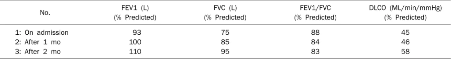
bilirubin, 0.3 mg/dL. The chest X-ray showed diffuse in- filtrative opacities in both lungs. High- resolution computed to- mography (HRCT) of the lung showed peripheral and peri- bronchovascular ground glass attenuation and consolidation associated with reticulation (Fig. 1A, B). Pulmonary function tests revealed a reduced forced vital capacity (Table 1-No.1).
IFN-α induced interstitial pneumonitis was suspected and the IFN-α therapy was discontinued. Bronchoalveolar lavage (BAL) was performed for further evaluation and showed a lym- phocyte level of 8.2%, an uncommon finding in IFN-α induced interstitial pneumonitis. Because there was no clinical im- provement after halting the IFN-α and the BAL findings were atypical for IFN-α induced interstitial pneumonitis, a wedge resection of the lesion in the right lower lung was performed via video-assisted thoracoscopy. On microscopic examina- tion, the lung showed patchy areas of organizing pneumonia
with polypoid plugs of loose connective tissue (Fig. 2A-C) and a chronic interstitial pneumonia pattern with interstitial thick- ening due to interstitial fibrosis and inflammation (Fig. 2D).
There were also focal areas of emphysematous change, bron- chiolization, and pleural thickening.
Three weeks after discontinuing the IFN-α therapy, fol- low-up HRCT showed improvement of the bilateral lung paren- chymal lesions (Fig. 1C, D). Although pulmonary function tests showed slight improvement in the FVC (Table 1-No.2), the pa- tient’s dry cough persisted. Prednisolone was started at a dose of 25 mg daily, and her symptoms improved after 1 week.
Over a 1-month period, the prednisolone was tapered to a dose of 7.5 mg daily. At last follow-up the patient’s FVC had im- proved markedly (Table 1-No.3). Although she initially showed a RVR to INF-α, the patient’s viral load increased again to 2.43×10⁷ IU/mL within 2 months of halting the INF-α therapy.
Table 1. The Cases of Interferon-induced Pulmonary Complications
No. FEV1 (L)
(% Predicted)
FVC (L) (% Predicted)
FEV1/FVC (% Predicted)
DLCO (ML/min/mmHg) (% Predicted) 1: On admission
2: After 1 mo 3: After 2 mo
93 100 110
75 85 95
88 84 83
45 46 58 FEV1, forced expiratory volume in one second; FVC, forced vital capacity; DLCO, carbon monoxide diffusing capacity.
Subsequently, we have monitored the patient’s liver function and viral load without IFN-α treatment. Five months after ceasing treatment, the AST and ALT levels returned to near normal levels.
DISCUSSION
The currently recommended regimen for the treatment of chronic hepatitis C is a combination of weekly subcutaneous peginterferon and twice daily oral ribavirin. In chronic hep- atitis C patients with renal failure, according to the KASL guidelines, low-dose peginterferon or conventional interferon is indicated instead of peginterferon.
Currently, IFN-α is prescribed for many viral and neoplastic diseases, such as chronic hepatitis B and C, chronic myeloge- nous leukemia, hairy cell leukemia, multiple myeloma, and non-Hodgkin’s lymphoma.11 Common acute side effects of in- terferon treatment include fever, chills, weight loss and myal- gia, which are typically transient. More serious conditions as- sociated with IFN-α treatment include hemolytic anemia, in- terstitial pneumonia, cholestatic liver dysfunction, immune thrombocytopenia, and autoimmune thyroid disease.12
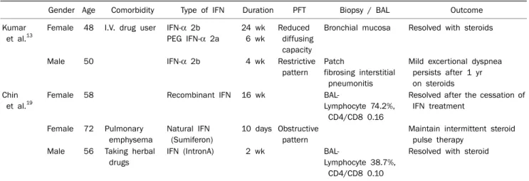
In 2002, Kumar et al. reported significant pulmonary tox- icity associated with interferon and ribavirin therapy in four hepatitis C patients.13 In four cases, lung biopsies revealed bronchiolitis obliterans organizing pneumonia (BOOP) in two cases, and interstitial pneumonitis in two cases.
The pathogenesis of autoantibody formation in IFN-in- duced autoimmunity is incompletely understood. A number of factors may alter the balance between self-tolerance and the activation of autoreactivity. IFN-α increases the activity of nat- ural killer cells and cytotoxic T lymphocytes, and it enhances the expression of HLA class I and II antigens.10,14 The in- creased expression of HLA-DR and CD 11b antigens in periph- eral blood lymphocytes may indicate the activation of T lym- phocytes and natural killer cells; furthermore, the increased
number of CD 8+ T-cells in BAL fluid also suggests that cyto- toxic T-cells in the lung were activated.10,12,14 Thus in this case, interstitial pneumonitis response to IFN-α therapy was medi- ated immunologically. In interstitial pneumonia, IFN-α is con- sidered an extrinsic antigen and potent immunomodulator and was responsible for the development and progression of IFN-α induced interstitial pneumonia.
Interferon toxicity is generally dose-dependent, increasing with the dose and duration of treatment. However, no clear re- lationship between the dose and likelihood of side effects has been demonstrated.15
Pulmonary complications due to interferon therapy for chronic hepatitis C typically occur within the first few weeks of therapy and can range from mild flu-like symptoms (e.g., fe- ver, cough lasting more than 2 weeks while on therapy) to life-threatening hypoxemia and respiratory failure.16-18 In our patient, a chest X-ray revealed bilateral diffuse retic- ulonodular infiltrates, pulmonary function tests showed a re- strictive pattern, and the diffusion capacity was reduced.
Based on these results, together with the mononuclear cell in- filtrate on lung biopsy, a diagnosis of interstitial pneumonitis was made. A typical finding of IFN-α induced interstitial pneu- monitis in BAL fluid is increased numbers of lymphocytes with elevated CD8+/CD4+. In this case, BAL showed a lymphocyte level of 8.2%, an uncommon finding in IFN-α induced inter- stitial pneumonitis. The lung biopsy showed patchy areas of organizing pneumonia and chronic interstitial pneumonia with interstitial thickening due to interstitial fibrosis and inflammation.
In most patients, IFN-α withdrawal or corticosteroid ther- apy alone is enough for spontaneous remission. However, per- sistent pneumonitis has been observed with high-dose corti- costeroid therapy.19 Most reported cases of interferon-asso- ciated pulmonary toxicity in patients with chronic hepatitis C were reversible; in many instances simply discontinuing the therapy or administering corticosteroids for a few months was
Table 2. Pulmonary Function Tests
Gender Age Comorbidity Type of IFN Duration PFT Biopsy / BAL Outcome
Kumar et al.13
Female 48 I.V. drug user IFN-α 2b
PEG IFN-α 2a 24 wk 6 wk
Reduced diffusing capacity
Bronchial mucosa Resolved with steroids
Male 50 IFN-α 2b 4 wk Restrictive
pattern Patch
fibrosing interstitial pneumonitis
Mild excertional dyspnea persists after 1 yr on steroids Chin
et al.19
Female 58 Recombinant IFN 16 wk BAL-
Lymphocyte 74.2%, CD4/CD8 0.16
Resolved after the cessation of IFN treatment
Female 72 Pulmonary emphysema
Natural IFN (Sumiferon)
10 days Obstructive pattern
Maintain intermittent steroid pulse therapy
Male 56 Taking herbal drugs
IFN (IntronA) 2 wk BAL-
Lymphocyte 38.7%, CD4/CD8 0.10
Resolved with steroid
FEV1, forced expiratory volume in one second; FVC, forced vital capacity; DLCO, carbon monoxide diffusing capacity; Aug, August; Sep, September; Oct, October.
sufficient. Kumar et al. reported two cases in which the symp- toms resolved after simply discontinuing the antiviral therapy.13 Another case improved after administering prednisolone. The final case showed persistent despite pre- dnisolone treatment. We summerized these cases (Table 2).13,19
We experienced a rare case of interstitial pneumonitis sec- ondary to interferon therapy in a 64-year-old female with chronic hepatitis C and chronic renal failure. Because no clin- ical improvement was apparent after ceasing IFN-α and BAL revealed an atypical finding in IFN-α induced interstitial pneu- monitis, we confirmed the diagnosis of interstitial pneumo- nitis secondary to IFN-α with a lung biopsy. Three months after stopping the IFN-α, the patient’s symptoms and liver function had improved (AST 41 IU/L; ALT 17 IU/L) and we stopped her prednisolone therapy.
This case illustrates potentially serious, but reversible, pul- monary toxicity associated with interferon therapy. Although rare, prompt investigation and discontinuation of medication is warranted if any sign of significant pulmonary involvement develops. We postulate that the usual dosage of interferon may have adverse effects in a patient diagnosed with chronic renal failure even without undergoing hemodialysis.
REFERENCES
1. Hoofnagle JH, Seeff LB. Peginterferon and ribavirin for chronic hepatitis C. N Engl J Med 2006;355:2444-2451.
2. Hoofnagle JH. Course and outcome of hepatitis C. Hepatology
2002;36(5 Suppl 1):S21-S29.
3. Rehermann B, Nascimbeni M. Immunology of hepatitis B virus and hepatitis C virus infection. Nat Rev Immunol 2005;5:215- 229.
4. Bowen DG, Walker CM. Adaptive immune responses in acute and chronic hepatitis C virus infection. Nature 2005;436:946- 952.
5. Strader DB, Wright T, Thomas DL, Seeff LB; American Association for the Study of Liver Diseases. Diagnosis, manage- ment, and treatment of hepatitis C. Hepatology 2004;39:1147- 1171.
6. Lauer GM, Walker BD. Hepatitis C virus infection. N Engl J Med 2001;345:41-52.
7. Davis GL, Balart LA, Schiff ER, et al. Treatment of chronic hep- atitis C with recombinant interferon alfa. A multicenter random- ized, controlled trial. Hepatitis Interventional Therapy Group. N Engl J Med 1989;321:1501-1506.
8. Onoda K, Takai K, Sanada M, Aoki S, Watanabe T. Autoimmune hemolytic anemia induced by alpha-interferon therapy in a case of IgG-kappa type multiple myeloma. Rinsho Ketsueki 1992;33:844-846.
9. Schilling PJ, Kurzrock R, Kantarjian H, Gutterman JU, Talpaz M.
Development of systemic lupus erythematosus after interferon therapy for chronic myelogenous leukemia. Cancer 1991;68:
1536-1537.
10. Schattner A. Interferons and autoimmunity. Am J Med Sci 1988;295:532-544.
11. Quesada JR, Talpaz M, Rios A, Kurzrock R, Gutterman JU. Clinical toxicity of interferons in cancer patients: a review. J Clin Oncol 1986;4:234-243.
12. Hizawa N, Kojima J, Kojima T, et al. A patient with chronic hep- atitis C who simultaneously developed interstitial pneumonia, hemolytic anemia and cholestatic liver dysfunction after al- pha-interferon administration. Intern Med 1994;33:337-341.
13. Kumar KS, Russo MW, Borczuk AC, et al. Significant pulmonary toxicity associated with interferon and ribavirin therapy for hep-
atitis C. Am J Gastroenterol 2002;97:2432-2440.
14. Silva MO, Reddy KR, Jeffers LJ, Hill M, Schiff ER. Interferon-in- duced chronic active hepatitis? Gastroenterology 1991;101:
840-842.
15. Okanoue T, Sakamoto S, Itoh Y, et al. Side effects of high-dose interferon therapy for chronic hepatitis C. J Hepatol 1996;25:
283-291.
16. Dumoulin FL, Leifeld L, Sauerbruch T, Spengler U. Autoimmunity induced by interferon-alpha therapy for chronic viral hepatitis.
Biomed Pharmacother 1999;53:242-254.
17. Wolf Y, Haddad R, Jossipov J, Werbin N. Alpha-interferon induced severe pneumonitis. J Toxicol Clin Toxicol 1997;35:113-114.
18. Bini EJ, Weinshel EH. Severe exacerbation of asthma: a new side effect of interferon-alpha in patients with asthma and chronic hepatitis C. Mayo Clin Proc 1999;74:367-370.
19. Chin K, Tabata C, Sataka N, Nagai S, Moriyasu F, Kuno K.
Pneumonitis associated with natural and recombinant interfer- on alfa therapy for chronic hepatitis C. Chest 1994;105:939- 941.