Article Info
Received June 21, 2020 Revised July 18, 2020 Accepted July 28, 2020 Corresponding Author Ki-song Kim
E-mail: [email protected]
https://orcid.org/0000-0002-7184-3027
Key Words Foot arch Insole Muscle activity
Background: The longitudinal arch is important for individuals with pes planus. The toe spread out exercise (TSO) has been widely used to continuously support the longitudinal arch by increasing the abductor hallucis (AbdH) muscle activation. However, the AbdH muscle is commonly lack of the sufficient activation during the TSO especially in individuals with pes planus.
Objects: This study was performed to investigate the effect of arch support on the muscle ac- tivity and strength of the AbdH during TSO in standing position in individuals with pes planus.
Methods: Twenty subjects with pes planus between 20 and 30 years of age participated in this study. The muscle activity and strength of the AbdH were measured using surface EMG system and the Smart KEMA tensiometer system. The AbdH muscle was evaluated during TSO between individuals with and without longitudinal arch support in standing position. The longitudinal arch was supported by using the insole. The paired t-test was used. The level of statistical significance was set at α = 0.05.
Results: The muscle activity and strength of the AbdH during TSO with arch support in standing position was significantly greater than that without arch support.
Conclusion: The muscle activity and strength of the AbdH during TSO in standing position can be influenced by the longitudinal arch support in individuals with pes planus. The AbdH strengthening during TSO in standing with arch support can be recommended especially in individuals with pes planus in the clinical settings.
Copyright ⓒ Korean Research Society of Physical Therapy
This is an Open Access article distributed under the terms of the Creative Commons Attribution Non-Commercial License (http://creativecommons.org/licenses/by-nc/4.0) which permits unrestricted non-commercial use, distribution, and reproduction in any medium, provided the original work is properly cited.
INTRODUCTION
Pes planus is the impairment of foot as flattening of the lon
gitudinal arch [1] and is misaligned foot condition as excessive pronation of the foot and abduction of the midfoot on the rear foot with valgus [1,2]. Pes planus is caused by overuse injuries, including patellofemoral pain syndrome [2], and plantar fasci
itis [1]. The consideration of normal alignment of foot is nec
essary to evaluate the longitudinal arch angle and to prevent overuse injuries related to pes planus. The longitudinal arch of foot is supported not only by intrinsic muscles such as the ab
ductor hallucis (AbdH), and extrinsic muscles such as the tibi
alis anterior/posterior but also by passive structure including the plantar fascia and spring ligament [1,3,4]. The longitudinal arch training has been recommended to prevent chronic pain in the foot and ankle [5], to improve balance in the standing
position [6], and to prevent damage of the musculoskeletal system [7]. Previous studies have reported that the strength of the dorsiflexion and AbdH of individuals with collapsed longi
tudinal arch was weaker than those of normal individuals [57].
In particular, the weakness of the AbdH along with pes planus due to the collapse of the longitudinal arch may contribute to foot or ankle joint damage or sprain [7,8].
The misaligned foot have the characteristic of collapsed lon
gitudinal arch in the standing position. The AbdH is one of the muscles that plays important roles in not only maintaining the stability of the foot in a standing position but also in improving the alignment of the foot [9,10]. The AbdH contraction, which originates in the calcaneus bone and inserts into the great toe, contribute to flexion and abduction of the first metatarsopha
langeal (MTP) joint [1], which moves the origin and insertion regions of AbdH close, and consequently increases and sup
Physical Therapy Korea
PTK https://doi.org/10.12674/ptk.2020.27.3.206 pISSN: 1225-8962 eISSN: 2287-982X Phys Ther Korea. 2020;27(3):206-211
Original Article
Effect of the Arch Support on the Strength of the Abductor Hallucis During the Toe Spread Out Exercise in Standing Position in Individuals With Pes Planus
In-cheol Jeon, PT, PhD, Ki-song Kim, PT, PhD
Department of Physical Therapy, College of Life and Health Science, Hoseo University, Asan, Korea
ports the longitudinal arch [1,5]. Following surgery of the foot and ankle joint because of arthritis and sprain, factors such as weakness of the intrinsic muscles of the foot and functional limitations of the dorsiflexion affect the weakness of the AbdH [11]. In the previous studies, to strengthen the AbdH muscle, the shortfoot exercise (SF), which pull the first metatarsal head toward the heel without toe flexion, and the toe spread out exercise (TSO), which perform abduction and flexion of the first and fifth metatarsophalangeal joints, are often investi
gated in the clinical rehabilitation [9].
However, the SF and TSO were considered without the fa
tigue of the AbdH muscle in individuals with pes planus, be
cause in individuals with pes planus, the foot structure could not support the longitudinal arch in standing position [10]. The AbdH muscle may be in insufficient activation during these exercises. The fatigue of the AbdH [12], and decreased muscle activity of the AbdH [13], exacerbated the longitudinal arch in the standing position, which implies the important role of the AbdH in supporting the longitudinal arch. The overactiva
tion of extrinsic muscles can be occurred to compensate for a weakened AbdH in individuals with pes planus [14,15]. The prolonged overactivation of extrinsic muscles including tibialis anterior and posterior results in muscle fatigue, consequently causing tendinitis, overuse injury [11,16]. Thus, AbdH strength
ening exercise is important to support the longitudinal arch and prevent secondary injuries in individuals with pes planus [15]. Therefore, to induce maximal effort in the AbdH, the arch support for the longitudinal arch of foot is required during TSO in standing position in individuals with pes planus.
In addition, sufficient understanding of the mechanism of such compensatory movements is essential to correctly evalu
ate and interpret the function of the AbdH [9,14]. Based on the studies conducted to date, there has been no study that has investigated the effect of the longitudinal arch support on the strength and muscle activity of individuals with pes planus while performing TSO in the standing position. To compre
hensively manage the AbdH, it is essential to investigate the effect of the support of the longitudinal arch on the strength and muscle activity of the AbdH while performing TSO in the state of supporting weight [15]. The longitudinal arch of the foot contributes to the stability and balance of the foot in the standing position. Furthermore, together with the longitudinal arch, the AbdH provides dynamic stability to the ankle joint and metatarsal joint during functional movements, such as
walking and running.
The purpose of this study is to investigate the effect of the support of the longitudinal arch using insole on the muscle activity of the AbdH while individuals with pes planus perform TSO in the standing position. The hypothesis of this study is that providing longitudinal arch support using insole to indi
viduals with pes planus while performing the TSO in the stand
ing position could improve the strength and muscle activity of the AbdH more than while it is not provided. The results of this study clinically suggest that the management of the AbdH is more effective while individuals with pes planus perform the TSO with the longitudinal arch using insole formed in the standing position.
MATERIALS AND METHODS
1. Subjects
For determining the number of the participants, power was set at 0.80, alpha level at 0.05, and effect size at 1.42, which are required elements of the G*Power program (ver. 3.1.2, Franz Faul; University of Kiel, Kiel, Germany). We found that a minimum of 15 participants were required for the experiment;
considering the possibility of dropout, a total of 20 partici
pants with pes planus (age: 25.1 ± 5.2 years, weight: 67.3 ± 8.6 kg, height: 170.1 ± 6.9 cm, males: 10, females: 10) were se
lected. The criterion of pes planus has a calcaneal stance angle during resting position with at least 4° and a navicular drop (ND) with at least 13 mm.
The inclusion criteria were as follows: (1) No limited range of motion of the foot and ankle joint, (2) no previous history of ankle joint surgery. The exclusion criteria were as follows: (1) history of musculoskeletal fracture, (2) pain in any part of the body experienced during the execution of the experiment.
The process of the experiment was explained in detail to the participants. All participants provided written consent ac
cording to the tenets of the Declaration of Helsinki. This study was approved by the Institutional Review Board (approval no.
1041231200218HR10301).
2. Instruments
1) Identification of the location of the AbdH using ultrasound
To confirm the precise muscle belly of the individual muscle,
a 7.5MHz linear head connected to the ultrasound (Q30; SG
Healthcare Co., Ltd., Seoul, Korea) was used. After marking the precise area identified via ultrasound with a pen, the ultra
sound gel was removed for attaching the electromyogram (EMG) patch.
2) Measurement of the muscle activity of the AbdH Muscle activity was measured using EMGfeedback tools (wireless EMG system 100RT; BTS, Millan, Italy). A software was used to analyze the muscle activity of the AbdH. Filter (60 Hz) and band pass filter type extraction rate (1,024 Hz) were set and the collected muscle activity was treated with rootmean
square. To minimize the resistance at the location where the EMG patch was attached, the location was shaved and disin
fected with alcohol cotton. Two electrodes were attached in parallel with the direction of the muscular fiber. The maximal voluntary isometric contraction (%MVIC) method was used to normalize the data. When the hallux was at its maximum level of abduction of the great toe from the axial line of the foot, a resisting force was exerted by the investigator’s hand to the center of its the medial side of the first metatarsal and proxi
mal phalanx in sitting position [14]. The mean obtained after a total of three measurements was used to determine %MVIC (Figure 1).
3) Measurement of the strength of the AbdH
The strength of the maximum isometric contraction of the AbdH was measured using the Smart KEMA Pulling Sensor (Factorial Holdings Co., Ltd., Seoul, Korea) (Figure 2). A thin strap connected to the pulling sensor was placed between the metatarsophalangeal joint and the interphalangeal joint of the hallux, i.e., at the center of its proximal phalanx. For the
measurement of the selective strength of the AbdH, the first metatarsal bone was fixed using a nonelastic fixation belt to prevent adduction of ankle joint. To minimize the errors in measurement, the area of the toe joints in contact with the strap was marked. The length of the strap was controlled in the neutral position of the hallux in the standing position. The maximum weight of measurement determined using the Smart KEMA Pulling Sensor was 100 kg, with disappearance and pre
cision of less than 0.1 kg. To provide the support for the longi
tudinal arch of foot, the insole formed with the same height as the height of the navicular bone measured in the sitting posi
tion was used [13].
3. Procedures
As all participants had both feet with pes planus, the maxi
mum isometric muscle contraction of the AbdH of the domi
nants was measured. The order for applying the longitudinal
Figure 2.
Figure 2. The strength measurement of abductor hallucis.
A B
Figure 1.
Figure 1. The muscle activity of the AbdH muscle in individuals with pes planus (A) without arch support, (B) with arch support by using the insole.
arch support was determined via randomization. Each partici
pant practiced TSO while the longitudinal arch support using insole was provided. The insole for longitudinal arch support was used according to the height of the navicular bone in the sitting position. Then, the participant rehearsed for the experiment in the exact same manner to precisely execute the procedure. The participant had weight support by stand
ing comfortably on both feet for measurement. Then, the strength and muscle activity of the AbdH in the dominant foot was measured via maximum isometric muscle contraction.
The participant performed the TSO with the support for the longitudinal arch. The spreading of the TSO was maintained for 5 seconds. The measurement of the muscle activity of the AbdH was executed together with the measurement of the strength. The strength and muscle activity were simultaneously measured using a tensiometer and EMG devices with three times, and the data was averaged. After onetime execution, a 10minute resting period was provided.
4. Statistical Analysis
The SPSS software (ver. 18.0; IBM Co., Armonk, NY, USA) was used for statistical analysis. The normal distribution of the data measured was confirmed via the onesample Kol
mogorov–Smirnov test. The paired ttest was used to confirm whether there are statistically significant differences between the conditions (longitudinal arch support vs. no support). The significance level of the statistics was set at p < 0.05.
RESULTS
There was a statistically significance increase in the strength and muscle activity of the AbdH while the longitudinal arch support was provided than that while the longitudinal arch support was not provided (Tables 1, 2) (p < 0.05).
DISCUSSION
Adequate strength of the AbdH is an important factor for rehabilitation of the pes planus. However, clinical evaluation of the strength and muscle activity of the individuals with pes planus required consideration of the longitudinal arch. This study aimed to determine the effect of the longitudinal arch support using insole on the strength and muscle activity of the AbdH in individuals with pes planus in the standing position.
While individuals with pes planus performed TSO, the strength and muscle activity of the AbdH showed a statistically signifi
cant increase with the longitudinal arch support (strength in
creased by 1.43 kg and muscle activity increased by 12.71%) (p
< 0.05). As a result, the muscle activity of the AbdH increased at the alignment, allowing its ideal contraction. This could be considered to have positively influenced the increased strength of the AbdH [17].
There are several reasons for the difference in the strength and muscle activity of the AbdH depending on the longitudinal arch support during the performance of TSO. First, it could be explained with regards to the length –tension relationship. The AbdH plays an important stabilizing role in the first metatarsal joint, particularly in the one leg standing position [13]. The AbdH contraction, which originates in the calcaneus bone and inserts into the great toe, contribute to flexion and abduction of the first MTP joint [1], which moves the origin and insertion regions of AbdH close, and consequently increases and sup
ports the longitudinal arch [1,5]. While the longitudinal arch support was not provided, pronation of ankle joint and the pes planus in the standing position were occurred because of fall
ing of the navicular bone. This became a lengthened position of the AbdH. However, while the longitudinal arch of the foot is formed in the standing position during TSO, the pronation of the ankle bone could be prevented via the longitudinal arch support against the weight [13,18]. As a result, the ideal align
ment for effective contraction of the AbdH in standing position was formed. The ideal alignment of the foot, which allows ef
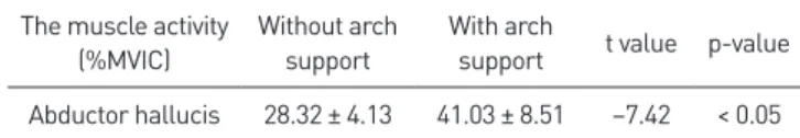
Table 1.
Table 1. The muscle activity of the abductor hallucis in individuals with
pes planus
The muscle activity (%MVIC)
Without arch support
With arch
support t value pvalue Abductor hallucis 28.32 ± 4.13 41.03 ± 8.51 –7.42 < 0.05 Values are presented as mean ± standard deviation. MVIC, maximal vol
untary isometric contraction.
Table 2.
Table 2. The strength of the abductor hallucis in individuals with pes pla