• 검색 결과가 없습니다.

Utility of Computed Tomography in a Differential Diagnosis for the Patients with an Initial Diagnosis of Chronic Obstructive Pulmonary Disease Exacerbation

N/A
N/A
Protected

Academic year: 2021

Share "Utility of Computed Tomography in a Differential Diagnosis for the Patients with an Initial Diagnosis of Chronic Obstructive Pulmonary Disease Exacerbation"

Copied!
8
0
0

로드 중.... (전체 텍스트 보기)

전체 글

(1)

Utility of Computed Tomography in a

Differential Diagnosis for the Patients with an Initial Diagnosis of Chronic Obstructive Pulmonary Disease Exacerbation

Hyung Jun Park, M.D.

1

, Soo Han Kim, M.D.

1

, Ho-Cheol Kim, M.D.

1

, Bo Young Lee, M.D., Ph.D.

1

, Sei Won Lee, M.D., Ph.D.

1

, Jae Seung Lee, M.D., Ph.D.

1

, Sang-Do Lee, M.D., Ph.D.

1

,

Joon Beom Seo, M.D., Ph.D.

2

and Yeon-Mok Oh, M.D., Ph.D.

1

Departments of

1

Pulmonary and Critical Care Medicine and

2

Radiology, Asan Medical Center, University of Ulsan College of Medicine, Seoul, Korea

Background: The utility of computed tomography (CT) in the differential diagnosis of patients with chronic obstructive pulmonary disease (COPD) exacerbation remains uncertain. However, due to the low cost associated with CT scan along with the impact of Koreas’ health insurance system, there has been a rise in the number of CT scans in the patients with initial diagnosis of COPD exacerbations. Therefore, the utility of CT in the differential diagnosis was investigated to determine whether performing CT scans affect the clinical outcomes of the patients with an initial diagnosis of COPD exacerbation.

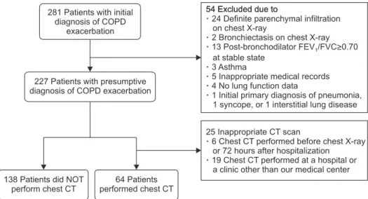
Methods: This study involved 202 COPD patients hospitalized with an initial diagnosis of COPD exacerbation. We evaluated the change in diagnosis or treatment after performing a CT scan, and compared the clinical outcomes of patient groups with vs. without performing CT (non-CT group vs. CT group).

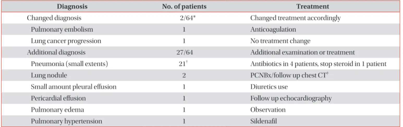
Results: After performing CT, the diagnosis was changed for two (3.0%) while additional diagnoses were made for 27 of the 64 patients (42.1%). However, the treatment changed for only one (1.5%), and six patients (9.3%) received supplementary medication. There were no difference in the median length of hospital stay (8 [6–13] days vs. 8 [6–12]

days, p=0.786) and intensive care unit care (14 [10.1%] vs. 11 [16.7%], p=0.236) between the CT and non-CT groups, respectively. These findings remained consistent even after the propensity score matching.

Conclusion: Utility of CT in patients with acute COPD exacerbation might not be helpful; therefore, we do not recommend chest CT scan as a routine initial diagnostic tool.

Keywords: Disease Exacerbation; Pulmonary Disease, Chronic Obstructive; Hospitalization; Tomography, X-Ray Computed

Address for correspondence: Yeon-Mok Oh, M.D., Ph.D.

Department of Pulmonary and Critical Care Medicine, Asan Medical Center, University of Ulsan College of Medicine, 88 Olympic-ro 43-gil, Songpa-gu, Seoul 05505, Korea

Phone: 82-2-3010-3136, Fax: 82-2-3010-4650, E-mail: [email protected] Address for co-correspondence: Joon Beom Seo, M.D., Ph.D.

Department of Radiology, Asan Medical Center, University of Ulsan College of Medicine, 88 Olympic-ro 43-gil, Songpa-gu, Seoul 05505, Korea Phone: 82-2-3010-4383, Fax: 82-2-476-0090, E-mail: [email protected]

Received: Nov. 22, 2018, Revised: Jan. 24, 2019, Accepted: Feb. 22, 2019, Published online: May. 31, 2019

cc

It is identical to the Creative Commons Attribution Non-Commercial License (http://creativecommons.org/licenses/by-nc/4.0/).

Copyright © 2019

The Korean Academy of Tuberculosis and Respiratory Diseases.

(2)

Introduction

Chronic obstructive pulmonary disease (COPD) is one of the leading causes of morbidity and mortality worldwide

1

. During the disease course, COPD patients can experience an exacerbation, which may necessitate hospitalization, increase subsequent risk of future exacerbations, and even lead to death particularly in cases requiring hospitalization

1,2

. During a COPD exacerbation, patients experience dyspnea, cough, and sputum, which may lead to a change in medication

3

. These symptoms may occur both during a COPD exacerba- tion and with other diseases such as pneumonia, pneumotho- rax, pulmonary embolism, arrhythmia, and congestive heart failure

1

. Therefore, chest X-ray and electrocardiography (ECG) has been routinely performed in the emergency department to check for these diseases.

Computed tomography (CT) may have a higher sensitivity to detect these lung diseases than conventional chest X-ray

4-6

. The CT scans is used to detect unnoticed finding in chest X- ray and to explain for other cause of desaturation in COPD exacerbation, especially for the ones severe enough to warrant hospitalization. The cost of a CT scan is relatively inexpensive in Korea compared to other countries. As a result, the number of CT scans for patients with an initial diagnosis of COPD ex- acerbation has increased. However, no study has yet proven that chest CT has higher diagnostic value to find these lung diseases compared to chest X-ray and whether it could affect clinical outcomes such as hospital stay and intensive care unit (ICU) care.

In this study, we investigated the utility of CT in diagnostic change or an additional diagnosis for patients who were hos- pitalized with an initial diagnosis of COPD exacerbation, and showed whether performing CT may affect treatment or clini- cal outcomes.

Materials and Methods

1. Study design and subjects

In this retrospective cohort study, we included COPD patients who were hospitalized with an initial diagnosis of a COPD exacerbation at the Asan Medical Center, a tertiary hospital located in Seoul, South Korea, from January 2010 to December 2012. COPD exacerbation was defined as aggra- vated respiratory symptoms in COPD patients and excluded patients in whom another disease, such as pneumonia, pneu- mothorax, interstitial lung disease, and arrhythmia, were di- agnosed by an initial chest X-ray or ECG. In these patients, we evaluated the change in the diagnosis or treatment following the chest CT scan. We also compared the clinical outcomes, including the length of hospital stay, ICU care and number of hospital deaths between patients who underwent CT (the CT

group) and those who did not (the non-CT group). The deci- sion to perform CT was at the discretion of the physician who first evaluated the COPD patient in the emergency depart- ment (ER) or by the respiratory specialist who was in charge of the patient during hospitalization.

The included patients were hospitalized both from the ER and the outpatient clinic. The initial physician diagnosis of a COPD exacerbation was made by an ER physician or a re- spiratory specialist. Irrespective of cigarette smoking history, we included patients whose stable-state spirometry revealed a ratio of the forced expiratory volume in 1 second (FEV

1

) to forced vital capacity of less than 0.70. We excluded definite asthma patients, however, included patients with broncho- dilator response positive. It might include patients as asthma and COPD overlap syndrome.

We also excluded patients whose medical records were in- complete for the evaluation of clinical outcomes, the length of hospital stay, hospital death, and ICU care. Additionally, of the patients who underwent a chest CT, we excluded any patients who underwent a chest CT before the initial chest X-ray or 72 hours after hospitalization. We also excluded patients who un- derwent a chest CT at a hospital or clinic other than our medi- cal center.

2. Ethics committee approval

This study was approved by the ethics committee of the Asan Medical Center (approval number, 2014-0907), and the need to obtain informed consent was waived due to the retro- spective observational nature of the study.

3. CT protocol

Chest CT was performed by using 16- or 64-detector row equipment (Somatom Sensation 16; Siemens Medical Solu- tions, Forchheim, Germany) and LightSpeed VCT (GE Medi- cal, Milwaukee, WI, USA). For the 16-detector row scanner, the parameters were 120 kV and 100 effective mA with dose modulation. For the 64-detector row scanner, the parameters were 120 kV and 100–400 mA with dose modulation. CT was performed with contrast enhancement for 40 patients and without contrast enhancement for 24 patients. Low-dose chest CT was performed two patients in contrast-enhanced chest CT group. For contrast-enhanced CT, images were obtained after the intravenous administration of 100 mL of iopromide with 300 mg/mL of iodine (Ultravist 300; Bayer Pharma, Ber- lin, Germany) at a rate of 2.5 mL/sec using a power injector.

CT was performed with a 50-second delay.

4. Clinical outcomes and statistical analysis

The baseline characteristics of the continuous variables

were compared using Student’s t test or Mann-Whitney U test

(3)

according to normality. The categorical variables were com- pared using a chi-square test while some categorical variables less than five were analyzed by Fisher exact test.

We evaluated the relationship between CT scan and the length of hospital stay by using a multiple linear regression analysis with an adjustment for the following covariates: age, sex, FEV

1

, the initial concentration of O

2

supply, body mass in- dex, history of hospitalization due to a COPD exacerbation in the previous year, baseline modified Medical Research Coun- cil (mMRC) dyspnea scale, increased or purulent sputum, and the initial arterial blood gases of oxygen and carbon dioxide.

In multiple linear regression, we chose the covariates through backward method which exclude covariate p-value above 0.2.

But we included CT scan as covariate to determine relation- ship with CT scan and length of hospital stay. To overcome the design of retrospective study, we performed propensity score matching to normalize baseline characteristics both CT group and non-CT group.

Statistical analyses were performed using SPSS software version 23.0 (IBM Corp., Armonk, NY, USA). p<0.05 was con- sidered to indicate a statistically significant difference.

Results

1. Baseline characteristics

After applying the inclusion/exclusion criteria to 281 pa- tients with an initial primary diagnosis of a COPD exacerba- tion, 54 were excluded (Figure 1). Another 25 who underwent a chest CT were excluded because they underwent CT scan at another hospital (n=19) or had the CT scan before the chest X-ray or 72 hours after hospitalization (n=6). Among the in- cluded 202 patients, 64 were in the CT group and 138 in the non-CT group. Table 1 represents that the patients were pre-

dominantly males, and the mean age was similar between the two groups. The non-CT group appeared to have lower lung function. There was no significant difference of the presence of bronchodilator response in two groups (Supplementary Table S1). All other characteristics were similar between the two groups.

2. Role of chest CT in a diagnostic change or in an additional diagnosis of COPD exacerbation

After the chest CT, the diagnosis was changed for two of 64 patients which was represented Table 2. One was from COPD exacerbation to pulmonary embolism (n=1) and the other one was to progression of lung cancer (n=1). Due to the change in diagnosis, anticoagulation was started for the patient with the pulmonary embolism. The other patient with lung cancer pro- gression was treated as for COPD exacerbation without any further treatment for the lung cancer.

Twenty-seven patients were revealed to have an additional diagnosis, including pneumonic infiltration in 21 (32.8%).

These pulmonary infiltrations were in small extent which are not obvious in chest X-ray but conspicuous in CT scan. In four of these 21 patients (19%), antibiotics were started after the CT, but the other 17 were already being treated with antibiot- ics for a COPD exacerbation before the CT. Among the 21 patients with pneumonic infiltration, 16 patients continued systemic corticosteroid; one patient discontinued systemic corticosteroid after detecting pneumonic infiltration by chest CT. One patient with pleural effusion was treated with diuret- ics and nitrate for heart failure. One patient with pericardial effusion was followed up with observation because the pericardial effusion was inaccessible to drainage by pericar- diocentesis due to loculation. One patient with pulmonary edema also has pulmonary artery enlargement at exacerba- tion of COPD. And, the patient was confirmed pulmonary hy-

Figure 1. Flow of patient selection. We excluded the patients with bronchiec- tasis, history of asthma and the patients with definite parenchymal infiltration on the initial chest X-ray. In addition, we ex- cluded the patients who performed chest computed tomography (CT) before the initial chest X-ray or 48 hours after hospi- talization and also excluded the patients who performed chest CT at a hospital or a clinic other than our medical center.

Initial diagnosis of chronic obstructive

pulmonary disease (COPD) exacerba-

tion is aggravated respiratory symptoms

in COPD patients and defined as no defi-

nite infiltration on initial chest X-ray.

(4)

pertension by echocardiography. Table 2 represents that chest CT was helpful in finding the cause of COPD exacerbation by characterizing the lesion.

3. Chest CT and clinical outcomes

We found no differences regarding the three clinical out-

comes between the non-CT and CT groups for: the median length of hospital stay in days (range) (8 [6–13] vs. 8 [6–12]

days, p=0.786); ICU care (14 patients [10.1%] vs. 11 patients [16.7%], p=0.236); hospital death (1 [1.4%] vs. 1 [1.5%], p≥0.999) were represented Table 3.

Additionally, we compared treatment courses of two group.

Treatment courses as duration of antibiotics, steroid, nebuliz- Table 1. Baseline patients’ characteristics

Characteristic

Unmatched group Matched group*

Non-CT group

(n=138) CT group

(n=64) p-value Non-CT group

(n=64) CT group

(n=64) p-value

Male sex, n (%) 120 (87.0) 52 (81.2) 0.396 55 (85.9) 52 (81.2) 0.633

Mean age, yr 72 (67.0–76.0) 73 (67.5–77.5) 0.277 71.5 (67.0–78.0) 73.0 (67.5–77.5) 0.518 Smoking status

(never/ex-smoker/current), %

23/56/21 23/56/20 0.993 22/58/20 23/56/20 0.976

Pre-bronchodilator FEV

1

, % of predicted value

§

38.0 (28.0–51.0) 43.5 (32.0–57.0) 0.063 42.5 (29.5–52.0) 43.5 (32.0–57.0) 0.410

mMRC dyspnea score

(0/1/2/3/4), % 7/21/27/23/20 3/23/26/34/12 0.302 8/17/30/31/14 3/23/27/34/13 0.705

Body mass index, kg/m

22.0±4.0 22.8±4.2 0.168 22.2±3.9 22.8±4.2 0.414

History of hospitalization due to COPD exacerbation in the previous year, n (%)

§

33 (24.0) 15 (23.4) >0.999 12 (18.8) 15 (23.4) 0.665

Charlson comorbidity index

4 (4–5) 4 (4–6) 0.365 5 (4–6) 4 (4–6) 0.600

Disease, n (%)

Myocardial infarction 8 (5.8) 9 (14.0) 0.059 3 (4.7) 9 (14.1) 0.129

Congestive heart failure 24 (17.4) 17 (26.5) 0.137 16 (25.0) 17 (26.6) 0.999

Peripheral disease 2 (1.4) 4 (6.2) 0.081 2 (3.1) 4 (6.3) 0.676

Cerebrovascular disease 3 (2.2) 3 (4.9) 0.384 1 (1.6) 3 (4.7) 0.611

Liver disease 4 (2.9) 2 (3.1) 0.999 3 (4.7) 2 (3.1) 0.999

Diabetes 21 (15.2) 8 (12.5) 0.672 7 (10.9) 8 (12.5) 0.999

Moderate or severe renal disease 9 (6.5) 4 (6.2) 0.999 5 (10.9) 4 (6.3) 0.999

Malignancy 17 (12.3) 11 (17.2) 0.518 12 (18.8) 11 (17.2) 0.999

Increased sputum, % 95 (68.8) 39 (60.9) 0.344 38 (59.4) 39 (60.9) 0.999

Purulent sputum, % 53 (38.4) 19 (29.7) 0.296 22 (34.4) 19 (29.7) 0.705

ABGA

pO

2

, mm Hg

§

58.5 (47.0–72.0) 62.0 (47.0–71.1) 0.823 60.0 (49.0–72.0) 62.0 (47.0–71.1) 0.924 pCO

2

, mm Hg

§

44.0 (36.0–62.0) 41.0 (36.5–47.5) 0.224 41.0 (35.0–50.0) 41.0 (36.5–47.5) 0.859 SpO

2

, %

§

91.0 (82.0–95.0) 91.0 (85.0–95.0) 0.904 91.0 (83.0–95.0) 91.0 (85.0–95.0) 0.881 Oxygen supply, FiO

2

, % 28.0 (20.0–36.0) 28.0 (20.0–30.0) 0.243 27.0 (20.0–30.0) 28.0 (20.0–30.0) 0.649

*Matched group: after propensity score matching, CT group and non-CT group was matched with baseline characteristics. The following

variables were matched; sex, age, smoking status, Charlson comorbidity index, mMRC dyspnea scale, FEV

1

%, initial concentration of O2 sup-

ply.

Non-CT group: patients with COPD exacerbation but didn’t performed chest CT.

CT group: patients with COPD exacerbation and per-

formed chest CT within 72 hours after ER visit.

§

These characteristics were not normal distribution, and described as median (interquartile)

scale.

The data of these characteristics were at the time in stable state of the COPD patients before hospitalization.

Charlson comorbidity

index predict one-year mortality for patients who have comorbid conditions, such as heart disease, cancer, diabetes mellitus. Each condition

is assigned a score of 1, 2, 3, or 6, depending on the risk of dying associated with each one.

(5)

er showed no difference in two groups (Supplementary Table S2).

The multivariate analysis revealed that the length of hospital stay was increased as if male, high initial concentration of O

2

supply or purulent sputum at admission, high mMRC dys- pnea score in the stable state and past hospitalization within 1 year due to COPD exacerbation. But Table 4 represents that chest CT does not affect the length of hospital stay in patients with COPD exacerbation.

4. Complications of contrast-enhanced chest CT

In this study, 40 patients performed contrast-enhanced CT.

Contrast induced nephropathy were founded in five patients (12.5%) among the patients; however, all of the five patients

recovered in a follow-up year. We used definition of contrast induced nephropathy as a 25% relative increase, or a 0.5 mg/

dL absolute increase, in serum creatinine within 72 hours of contrast exposure. There were no other complications such as agitation, red skin, and hypersensitivity to contrast media or any side effect wihin the patients.

5. Propensity score matching

The baseline FEV

1

differed between the non-CT and CT groups, and some other variables were mismatched. Some variables might influence clinical outcomes such as hospital stay. Thus, the patients were matched based on the logit of the propensity score. Matching variables were followed; sex, age, smoking status, Charlson comorbidity index, purulent Table 2. Changed or additional diagnosis and treatment after chest CT

Diagnosis No. of patients Treatment

Changed diagnosis 2/64* Changed treatment accordingly

Pulmonary embolism 1 Anticoagulation

Lung cancer progression 1 No treatment change

Additional diagnosis 27/64 Additional examination or treatment

Pneumonia (small extents) 21

Antibiotics in 4 patients, stop steroid in 1 patient

Lung nodule 2 PCNBx/follow up chest CT

Small amount pleural effusion 1 Diuretics use

Pericardial effusion 1 Follow up echocardiography

Pulmonary edema 1 Observation

Pulmonary hypertension 1 Sildenafil

*After computed tomography (CT) diagnosis was changed in two patients out of 64 patients who performed CT.

17 out of 21 patients already used antibiotics before CT was performed; to only 4 patients, antibiotics was added.

One patient with lung nodule performed percutaneous needle biopsy for lung nodule and squamous cell carcinoma was diagnosed but no further treatment for poor performance status. The other patient with lung nodule was examined 6 months later, and the lung nodule disappeared at follow up chest CT.

Table 3. Clinical outcomes according to CT group vs. non-CT group  

Unmatched group Matched group

Non-CT group (n=138)

CT group

(n=64) p-value Non-CT group (n=64)

CT group

(n=64) p-value Primary outcome

Length of hospital stay, day 8 (6.0–13.0) 8 (6.0–12.0) 0.786 8 (6.0–11.0) 8 (6.0–12.0) 0.340

Hospital death, n (%) 1 (1.4) 1 (1.5) 0.999 0 (0) 1 (1.6) 0.999

Secondary outcome

ICU care, n (%) 14 (10.1) 11 (16.7) 0.236 4 (6.2) 11 (17.2) 0.099

ICU stay, day 2.0 (1.0–4.0) 3.0 (1.0–4.0) 0.978 3.0 (2.0–4.0) 3.0 (1.0–4.0) 0.735 Matching variables were selected through a multivariate regression model of ICU care and hospital stay: sex, age, smoking status, Charlson comorbidity index, purulent sputum, history of hospitalization due to COPD exacerbation in the previous year, mMRC dyspnea scale, FEV

1

%, and initial concentration of O

2

supply.

CT: computed tomography; ICU: intensive care unit; COPD: chronic obstructive pulmonary disease; mMRC: modified Medical Round Coun-

cil Dyspnea Scale; FEV

1

: forced expiratory volume in 1 second.

(6)

sputum, history of hospitalization due to COPD exacerbation in the previous year, mMRC dyspnea scale, FEV

1

%, and initial concentration of O

2

supply.

After matching, 64 patients were selected from each group.

Hospital stay and ICU care were compared between the two groups. However, there were no differences between the non-CT and CT groups for the following: length of hospital stay (days) (8 days [interquartile range, 6.0–11.0] vs. 8 days [6.0–12.0], p=0.340); ICU care (4 patients [6.2%] vs. 11 pa- tients [17.2%], p=0.099); hospital death (0 [0.0%] vs. 1 [1.6%], p=0.999).

Discussion

In the present study, CT was helpful for a changed and addi- tional diagnosis in patients hospitalized with an initial diagno- sis of a COPD exacerbation, but its use did not appear to affect the clinical outcomes.

Previously, physicians have increased the number of chest CT performed on patients with COPD exacerbation. CT scans are perpetuated even on those with no definite chest X-ray in- filtration or other abnormalities. Some studies demonstrated the ability of CT scan to identify the main pathological chang- es of a COPD exacerbation, including emphysema, airway wall thickening, and reduced lumen caliber

7,8

. A pilot study reported that the non-enhanced chest CT exhibited bronchial infiltration in 61.5% of COPD patients during an exacerbation

9

. However, the previous studies did not focus on the changed or additional diagnoses of COPD exacerbation. They had focused on the changes in the image of the COPD exacerba- tion itself. To the best of our knowledge, this is the first study to provide the evidence with supports the role of CT in regards to other possible diagnoses that should be considered in COPD exacerbation patients.

We found that chest CT changed the diagnoses in two of 64

patients and identified additional diagnoses 27 patients. How- ever, there was no difference in clinical outcomes between the two groups, possibly because the treatment was changed in only 10.9% of the CT group. Although the most common additional diagnoses were pneumonia, 81% of patients had already been started with antibiotics before the CT; also, those who received antibiotics after the CT presented with relatively milder symptoms or signs of infection: mild or no fever, no purulent sputum, or low serum level of C-reactive protein. The treatment remained unchanged within patients with a lung nodule, pericardial effusion, or pulmonary edema.

Respiratory infections trigger approximately 70% of COPD exacerbations

10

. Pulmonary infiltration on the CT scan was found within one-third of the patients in the CT group, in ac- cordance with the evidence indicating that infection may be a triggering factor in patients with no definite chest X-ray infiltration. In a recent study, patients with clinically suspected community-acquired pneumonia underwent a chest CT, which revealed parenchymal infiltration in 33% of the patients without infiltration via chest X-ray

4

. This suggests that in pa- tients suspected of having a community-acquired pneumonia, a chest CT could lead to a change of treatment. In the present study, however, most of the patients with the pneumonic infil- tration continued corticosteroid. Because it was small extent of pneumonic infiltration that clinicians thought the risk of infection was not high enough to consider steroid-induced immunosuppression.

In the present study, 40 patients underwent contrast- enhanced CT, and one patient (2.5%) was diagnosed with a pulmonary embolism. In literature reviews, the prevalence of pulmonary embolism in patients admitted for an acute COPD exacerbation ranged from 3% to 20%

11-13

. In a Korean study, the prevalence of a pulmonary embolism in cases of COPD exacerbation was 5%

14

, suggesting a relatively low prevalence of pulmonary embolism in Korea

11-13,15,16

. In this study, we also found a low prevalence of pulmonary embolism. This may not Table 4. Length of hospital stay and performance of CT were not related significantly

β±standard error Standardized β Partial R p-value

Sex (male/female) –2.050±1.149 –0.115 –0.127 0.076

FiO

2

, % 0.109±0.030 0.239 0.250 <0.001

mMRC 1.054±0.357 0.194 0.207 0.004

Past hospitalization within 1 year 2.697±0.968 0.181 0.196 0.006

Purulent sputum –1.642±0.862 –0.124 –0.135 0.058

CT scan 0.525±0.887 0.039 0.042 0.554

Multiple linear regression analysis was performed with an adjustment of covariates; age, sex, FEV

1

, initial concentration of O

2

supply, smok- ing, charlson comorbidity index, body mass index, history of hospitalization due to COPD exacerbation in the previous year, baseline mMRC dyspnea scale, increased or purulent sputum, initial pO

2

, pCO

2

results.

CT: computed tomography; β: regression coefficient; FiO

2

: fraction of inspired oxygen; mMRC: modified Medical Research Council Dyspnea

Scale; FEV

1

: forced expiratory volume in 1 second; COPD: chronic obstructive pulmonary disease; pO

2

: partial pressure of oxygen; pCO

2

: par-

tial pressure of carbon dioxide.

(7)

significantly affect the diagnostic change and clinical outcome in patients with contrast-enhanced chest CT.

In the present study, we found a contrast-induced nephrop- athy in five patients among the 40 patients who performed contrast-enhanced CT. These finding may reflect that the risk of contrast induced nephropathy is greather than the benefit from contrast enhanced chest CT in patients with acute exac- erbation of COPD.

In the present study, the clinical outcomes of COPD exac- erbation exhibited risk factors or predictive factors similar to other studies

17-19

. Current smokers were less likely to be admit- ted to the ICU than ex-smokers or non-smokers. As a support- ing evidence, in an Australian study of COPD exacerbation, ex- smokers exhibited a longer length of hospital stay than current smokers

20

.

Lung function, represented by FEV

1

, appeared not to be related to clinical outcomes when corrected with covariates in this study, even after propensity score matching. In a Greek report, lower FEV

1

values were associated with a longer hos- pital stay in patients with COPD exacerbation

19

. In contrast, in a study by Soltani et al.

20

, FEV

1

was not related to the hospital mortality and length of the hospital stay. Thus, lung function should be studied further in order to determine whether it is a significant factor for clinical outcomes in patients with COPD exacerbation.

There are a few additional limitations to note. First, it may have a selection bias as it is a retrospective study. Chest CT could be performed in patients who were clinically less respi- ratory infection symptoms or whose initial symptoms were not explained by COPD exacerbation. To overcome the bias, we have performed propensity score matching to evaluate clinically similar patients in COPD exacerbation. This means that design of study did not affect the outcome. Second, chest CT scans were not interpreted by a single radiologist, but by eight different radiologists; which may have led to inadequate conclusions. However, this study reflects the conditions in the actual clinical practice. and it is unlikely that it will be difficult for radiologists to detect for such diseases; pneumonia, lung nodules, lung cancer progression, pericardial effusion, pleural effusion.

Third, contrast enhanced CT was performed for 31% pa- tients in the CT group, indicating that protocol of chest CT might cause change in diagnosis and clinical outcomes. In the case of a pulmonary embolism, it is only checked within enhanced chest CT. If an unnoticed pulmonary embolism or other disease is in non-enhance CT group, it can affect clinical outcomes. In sub-analysis group, we compared the results be- tween non-CT group and contrast-enhanced CT group. And there was no significant difference between two group (Sup- plementary Tables S3, S4). Fourth, the reason why a physician performed a CT in this study was not obtained because most the reason was not described accurately in medical charts.

All physicians might not have the same indication with which

CTs were performed in the COPD patients at emergency department. Some physicians might have reasonable indica- tions, and other physicians might have vague indications.

However, this study could reflect actual practice of performing chest CT in patients with COPD exacerbation.

Utility of CT in patient with acute COPD exacerbation might not be beneficial, and the present study did not provide enough rational evidence to recommend chest CT scan as a routine initial diagnostic tool.

Authors’ Contributions

Conceptualization: Oh YM. Methodology: Park HJ, Oh YM.

Formal analysis: Park HJ, Kim SH, Kim HC. Data curation: Park HJ, Kim HC. Validation: Seo JB, Lee BY, Lee SW, Lee JS, Lee SD.

Writing - original draft preparation: Park HJ, Kim SH. Writing - review and editing: Park HJ, Oh YM. Approval of final manu- script: all authors.

Conflicts of Interest

No potential conflict of interest relevant to this article was reported.

Acknowledgments

This study was supported by Obstructive Lung Disease Re- search Foundation, Seoul, Korea (www.oldrf.org/En/HOME/).

Supplementary Material

Supplementary material can be found in the journal homepage (https://e-trd.org/).

Supplementary Table S1. Bronchodilator response in CT vs.

non-CT group.

Supplementary Table S2. The duration of drugs use for acute exacerbation of COPD.

Supplementary Table S3. Baseline characteristics of non-CT group and contrast enhanced CT group.

Supplementary Table S4. Clinical outcomes according to contrast enhanced CT group vs. non-CT group.

References

1. Global Initiative for Chronic Obstructive Lung Disease.

Global strategy for the diagnosis, management and preven-

tion of COPD. Global Initiative for Chronic Obstructive Lung

Disease (GOLD); 2016. p. 111.

(8)

2. Abudagga A, Sun SX, Tan H, Solem CT. Exacerbations among chronic bronchitis patients treated with maintenance medi- cations from a US managed care population: an administra- tive claims data analysis. Int J Chron Obstruct Pulmon Dis 2013;8:175-85.

3. Dixit D, Bridgeman MB, Andrews LB, Narayanan N, Radbel J, Parikh A, et al. Acute exacerbations of chronic obstructive pulmonary disease: diagnosis, management, and prevention in critically ill patients. Pharmacotherapy 2015;35:631-48.

4. Claessens YE, Debray MP, Tubach F, Brun AL, Rammaert B, Hausfater P, et al. Early chest computed tomography scan to assist diagnosis and guide treatment decision for suspected community-acquired pneumonia. Am J Respir Crit Care Med 2015;192:974-82.

5. Cardinale L, Priola AM, Moretti F, Volpicelli G. Effectiveness of chest radiography, lung ultrasound and thoracic computed tomography in the diagnosis of congestive heart failure.

World J Radiol 2014;6:230-7.

6. Nagarsheth K, Kurek S. Ultrasound detection of pneumotho- rax compared with chest X-ray and computed tomography scan. Am Surg 2011;77:480-4.

7. Cheng T, Wan HY, Cheng QJ, Guo Y, Qian YR, Fan L, et al.

Obvious emphysema on computed tomography during an acute exacerbation of chronic obstructive pulmonary disease predicts a poor prognosis. Intern Med J 2015;45:517-26.

8. Hackx M, Ghaye B, Coche E, Muylem AV, Gevenois PA. Severe COPD exacerbation: CT features. COPD 2015;12:38-45.

9. Cheng T, Wan H, Cheng Q, Guo YI, Qian Y, Fan L, et al. Com- puted tomography manifestation of acute exacerbation of chronic obstructive pulmonary disease: a pilot study. Exp Ther Med 2016;11:519-29.

10. Sogaard M, Madsen M, Lokke A, Hilberg O, Sorensen HT, Thomsen RW. Incidence and outcomes of patients hospital- ized with COPD exacerbation with and without pneumonia.

Int J Chron Obstruct Pulmon Dis 2016;11:455-65.

11. Shapira-Rootman M, Beckerman M, Soimu U, Nachtigal A, Zeina AR. The prevalence of pulmonary embolism among patients suffering from acute exacerbations of chronic ob- structive pulmonary disease. Emerg Radiol 2015;22:257-60.

12. Rizkallah J, Man SFP, Sin DD. Prevalence of pulmonary em- bolism in acute exacerbations of COPD: a systematic review and metaanalysis. Chest 2009;135:786-93.

13. Rutschmann OT, Cornuz J, Poletti PA, Bridevaux PO, Hugli OW, Qanadli SD, et al. Should pulmonary embolism be sus- pected in exacerbation of chronic obstructive pulmonary disease? Thorax 2007;62:121-5.

14. Choi KJ, Cha SI, Shin KM, Lee J, Hwangbo Y, Yoo SS, et al.

Prevalence and predictors of pulmonary embolism in Korean patients with exacerbation of chronic obstructive pulmonary disease. Respiration 2013;85:203-9.

15. Hartmann IJ, Hagen PJ, Melissant CF, Postmus PE, Prins MH.

Diagnosing acute pulmonary embolism: effect of chronic ob- structive pulmonary disease on the performance of D-dimer testing, ventilation/perfusion scintigraphy, spiral computed tomographic angiography, and conventional angiography.

ANTELOPE Study Group. Advances in New Technologies Evaluating the Localization of Pulmonary Embolism. Am J Respir Crit Care Med 2000;162:2232-7.

16. Akpinar EE, Hosgun D, Doganay B, Atac GK, Gulhan M.

Should the cut-off value of D-dimer be elevated to exclude pulmonary embolism in acute exacerbation of COPD? J Tho- rac Dis 2013;5:430-4.

17. Garcia-Sanz MT, Canive-Gomez JC, Garcia-Couceiro N, Senin-Rial L, Alonso-Acuna S, Barreiro-Garcia A, et al. Factors associated with the incidence of serious adverse events in pa- tients admitted with COPD acute exacerbation. Ir J Med Sci 2017;186:477-83.

18. Ramaraju K, Kaza AM, Balasubramanian N, Chandrasekaran S. Predicting healthcare utilization by patients admitted for COPD exacerbation. J Clin Diagn Res 2016;10:OC13-7.

19. Diamantea F, Kostikas K, Bartziokas K, Karakontaki F, Tsi- krika S, Pouriki S, et al. Prediction of hospitalization stay in COPD exacerbations: the AECOPD-F score. Respir Care 2014;59:1679-86.

20. Soltani A, Reid D, Wills K, Walters EH. Prospective outcomes

in patients with acute exacerbations of chronic obstructive

pulmonary disease presenting to hospital: a generalisable

clinical audit. Intern Med J 2015;45:925-33.

수치

Figure 1. Flow of patient selection. We  excluded the patients with  bronchiec-tasis, history of asthma and the patients  with definite parenchymal infiltration on  the initial chest X-ray
Table 3. Clinical outcomes according to CT group vs. non-CT group  

참조

관련 문서

Background: Osteoporosis is a common disease that occurs comorbidly in patients with chronic inflammatory airway diseases, including asthma, chronic obstructive

However, serum levels of osteopontin have not been evaluated in patients with chronic obstructive pulmonary disease (COPD).. Thus, the aim of this study was

Background: Due to the irreversible nature of chronic obstructive pulmonary disease (COPD), the treatment aim in patients with COPD is not to cure but to

Impact of nutritional support on functional status during an acute exacerbation of chronic obstructive pulmonary disease. Hypercapnic - hypoxemic

Bone metabolism status and associated risk factors in elderly patients with chronic obstructive pulmonary disease (COPD).. Romme EA, Smeenk FW, Rutten EP,

Purpose: This study compares the characteristics of patients with Chronic Obstructive Pulmonary Disease (COPD) according to hospitalization frequency due to Acute Exacerbation

The aim of this study was to assess the prevalence of colorectal adenomatous polyps in chronic obstructive pulmonary disease (COPD) patients and determine whether COPD is associated

A reduction in diaphragm mobility has been identified in patients with chronic obstructive pulmonary disease (COPD) and has been associated with a decline in pulmonary function