Influence of Diaphragmatic Mobility on Hypercapnia in Patients with Chronic Obstructive Pulmonary Disease
A reduction in diaphragm mobility has been identified in patients with chronic obstructive pulmonary disease (COPD) and has been associated with a decline in pulmonary function parameters. However, little information exists regarding the potential role of diaphragm mobility on hypercapnia in COPD. A new method of assessing the mobility of the diaphragm, using ultrasound, has recently been validated. The purpose of the present study was to investigate the relationship between diaphragm mobility and pulmonary function parameters, as well as that between arterial blood gas values and diaphragm mobility, in COPD patients. Thirty seven COPD patients were recruited for pulmonary function test, arterial blood gas analysis and diaphragm mobility using ultrasound to measure the craniocaudal displacement of the left branch of the portal vein. There were significant negative correlations between diaphragmatic mobility and PaCO2 (r = -0.373, P = 0.030). Diaphragmatic mobility correlated with airway obstruction (FEV1, r = 0.415, P = 0.011) and with ventilatory capacity (FVC, r = 0.302, P = 0.029; MVV, r = 0.481, P = 0.003). Diaphragmatic mobility also correlated significantly with pulmonary hyperinflation. No relationship was observed between diaphragm mobility and PaO2 (r = -0.028, P = 0.873). These findings support a possibility that the reduction in diaphragm mobility relates to hypercapnia in COPD patients.
Key Words: Pulmonary Disease, Chronic Obstructive; Diaphragm mobility; Hypercapnia;
Ultrasonography Hyun Wook Kang, Tae Ok Kim,
Bo Ram Lee, Jin Yeong Yu, Su Young Chi, Hee Jung Ban, In Jae Oh, Kyu Sik Kim, Yong Soo Kwon, Yu Il Kim,
Young Chul Kim and Sung Chul Lim Department of Internal Medicine, Chonnam National University Hospital, Gwangju, Korea
Received: 16 May 2011 Accepted: 17 July 2011 Address for Correspondence:
Sung Chul Lim, MD
Department of Internal Medicine, Chonnam National University Hospital, 671 Jaebong-ro, Dong-gu, Gwangju 501-757, Korea Tel: +82.62-220-6570, Fax: +82.62-225-8578
E-mail: [email protected]
This work was supported by the Chonnam National University Hospital Research Institute of Clinical Medicine Grant CRI09047-1.
http://dx.doi.org/10.3346/jkms.2011.26.9.1209 • J Korean Med Sci 2011; 26: 1209-1213 Respiratory Diseases
INTRODUCTION
Chronic obstructive pulmonary disease (COPD) is character- ized by airflow limitation that is not fully reversible and abnor- mal inflammatory response of the lungs to noxious particles or gases (1). A number of pathophysiological mechanisms of COPD such as airway obstruction, pulmonary hyperinflation and air trapping, might be involved in this process of impairment of di- aphragmatic function (2).
The respiratory muscles constitute a vital component of the respiratory pump. Their contraction during part of the breath- ing cycle changes the anatomic configuration of the thorax and displaces its components, so that air moves into and out of the gas exchanging portion of the lungs (3, 4). Diaphragm is the prin- cipal generator of tidal volume in normal subjects at rest. Studies have shown that the impairment of diaphragm mobility might be associated with alterations in the principal pulmonary func- tion parameters (5, 6). Several methods are used to quantify the diaphragmatic mobility using chest radiographs (7), computed tomography scan (8), and dynamic magnetic resonance imag- ing (9). Radiological evaluation of diaphragm mobility has tra- ditionally been performed through fluoroscopy (10), but recent- ly ultrasound measurement of the craniocaudal displacement
of the left branch of the portal vein has validated as a new meth- od for assessing diaphragm mobility (11). Ultrasonography has several advantages over fluoroscopy, including lack of risk from ionizing radiation, equipment portability, and direct quantita- tive information (12).
The mechanisms which lead to chronic hypercapnia in pa- tients with COPD are not completely understood. Several fac- tors such as airway obstruction, ventilation-perfusion mismatch, abnormalities in ventilatory control, respiratory muscle weak- ness, the pattern of breathing, and dynamic pulmonary hyper- inflation have been reported to contribute to chronic carbon dioxide retention in patients with COPD (13-15). To our knowl- edge, there are few reports available concerning the relationships between the diaphragm mobility and hypercapnia in patients with COPD. Therefore, the purpose of this study was designed to investigate the relationship between diaphragm mobility and pulmonary function parameters, as well as that between arteri- al blood gas values and diaphragm mobility, in COPD patients.
MATERIALS AND METHODS Subjects
Patient with a diagnosis of COPD, determined according to the
American Thoracic Society (ATS) guidelines (1), were recruited from January to December 2010 in the study. The inclusion cri- teria were a history of smoking, an FEV1 (forced expiratory vol- ume in 1 second)/FVC (forced vital capacity) ratio < 70%, and an FEV1 < 80% of the predicted values. Exclusion criteria were respiratory disorders other than COPD, pulmonary embolism, infectious diseases, malignancy, left ventricular dysfunction, recent surgery, and severe endocrine, hepatic, or renal diseases.
All patients were clinically stable COPD with no exacerbation of symptoms and no hospitalization with the preceding 2 months.
At recruitment, height and weight were measured and the body mass index (BMI) was calculated (weight [kg]/height [m2]).
Pulmonary function test
All pulmonary function tests were evaluated using a Sensormed- ics 2400 Unit (Sensormedics; San Diego, CA, USA) that met the standards of the ATS (1). Most pulmonary function tests were obtained on the same day as the ultrasonography and/or arte- rial blood gas analysis. Three technically acceptable measure- ments were performed in each patient, and highest one was in- cluded to the analyses. Acceptable maneuvers were defined as those with sharp peak in the flow curve and an expiratory dura- tion greater than six seconds. FEV1, FVC, and lung volume were expressed as percentage of predicted values.
Arterial blood gas analysis
Arterial blood samples taken with the patients at rest and breath- ing room air were analyzed for oxygen (PaO2), carbon dioxide tension (PaCO2), and pH with a standard blood gas analyzer (Ra- diometer ABL 700 Series; Kopenhagen, Denmark).
Diaphragm mobility
Assessment of the mobility of the right hemidiaphragm was per- formed by ultrasound measurement of the craniocaudal dis- placement of the left branch of the portal vein, using an ultra- sound scanner (ALOKA KEC-620; Tokyo, Japan) in mode B (16).
All evaluations were made by the same pulmonologist who was blinded to the pulmonary function status of each patient. The patients were evaluated in the dorsal decubitus position, using a 3.5-MHz convex transducer positioned over the right subcos- tal region, at an angle of incidence perpendicular to the cranio- caudal axis. The position of the left branch was then demarcat- ed with the cursor during forced expiration and inspiration. The mobility of the right hemidiaphragm was defined as the cranio- caudal displacement of this branch, in millimeters (Fig. 1). Three measurements were recorded for each patient, and the greatest value was used for analysis.
Statistical analysis
A statistical software package (SPSS, version 18.0; SPSS Inc; Chi- cago, IL, USA) was used for all statistical analyses. The data are reported as mean ± standard deviation. Correlations between diaphragmatic mobility and other parameters were assessed using Spearman’s correlation test. A P value less than 0.05 was considered statistically significant.
Ethics statement
This study protocol was approved by the institutional review boards of Chonnam National University Hospital (CRI09047-1).
All subjects provided written informed consent before partici- pation.
RESULTS
The 37 patients with COPD (31 male and 6 female) were partici- pated in this study. The mean age of the patients was 69.1 ± 9.0 Table 1. Clinical characteristics and pulmonary function values of the subject studied
Variables Observed values
Gender Male
Female 31
6
Age (yr) 69.1 ± 9.0
BMI (kg/m2) 21.2 ± 4.1
Smoking history (pack-years) 30.7 ± 32.2
FEV1 (% predicted) 49.9 ± 26.1
FVC (% predicted) 69.1 ± 21.5
FEV1/FVC ratio (%) 48.4 ± 15.7
RV (% predicted) 145.7 ± 29.7
TLC (% predicted) 119.4 ± 26.5
MVV (L/min) 38.5 ± 21.5
Diaphragm mobility (mm) 19.8 ± 7.5
Data are expressed as mean ± standard deviation. BMI, body mass index; FEV1, forced expiratory volume in 1 second; FVC, forced vital capacity; RV, residual volume; TLC, total lung capacity; MVV, maximal voluntary ventilation.
Fig. 1. B-mode ultrasonography measurement of craniocaudal displacement of the left intrahepatic portal vein branch. The line shows displacement of the left branch of the portal vein during maximal inspiratory and expiratory breathing in the sagittal plane.
The P cursor marks the initial position of this vessel during maximal expiration, and another cursor (+) marks the position of the vessel during maximal inspiration. The craniocaudal displacement of this branch was recorded in millimeters.
Expiration
P +
Inspiration
yr, and the mean BMI was 21.2 ± 4.1 kg/m2. All patients present- ed airflow limitation (mean FEV1/FVC, 48.4% ± 15.7%) and pul- monary hyperinflation (mean total lung capacity [TLC], 119.4%
± 26.5% of predicted; mean residual volume [RV], 145.7% ± 29.7%
of predicted) (Table 1).
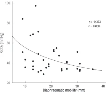
The mean values of diaphragmatic mobility were 19.8 ± 7.5 mm, and the mean PaCO2 was 45.2 ± 16.0 mmHg. There were significant negative correlations between the diaphragm mobil- ity and PaCO2 in patients with COPD (r = -0.373, P = 0.030) (Fig.
2). However, there was no correlation between diaphragm mo- bility and PaO2 (r = 0.028, P = 0.873).
Diaphragm mobility correlated with the pulmonary function parameters that quantify airway obstruction (FEV1: r = 0.415, P = 0.011) and pulmonary hyperinflation (RV: r = -0.501, P = 0.021;
TLC: r = -0.281, P = 0.030; RV/TLC: r = -0.527, P = 0.001). Dia- phragmatic mobility also correlated significantly with ventilator capacity (FVC: r = 0.302, P = 0.029; MVV: r = 0.481, P = 0.003).
However, there was no correlation between diaphragm mobili- ty and FEV1/FVC (r = 0.233, P = 0.166) (Table 2). In the multivar- iate analyses, PaCO2 and RV/TLC were independent factors for the diaphragm mobility.
DISCUSSION
The present study has shown that COPD patients with reduction in diaphragmatic mobility are associated with PaCO2 values. In addition, our result show that reduced diaphragmatic mobility is associated with airway obstruction and pulmonary hyperin- flation.
Expiratory flow limitation is the pathophysiological hallmark of COPD. The disease is associated with systemic manifestations
that can result in worsening dyspnea, impaired functional ca- pacity, reduced healthy-related quality of life and increased mor- tality. Dyspnea could result from a decreased capacity of the re- spiratory muscle to meet an increased mechanical load. In ad- dition, COPD patients commonly present increased airflow re- sistance as well as air trapping and pulmonary hyperinflation.
Pulmonary hyperinflation reduces the flow and pressure-gen- erating capacity of the diaphragm (17). As COPD patients devel- op hyperinflation, their diaphragm becomes flatter and shorter.
COPD patients have decreased diaphragm mobility when com- pared with healthy humans (6, 9). The reduced diaphragmatic mobility observed in COPD patients might be attributable to ex- cessive actin–myosin filament overlap and sarcomere remodel- ing (18, 19). The main cause of the reduction in diaphragmatic mobility is shortening of the apposition zone resulting from the absence of the piston-like movement of the diaphragm (20). A recent study demonstrated that reduction in diaphragm mobil- ity correlated with airway obstruction, pulmonary hyperinfla- tion and ventilator capacity (5). These findings are consistency with our results showing that diaphragm mobility is positively correlated with airway obstruction and ventilator capacity. Re- duction in diaphragm mobility in patients with COPD is nega- tively correlated with pulmonary function parameters related to lung hyperinflation. Although FEV1 is essential to diagnosis and quantifying COPD-related ventilator impairment (1), it does not adequately reflect the systemic manifestation of COPD (21).
Therefore, it has been proposed that other parameters should be evaluated in order to measure changes in the functional sta- tus of COPD patients. The results of the present study suggest that diaphragmatic mobility is another parameter that could provide information on respiratory mechanics and functional capacity in COPD patients.
Several factors such as airway obstruction, ventilation-perfu- sion mismatch, abnormalities in ventilatory control, respiratory muscle weakness, pattern of breathing, and dynamic pulmo- nary hyperinflation have been reported to contribute to carbon dioxide retention in patients with COPD (13-15). A previous study demonstrated that hypercapnia increases with the severity of the obstruction, obesity, and inspiratory muscle weakness (22).
Hypercapnic respiratory failure due to inspiratory muscle weak- ness is associated with morbidity in patients with COPD (23).
Table 2. Correlation between diaphragm mobility with pulmonary function variables
Variables Linear correlation
coefficient (r-value) P value
FEV1 (% predicted) 0.415 0.011
FVC (% predicted) 0.302 0.029
FEV1/FVC ratio (%) 0.233 0.166
RV (% predicted) -0.501 0.021
TLC (% predicted) -0.281 0.030
RV/TLC (%) -0.527 0.001
MVV (L/min) 0.481 0.003
Fig. 2. The relationship between diaphragmatic mobility and the PaCO2. There was a negative linear correlation between the two measurements (r = -0.373, P = 0.030).
PaCO2, arterial carbon dioxide tension.
PaCO2 (mmHg)
Diaphragmatic mobility (mm)
r = -0.373 P = 0.030
10 20 30 40
100
80
60
40
20
Rapid and shallow breathing in patients with COPD is the con- sequence of an excessive load imposed on the inspiratory mus- cles. The shallow breathing reduces alveolar ventilation and pre- disposes to hypercapnia. A study demonstrated that, in patients with stable COPD, PaCO2 was significantly related to positive end expiratory alveolar pressure which suggests that dynamic pul- monary hyperinflation could play a part in chronic carbon diox- ide retention (24). To our knowledge, however, no previous stud- ies have specifically examined the correlations between dia- phragm mobility and PaCO2 values. Our results show that sig- nificant negative correlations between the diaphragm mobility and PaCO2 levels in patients with COPD.
In conclusion, ultrasonography was used to determine the diaphragm mobility in this study. Our study demonstrates that the reduction of diaphragm mobility in patients with COPD is associated with carbon dioxide retention. In addition, diaphrag- matic mobility correlates with airway obstruction, ventilatory capacity and pulmonary hyperinflation. These findings support a possibility that the reduction in diaphragm mobility relates to hypercapnia in COPD patients. Further studies are required to better understand the diaphragm mobility on hypercapnia in patients with COPD.
REFERENCES
1. Rabe KF, Hurd S, Anzueto A, Barnes PJ, Buist SA, Calverley P, Fukuchi Y, Jenkins C, Rodriguez-Roisin R, van Weel C, Zielinski J. Global strategy for the diagnosis, management, and prevention of chronic obstructive pulmonary disease. Am J Respir Crit Care Med 2007; 176: 532-55.
2. Iwasawa T, Kagei S, Gotoh T, Yoshiike Y, Matsushita K, Kurihara H, Saito K, Matsubara S. Magnetic resonance analysis of abnormal diaphragmat- ic motion in patients with emphysema. Eur Respir J 2002; 19: 225-31.
3. Roussos C, Macklem PT. The respiratory muscles. N Engl J Med 1982;
307: 786-97.
4. Rochester DF. The diaphragm: contractile properties and fatigue. J Clin Invest 1985; 75: 1397-402.
5. Dos Santos Yamaguti WP, Paulin E, Shibao S, Chammas MC, Salge JM, Ribeiro M, Cukier A, Carvalho CR. Air trapping: The major factor limit- ing diaphragm mobility in chronic obstructive pulmonary disease pa- tients. Respirology 2008; 13: 138-44.
6. Unal O, Arslan H, Uzun K, Ozbay B, Sakarya ME. Evaluation of diaphrag- matic movement with MR fluoroscopy in chronic obstructive pulmonary disease. Clin Imaging 2000; 24: 347-50.
7. Singh B, Eastwood PR, Finucane KE. Volume displaced by diaphragm motion in emphysema. J Appl Physiol 2001; 91: 1913-23.
8. Cassart M, Pettiaux N, Gevenois PA, Paiva M, Estenne M. Effect of chron- ic hyperinflation on diaphragm length and surface area. Am J Respir Crit
Care Med 1997; 156: 504-8.
9. Suga K, Tsukuda T, Awaya H, Takano K, Koike S, Matsunaga N, Sugi K, Esato K. Impaired respiratory mechanics in pulmonary emphysema:
evaluation with dynamic breathing MRI. J Magn Reson Imaging 1999;
10: 510-20.
10. Harris RS, Giovannetti M, Kim BK. Normal ventilatory movement of the right hemidiaphragm studied by ultrasonography and pneumotachog- raphy. Radiology 1983; 146: 141-4.
11. Toledo NS, Kodaira SK, Massarollo PC, Pereira OI, Mies S. Right hemidi- aphragmatic mobility: assessment with US measurement of craniocaudal displacement of left branches of portal vein. Radiology 2003; 228: 389-94.
12. Houston JG, Fleet M, Cowan MD, McMillan NC. Comparison of ultra- sound with fluoroscopy in the assessment of suspected hemidiaphrag- matic movement abnormality. Clin Radiol 1995; 50: 95-8.
13. West JB. Causes of carbon dioxide retention in lung disease. N Engl J Med 1971; 284: 1232-6.
14. Rochester DF, Braun NM. Determinants of maximal inspiratory pres- sure in chronic obstructive pulmonary disease. Am Rev Respir Dis 1985;
132: 42-7.
15. Gorini M, Spinelli A, Ginanni R, Duranti R, Gigliotti F, Scano G. Neural respiratory drive and neuromuscular coupling in patients with chronic obstructive pulmonary disease (COPD). Chest 1990; 98: 1179-86.
16. Toledo NS, Kodaira SK, Massarollo PC, Pereira OI, Mies S. Right hemidi- aphragmatic mobility: assessment with US measurement of craniocaudal displacement of left branches of portal vein. Radiology 2003; 228: 389-94.
17. McKenzie DK, Butler JE, Gandevia SC. Respiratory muscle function and activation in chronic obstructive pulmonary disease. J Appl Physiol 2009;
107: 621-9.
18. Hoppin FG Jr. Theoretical basis for improvement following reduction pneumoplasty in emphysema. Am J Respir Crit Care Med 1997; 155:
520-5.
19. Orozco-Levi M, Gea J, Lloreta JL, Félez M, Minguella J, Serrano S, Bro- quetas JM. Subcelullar adaptation of the human diaphragm in chronic obstructive pulmonary disease. Eur Respir J 1999; 13: 371-8.
20. Gauthier AP, Verbanck S, Estenne M, Segebarth C, Macklem PT, Paiva M. Three-dimensional reconstruction of the in vivo human diaphragm shape at different lung volumes. J Appl Physiol 1994; 76: 495-506.
21. Gross NJ. Extrapulmonary effects of chronic obstructive pulmonary dis- ease. Curr Opin Pulm Med 2001; 7: 84-92.
22. Bégin P, Grassino A. Inspiratory muscle dysfunction and chronic hyper- capnia in chronic obstructive pulmonary disease. Am Rev Respir Dis 1991; 143: 905-12.
23. Zielinski J, MacNee W, Wedzicha J, Ambrosino N, Braghiroli A, Dolen- sky J, Howard P, Gorzelak K, Lahdensuo A, Strom K, Tobiasz M, Weitzen- blum E. Causes of death in patients with COPD and chronic respiratory failure. Monaldi Arch Chest Dis 1997; 52: 43-7.
24. Haluszka J, Chartrand DA, Grassino AE, Milic-Emili J. Intrinsic PEEP and arterial PCO2 in stable patients with chronic obstructive pulmonary dis- ease. Am Rev Respir Dis 1990; 141: 1194-7.
AUTHOR SUMMARY
Influence of Diaphragmatic Mobility on Hypercapnia in Patients with Chronic Obstructive Pulmonary Disease
Hyun Wook Kang, Tae Ok Kim, Bo Ram Lee, Jin Yeong Yu, Su Young Chi, Hee Jung Ban, In Jae Oh, Kyu Sik Kim, Yong Soo Kwon, Yu Il Kim, Young Chul Kim and Sung Chul Lim
A reduction in diaphragm mobility has been identified in patients with chronic obstructive pulmonary disease (COPD) and has been associated with a decline in pulmonary function parameters. However, little information exists regarding the potential role of diaphragm mobility on hypercapnia in COPD. We investigated the relationship between diaphragm mobility and arterial blood gas values in COPD patients. There were significant negative correlations between diaphragmatic mobility and PaCO2. Also,
diaphragmatic mobility correlated with airway obstruction, ventilatory capacity and hyperinflation of lungs.