• 검색 결과가 없습니다.

Correlation between Telomere Length and Chronic Obstructive Pulmonary Disease-Related Phenotypes: Results from the Chronic Obstructive Pulmonary Disease in Dusty Areas (CODA) Cohort

N/A
N/A
Protected

Academic year: 2021

Share "Correlation between Telomere Length and Chronic Obstructive Pulmonary Disease-Related Phenotypes: Results from the Chronic Obstructive Pulmonary Disease in Dusty Areas (CODA) Cohort"

Copied!
12
0
0

로드 중.... (전체 텍스트 보기)

전체 글

(1)

Correlation between Telomere Length and Chronic Obstructive Pulmonary Disease–

Related Phenotypes: Results from the

Chronic Obstructive Pulmonary Disease in Dusty Areas (CODA) Cohort

Da Hye Moon, M.D., Ph.D.

1

, Jeeyoung Kim, Ph.D.

2

, Myoung Nam Lim, Ph.D.

3

, So Hyen Bak, M.D., Ph.D.

4

and Woo Jin Kim, M.D., Ph.D.

1,2

1

Department of Internal Medicine, Kangwon National University Hospital, Chuncheon,

2

Department of Internal Medicine and Environmental Health Center, Kangwon National University School of Medicine, Chuncheon,

3

Biomedical Research Institute, Kangwon National University, Chuncheon,

4

Department of Radiology, Kangwon National University Hospital, Kangwon National University of School of Medicine, Chuncheon, Republic of Korea

Background: Chronic obstructive pulmonary disease (COPD) is a common chronic respiratory disease with increased prevalence in the elderly. Telomeres are repetitive DNA sequences found at the end of the chromosome, which progressively shorten as cells divide. Telomere length is known to be a molecular marker of aging. This study aimed to assess the relationship between telomere length and the risk of COPD, lung function, respiratory symptoms, and emphysema index in Chronic Obstructive Pulmonary Disease in Dusty Areas (CODA) cohort.

Methods: We extracted DNA from the peripheral blood samples of 446 participants, including 285 COPD patients and 161 control participants. We measured absolute telomere length using quantitative real-time polymerase chain reaction. All participants underwent spirometry and quantitative computed tomography scan. Questionnaires assessing respiratory symptoms and the COPD Assessment Test was filled by all the participants.

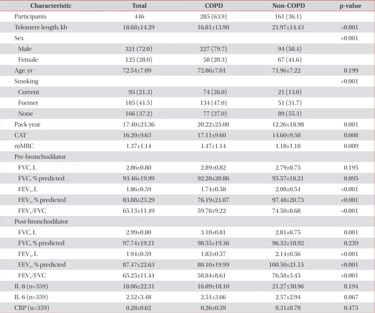
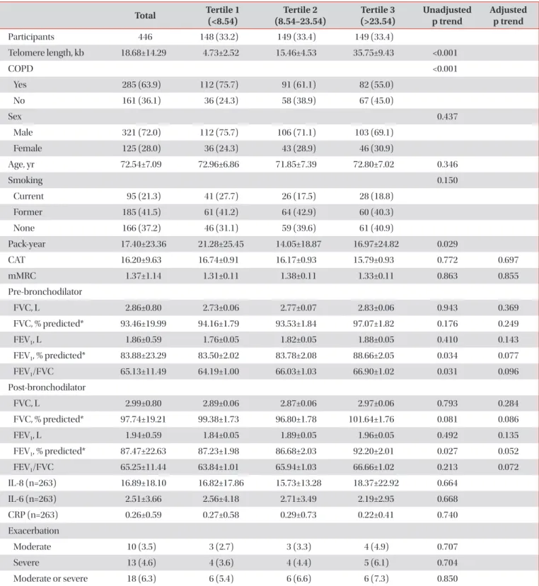
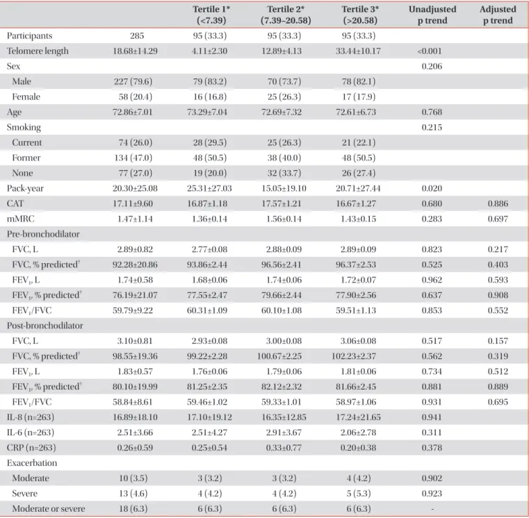
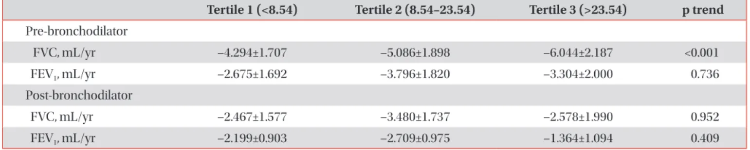
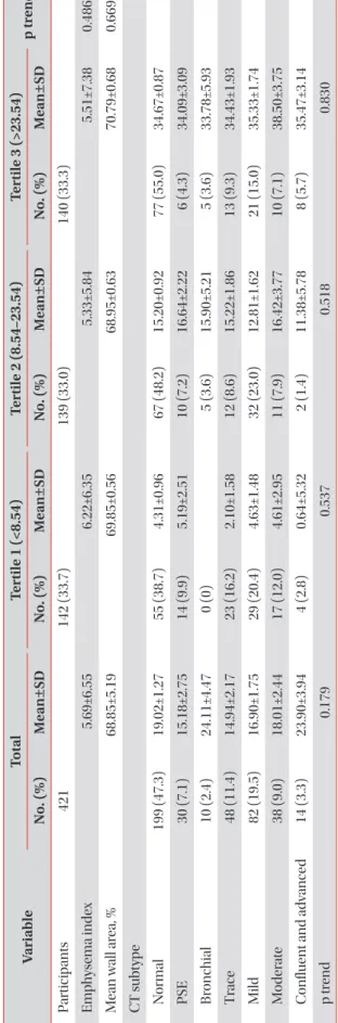
Results: The mean age of participants at the baseline visit was 72.5±7.1 years. Males accounted for 72% (321 participants) of the all participants. The mean telomere length was lower in the COPD group compared to the non-COPD group (COPD, 16.81±13.90 kb; non-COPD, 21.97±14.43 kb). In COPD patients, 112 (75.7%) were distributed as tertile 1 (shortest), 91 (61.1%) as tertile 2 and 82 (55%) as tertile 3 (longest). We did not find significant associations between telomere length and lung function, exacerbation, airway wall thickness, and emphysema index after adjusting for sex, age, and smoking status.

Conclusion: In this study, the relationship between various COPD phenotypes and telomere length was analyzed, but no significant statistical associations were shown.

Keywords: Telomere Length; Chronic Obstructive Pulmonary Disease; Phenotype

Address for correspondence: Woo Jin Kim, M.D., Ph.D.

Department of Internal Medicine, Kangwon National University School of Medicine, 1 Gangwondaehak-gil, Chuncheon 24341, Republic of Korea Phone: 82-33-258-9364, Fax: 82-33-255-6567, E-mail: pulmo2@kangwon.ac.kr

Received: Jan. 24, 2021, Revised: Apr. 10, 2021, Accepted: May. 12, 2021, Published online: May. 13, 2021

cc It is identical to the Creative Commons Attribution Non-Commercial License (http://creativecommons.org/licenses/by-nc/4.0/).

Copyright © 2021

The Korean Academy of Tuberculosis and Respiratory Diseases.

(2)

Introduction

Chronic obstructive pulmonary disease (COPD) is a major and increasing global health problem with large healthcare costs

1

. COPD is a heterogeneous disease, it comprises of emphysema in the lungs parenchyma, large central airway inflammation, mucociliary dysfunction, bronchiolitis, and small airway structural changes

2

. Airflow limitation, measured by reduced forced expiratory volume in 1 second (FEV

1

), progresses very slowly over several decades. Therefore most patients with symptomatic COPD are in the late middle-aged or elderly. An accelerated rate of lung function decline with age is one of the central pathophysiological characteristics of COPD. Therefore COPD prevalence increases with age

1

.

Telomeres are repetitive DNA sequences, with high G-C content, at the end of chromosome protection. They protect chromosomal ends from being recognized as double-strand breaks and therefore protecting them from end-to-end fusion and degradation

3

. DNA polymerases cannot fully replicate chromosomes, because one RNA primer remains on each daughter DNA strand. This loss of base pairs is a partial con- sequence of the end-replication problem

4

. Therefore, DNA cannot be duplicated at the end of the chromosome. Each duplication results in a gradual shortening of telomeres, until a critical length is reached at which point cells undergo apopto- sis

5

. In addition, exposure to oxidative stress and inflammation aggravates this shortening

6

. Based on this, some studies have shown that telomere length is a biomarker of cellular aging

7,8

.

Several studies have shown a significant relationship be- tween reduced telomere length in peripheral blood leukocytes and increased risk of malignancy, cardiovascular disease, and diabetes mellitus

9-11

. In these studies, leukocyte telomere length was used as a biomarker of aging based on the hypoth-

esis that it reflects the physiological age of individuals. Since COPD is an age-dependent process, assessment of telomere length may be useful for better understanding of the disease pathogenesis.

Since COPD is a heterogeneous disease with variable phe- notypes

12

, in the present study, we assumed that the shorter the telomere length, the more depressed the lung function, and the higher the exacerbation rate and degree of emphy- sema. Accordingly, this study aimed to assess the relationship between telomere length and the lungs function, exacerba- tion, and visual assessments of emphysema as well as smok- ing exposure in a Korean COPD cohort residing near cement plants.

Materials and Methods

1. Study design and population

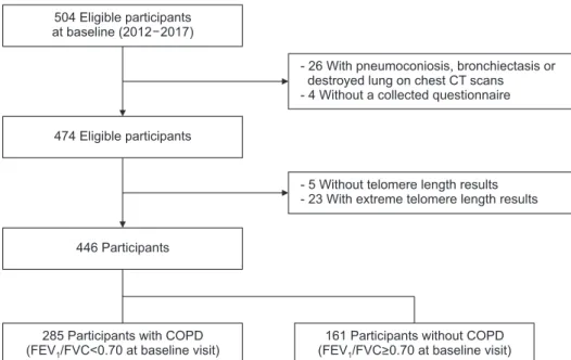
The Participants of the Chronic Obstructive Pulmonary Disease in Dusty Areas (CODA) cohort were analyzed

13

. The CODA study enrolled participants living near six cement plants in the Kangwon and Chungbuk provinces of South Ko- rea. Overall, 504 participants (362 men and 142 women) were enrolled for baseline examinations between 2012 and 2017 (Figure 1). Twenty-five participants whose chest computed to- mography (CT) scans showed signs of pneumoconiosis, bron- chiectasis or destroyed lung were excluded, four were without completed questionnaire, five without telomere length results, and 23 with extreme telomere length results. Finally, a total of 446 participants were enlisted as eligible participants.

At baseline examinations, a medical interview and survey questionnaire were administered, and spirometry, physical

504 Eligible participants at baseline (2012 2017)

474 Eligible participants

446 Participants

285 Participants with COPD (FEV /FVC<0.70 at baseline visit)1

- 26 With pneumoconiosis, bronchiectasis or destroyed lung on chest CT scans - 4 Without a collected questionnaire

- 5 Without telomere length results - 23 With extreme telomere length results

161 Participants without COPD (FEV /FVC>0.70 at baseline visit)1

Figure 1. Flowchart of the study popula- tion. CT: computed tomography; COPD:

chronic obstructive pulmonary disease;

FEV

1

: forced expiratory volume in 1 sec-

ond; FVC: forced vital capacity.

(3)

examination, blood/urine sampling, and CT were performed for all participants. The questionnaire evaluated demographic factors, lifestyle factors, medical history, exacerbation history, and respiratory symptoms during the past year. We defined moderate exacerbation as a history of antibiotics or steroid use for more than two times, and severe exacerbation as more than one hospitalization due to respiratory symptoms within a year.

A written informed consent was given by each participant.

This study also received ethical approval from the Institu- tional Review Board of Kangwon National University Hospital (KNUH 2020-06-007).

2. Measurement

Dyspnea was evaluated using the modified Medical Re- search Council (mMRC) scoring system

14

. Quality of life was assessed using a patients-reported COPD Assessment Test (CAT).

Spirometry was measured yearly using the Easy One Kit (NDD Medizintechnik AG, Zurich, Switzerland), before and after inhalation of 400-μg salbutamol. All pulmonary func- tion tests were performed according to the guidelines of the American Thoracic Society/European Respiratory Society

15

. 3. CT image analysis

All participants underwent volumetric, thin-section, chest CT at full inspiration and expiration in the supine position.

CT images were acquired using a first-generation, dual- source scanner (Somatom Definition, Siemens Healthcare, Forchheim, Germany) in the caudocranial direction using the following parameters: 140 kVp, 100 mA, 0.9–1 beam pitch, and slice thickness of 0.6 mm and 3 mm. The CT data were reconstructed using a soft convolution kernel (B30f)

16

. Em- physema was evaluated by automatically extracting all lung images from the chest wall, mediastinum, and large airways.

The attenuation coefficients of pixels in these images were then measured. The emphysema index was defined as the volume fraction (%) of the lung below −950 HU in full inspira- tion

17

. Mean wall area percentage was calculated as a percent- age of the mean values measured in two segmental bronchi (wall area/[wall area+lumen area]×100)

18

. The visually defined subtype of COPD were evaluated by two radiologists (with 3 and 11 years of experience) based on the Fleischner Society classification system

19

. Differences in interpretation were re- solved through consensus. The subtype were classified into the following seven types: (1) normal, (2) paraseptal emphy- sema (subjects with substantial paraseptal emphysema), (3) bronchial airway disease, (4) trace centrilobular emphysema, (5) mild centrilobular emphysema, (6) moderate centrilobu- lar emphysema, and (7) confluent and advanced destructive emphysema

20

.

4. Telomere length measurement

Venous blood samples were obtained at baseline and DNA was extracted from the buffy coat. Telomere length was measured in DNA isolated from leukocytes. We modified the Cawthon method for relative measurement of telomere length by introducing an oligomer standard to measure absolute telomere length (aTL). In this approach, aTL was calculated by quantitative polymerase chain reaction according to by O’Callaghan and Fenech method

21

, where a standard curve was generated from the fluorescent signals obtained from a series of known concentrations of telomere oligomer DNA (TTAGG ×14). The concentration of each test sample was pre- dicted by plotting the fluorescence signal of the sample onto a standard curve. To serve as a reference gene, the concentra- tion of a single copy gene (36B4) DNA in each sample was measured using the same method. Telomere length was cal- culated as the ratio of telomere DNA length from the standard curve to the 36B4 DNA length.

The telomere lengths of all samples were normalized to a reference cell line control sample, which was evaluated on each plate. Telomere length measurements (kb/genome) were normalized by natural log-transformation. For analysis, we divided the participants into three tertiles according to their telomere lengths.

5. Statistical analysis

Four hundred and forty-six participants were divided into were divided into two groups; COPD and non-COPD groups.

COPD was defined according to GOLD (Global Initiative for Chronic Obstructive Lung Disease) with post-bronchodilator FEV

1

/forced vital capacity (FVC) <0.70. For analysis, the telo- mere length for each group (all participants, COPD group and non-COPD group) were divided into tertile groups.

Comparison of baseline characteristics between COPD group

and non-COPD group were performed using a Student t

test and chi-square test. Categorical variable were described

as number (%). Continuous variables were reported as the

mean±standard deviation. The lung function, mMRC, and

CAT score were evaluated using a general linear model adjust-

ing for age, sex, smoking status, and height. To compare the

trend of lung function decline according to the tertile groups,

we used a mixed model adjusting for age, sex, and smoking

status. Logistic regression was used to find the association be-

tween telomere length and COPD exacerbation. Odds ratios

(ORs) and 95% confidence intervals (CI) were calculated us-

ing logistic regression model after adjusting for sex, age, and

smoking status. For trend tests, individuals were categorized

according to telomere length tertile (coded 1–3) with the first

tertile consisting of individuals with the shortest telomere

lengths. Those with p-values less than 0.05 were considered

statistically significant. All analyses were performed using SAS

(4)

version 9.4 (SAS Institute Inc., Cary, NC, USA).

Results

1. Clinical characteristics, respiratory symptoms and lung functions of the all participants and COPD patients

The clinical characteristics, respiratory symptoms, and lung functions of the 446 participants at baseline are summarized in Table 1. There were 285 participants (63.9%) in the COPD

group and 161 (36.1%) in the non-COPD group. The aver- age telomere length was 18.68±14.29 kb. The mean telomere length was shorter in the COPD group than in the non-COPD group (COPD, 16.81±13.90 kb; non-COPD, 21.97±14.43 kb).

The mean age of participants at the baseline was 72.5±7.1 years and 72% (321 participants) of the cohort were men (Table 1).

We divided the participants into three groups depending on the telomere length (tertile 1, <8.54; tertile 2, 8.54–23.54; ter- tile 3, >23.54). A total of 285 participants (63.9%) had a FEV

1

/ FVC <0.70. In COPD patients, 112 (75.7%) were distributed as tertile 1, 91 (61.1%) as tertile 2, and 82 (55%) as tertile 3 (Table

Table 1. Baseline characteristics of the study participants

Characteristic Total COPD Non-COPD p-value

Participants 446 285 (63.9) 161 (36.1)

Telomere length, kb 18.68±14.29 16.81±13.90 21.97±14.43 <0.001

Sex <0.001

Male 321 (72.0) 227 (79.7) 94 (58.4)

Female 125 (28.0) 58 (20.3) 67 (41.6)

Age, yr 72.54±7.09 72.86±7.01 71.96±7.22 0.199

Smoking <0.001

Current 95 (21.3) 74 (26.0) 21 (13.0)

Former 185 (41.5) 134 (47.0) 51 (31.7)

None 166 (37.2) 77 (27.0) 89 (55.3)

Pack-year 17.40±23.36 20.22±25.08 12.26±18.98 0.001

CAT 16.20±9.63 17.11±9.60 14.60±9.50 0.008

mMRC 1.37±1.14 1.47±1.14 1.18±1.10 0.009

Pre-bronchodilator

FVC, L 2.86±0.80 2.89±0.82 2.79±0.75 0.195

FVC, % predicted 93.46±19.99 92.28±20.86 95.57±18.21 0.095

FEV

1

, L 1.86±0.59 1.74±0.58 2.08±0.54 <0.001

FEV

1

, % predicted 83.88±23.29 76.19±21.07 97.48±20.73 <0.001

FEV

1

/FVC 65.13±11.49 59.78±9.22 74.58±8.68 <0.001

Post-bronchodilator

FVC, L 2.99±0.80 3.10±0.81 2.81±0.75 0.001

FVC, % predicted 97.74±19.21 98.55±19.36 96.32±18.92 0.239

FEV

1

, L 1.94±0.59 1.83±0.57 2.14±0.56 <0.001

FEV

1

, % predicted 87.47±22.63 80.10±19.99 100.50±21.15 <0.001

FEV

1

/FVC 65.25±11.44 58.84±8.61 76.58±5.45 <0.001

IL-8 (n=359) 18.06±22.31 16.89±18.10 21.27±30.96 0.194

IL-6 (n=359) 2.52±3.48 2.51±3.66 2.57±2.94 0.867

CRP (n=359) 0.28±0.62 0.26±0.59 0.31±0.70 0.475

Values are presented as number (%) or mean±SD and analyzed with the t test or chi-square test.

COPD: chronic obstructive pulmonary disease; CAT: COPD Assessment Test; mMRC: modified Medical Research Council; FVC: forced vital

capacity; FEV

1

: forced expi ratory volume in 1 second; IL-8: interleukin-8; IL-6: interleukin-6; CRP: C-reactive protein.

(5)

Table 2. Clinical demographic characteristics, respiratory symptom and lung function of all participants (n=446)

Total Tertile 1

(<8.54) Tertile 2

(8.54–23.54) Tertile 3

(>23.54) Unadjusted

p trend Adjusted p trend

Participants 446 148 (33.2) 149 (33.4) 149 (33.4)

Telomere length, kb 18.68±14.29 4.73±2.52 15.46±4.53 35.75±9.43 <0.001

COPD <0.001

Yes 285 (63.9) 112 (75.7) 91 (61.1) 82 (55.0)

No 161 (36.1) 36 (24.3) 58 (38.9) 67 (45.0)

Sex 0.437

Male 321 (72.0) 112 (75.7) 106 (71.1) 103 (69.1)

Female 125 (28.0) 36 (24.3) 43 (28.9) 46 (30.9)

Age, yr 72.54±7.09 72.96±6.86 71.85±7.39 72.80±7.02 0.346

Smoking 0.150

Current 95 (21.3) 41 (27.7) 26 (17.5) 28 (18.8)

Former 185 (41.5) 61 (41.2) 64 (42.9) 60 (40.3)

None 166 (37.2) 46 (31.1) 59 (39.6) 61 (40.9)

Pack-year 17.40±23.36 21.28±25.45 14.05±18.87 16.97±24.82 0.029

CAT 16.20±9.63 16.74±0.91 16.17±0.93 15.79±0.93 0.772 0.697

mMRC 1.37±1.14 1.31±0.11 1.38±0.11 1.33±0.11 0.863 0.855

Pre-bronchodilator

FVC, L 2.86±0.80 2.73±0.06 2.77±0.07 2.83±0.06 0.943 0.369

FVC, % predicted* 93.46±19.99 94.16±1.79 93.53±1.84 97.07±1.82 0.176 0.249

FEV

1

, L 1.86±0.59 1.76±0.05 1.82±0.05 1.88±0.05 0.410 0.143

FEV

1

, % predicted* 83.88±23.29 83.50±2.02 83.78±2.08 88.66±2.05 0.034 0.077

FEV

1

/FVC 65.13±11.49 64.19±1.00 66.03±1.03 66.90±1.02 0.031 0.096

Post-bronchodilator

FVC, L 2.99±0.80 2.89±0.06 2.87±0.06 2.97±0.06 0.793 0.284

FVC, % predicted* 97.74±19.21 99.38±1.73 96.80±1.78 101.64±1.76 0.081 0.086

FEV

1

, L 1.94±0.59 1.84±0.05 1.89±0.05 1.96±0.05 0.492 0.135

FEV

1

, % predicted* 87.47±22.63 87.23±1.98 86.68±2.03 92.20±2.01 0.027 0.052

FEV

1

/FVC 65.25±11.44 63.84±1.01 65.94±1.03 66.66±1.02 0.213 0.072

IL-8 (n=263) 16.89±18.10 16.82±17.86 15.73±13.28 18.37±22.92 0.664

IL-6 (n=263) 2.51±3.66 2.56±4.18 2.71±3.49 2.19±2.95 0.668

CRP (n=263) 0.26±0.59 0.27±0.58 0.29±0.73 0.22±0.41 0.740

Exacerbation

Moderate 10 (3.5) 3 (2.7) 3 (3.3) 4 (4.9) 0.707

Severe 13 (4.6) 4 (3.6) 4 (4.4) 5 (6.1) 0.704

Moderate or severe 18 (6.3) 6 (5.4) 6 (6.6) 6 (7.3) 0.850

Values are presented as number (%) or mean±SD and analyzed with the t test or chi-square test.

Study participants were divided into three groups based on telomere length, with the first tertile being the shortest and the third tertile being the longest.

Adjusted variables: sex, age, smoking status, height.

*Adjusted variables: sex, age, smoking status.

COPD: chronic obstructive pulmonary disease; CAT: COPD Assessment Test; mMRC: modified Medical Research Council; FVC: forced vital

capacity; FEV

1

: forced expiratory volume in 1 second; IL-8: interleukin-8; IL-6: interleukin-6; CRP: C-reactive protein.

(6)

2). There were no statistically significant associations between lung function and telomere length after adjusting for sex, age, and smoking status. However, shorter telomere length showed

the tendencies that decreased FVC at pre-bronchodilator and FVC, FVC % predicted at post-bronchodilator in COPD patients (p trend, 0.217, 0.157, and 0.319, respectively) (Table

Table 3. Clinical demographic characteristics, respiratory symptom and pulmonary function of COPD patients (n=285) Tertile 1*

(<7.39) Tertile 2*

(7.39–20.58) Tertile 3*

(>20.58) Unadjusted

p trend Adjusted

p trend

Participants 285 95 (33.3) 95 (33.3) 95 (33.3)

Telomere length 18.68±14.29 4.11±2.30 12.89±4.13 33.44±10.17 <0.001

Sex 0.206

Male 227 (79.6) 79 (83.2) 70 (73.7) 78 (82.1)

Female 58 (20.4) 16 (16.8) 25 (26.3) 17 (17.9)

Age 72.86±7.01 73.29±7.04 72.69±7.32 72.61±6.73 0.768

Smoking 0.215

Current 74 (26.0) 28 (29.5) 25 (26.3) 21 (22.1)

Former 134 (47.0) 48 (50.5) 38 (40.0) 48 (50.5)

None 77 (27.0) 19 (20.0) 32 (33.7) 26 (27.4)

Pack-year 20.30±25.08 25.31±27.03 15.05±19.10 20.71±27.44 0.020

CAT 17.11±9.60 16.87±1.18 17.57±1.21 16.67±1.27 0.680 0.886

mMRC 1.47±1.14 1.36±0.14 1.56±0.14 1.43±0.15 0.283 0.697

Pre-bronchodilator

FVC, L 2.89±0.82 2.77±0.08 2.88±0.09 2.89±0.09 0.823 0.217

FVC, % predicted

92.28±20.86 93.86±2.44 96.56±2.41 96.37±2.53 0.525 0.403

FEV

1

, L 1.74±0.58 1.68±0.06 1.74±0.06 1.72±0.07 0.962 0.593

FEV

1

, % predicted

76.19±21.07 77.55±2.47 79.66±2.44 77.90±2.56 0.637 0.908

FEV

1

/FVC 59.79±9.22 60.31±1.09 60.10±1.08 59.51±1.13 0.853 0.552

Post-bronchodilator

FVC, L 3.10±0.81 2.93±0.08 3.00±0.08 3.06±0.08 0.517 0.157

FVC, % predicted

98.55±19.36 99.22±2.28 100.67±2.25 102.23±2.37 0.562 0.319

FEV

1

, L 1.83±0.57 1.76±0.06 1.79±0.06 1.81±0.06 0.734 0.512

FEV

1

, % predicted

80.10±19.99 81.25±2.35 82.12±2.32 81.66±2.45 0.881 0.889

FEV

1

/FVC 58.84±8.61 59.46±1.02 59.33±1.01 58.97±1.06 0.931 0.695

IL-8 (n=263) 16.89±18.10 17.10±19.12 16.35±12.85 17.24±21.65 0.941

IL-6 (n=263) 2.51±3.66 2.51±4.27 2.91±3.67 2.06±2.78 0.311

CRP (n=263) 0.26±0.59 0.25±0.54 0.33±0.77 0.20±0.38 0.378

Exacerbation

Moderate 10 (3.5) 3 (3.2) 3 (3.2) 4 (4.2) 0.902

Severe 13 (4.6) 4 (4.2) 4 (4.2) 5 (5.3) 0.923

Moderate or severe 18 (6.3) 6 (6.3) 6 (6.3) 6 (6.3) -

Values are presented as number (%) or mean±SD and analyzed with the t test or chi-square test.

Adjusted variables: sex, age, smoking status, height.

*COPD patients were divided into three groups based on telomere length, with the first tertile being the shortest and the third tertile being the longest.

Adjusted variables: sex, age, smoking status.

COPD: chronic obstructive pulmonary disease; CAT: COPD Assessment Test; mMRC: modified Medical Research Council; FVC: forced vital

capacity; FEV

1

: forced expiratory volume in 1 second; IL-8: interleukin-8; IL-6: interleukin-6; CRP: C-reactive protein.

(7)

3). In the non-COPD group, shorter telomere length in tertile showed tendencies of decreased FEV

1

/FVC at pre-bronchodi- lator and post-bronchodilator (p trend, 0.03 and 0.005, respec- tively) (Supplementary Table S1).

2. The relationship between telomere length and lung function decline

In all participants and COPD patients, shorter telomere length in tertile was associated with decreased FVC L at pre- bronchodilator (p trend, <0.001 and <0.001, respectively) (Tables 4, 5). In non-COPD group, longer telomere length in tertile showed tendencies of increased FVC L at post-bron- chodilator (p trend, <0.001) (Supplementary Table S2).

3. The relationship between telomere length and visual and quantitative CT imaging features

Of the 446 participants, 25 were excluded from the visual assessment of the CT scans due to severe lung distortion and other lung morbidity. Finally, we analyzed 421 participants.

Among the 285 patients with COPD, 13 were excluded from the visual assessment of CT scans; only 272 patients were

included in the analysis. There were no statistically significant associations between telomere length and emphysema index, mean wall area, and CT subtypes in all participants and COPD patients (Table 6, Supplementary Table S3). In non-COPD participants, decreasing telomere length was associated with increased mean wall area (p trend, 0.415) (Supplementary Table S4).

4. The relationship between telomere length and acute exacerbation

We did not establish a significant association between telo- mere length and COPD exacerbation in COPD patients. In multivariable analyses, OR for the shortest versus the longest telomere tertile was 0.707 (95% CI, 0.153–3.274) for moderate exacerbation and 0.788 (0.203–3.057) for severe exacerbation (Table 7).

Discussion

We investigated the relationship between telomere length and the lung function, respiratory symptoms, emphysema

Table 4. Change in FVC and FEV

1

according to telomere length (all participants, n=446)

Tertile 1 (<8.54) Tertile 2 (8.54–23.54) Tertile 3 (>23.54) p trend Pre-bronchodilator

FVC, mL/yr –4.294±1.707 –5.086±1.898 –6.044±2.187 <0.001

FEV

1

, mL/yr –2.675±1.692 –3.796±1.820 –3.304±2.000 0.736

Post-bronchodilator

FVC, mL/yr –2.467±1.577 –3.480±1.737 –2.578±1.990 0.952

FEV

1

, mL/yr –2.199±0.903 –2.709±0.975 –1.364±1.094 0.409

Study participants were divided into three groups based on telomere length, with the first tertile being the shortest and the third tertile being the longest.

Adjusted variables: sex, age, smoking status, height.

FVC: forced vital capacity; FEV

1

: forced expiratory volume in 1 second.

Table 5. Change in FVC and FEV

1

according to telomere length (COPD patients, n=285)

Tertile 1* (<7.39) Tertile 2* (7.39–20.58) Tertile 3* (>20.58) p trend Pre-bronchodilator

FVC, mL/yr –5.652±2.004 –6.358±2.248 –7.346±2.706 <0.001

FEV

1

, mL/yr –3.297±2.005 –5.225±2.197 –4.206±2.477 0.679

Post-bronchodilator

FVC, mL/yr –5.003±1.900 –5.368±2.114 –4.606±2.518 0.856

FEV

1

, mL/yr –2.463±0.975 –2.958±1.078 –1.998±1.269 0.674

Adjusted variables: sex, age, smoking status, height.

*COPD patients were divided into three groups based on telomere length, with the first tertile being the shortest and the third tertile being the longest.

FVC: forced vital capacity; FEV

1

: forced expiratory volume in 1 second; COPD: chronic obstructive pulmonary disease.

(8)

Ta ble 6. T h e as so ci ation b etw een t elom er e len gth an d v is ual an d q uan tit ativ e C T im ag in g fe at ur es in all p ar tic ip an ts (n=421) Var ia ble Tot al Ter tile 1 (<8.54) Ter tile 2 (8.54–23.54) Ter tile 3 (>23.54) p tr en d N o. (%) M ean±SD N o. (%) M ean±SD N o. (%) M ean±SD N o. (%) M ean±SD Particip ants 421 142 (33.7) 139 (33.0) 140 (33.3) Emph ysema index 5.69±6.55 6.22±6.35 5.33±5.84 5.51±7.38 0.486 M ean w all ar ea, % 68.85±5.19 69.85±0.56 68.95±0.63 70.79±0.68 0.669 C T s ubt yp e N ormal 199 (47.3) 19.02±1.27 55 (38.7) 4.31±0.96 67 (48.2) 15.20±0.92 77 (55.0) 34.67±0.87 P SE 30 (7.1) 15.18±2.75 14 (9.9) 5.19±2.51 10 (7.2) 16.64±2.22 6 (4.3) 34.09±3.09 Br onc hi al 10 (2.4) 24.11±4.47 0 (0) 5 (3.6) 15.90±5.21 5 (3.6) 33.78±5.93 Tr ace 48 (11.4) 14.94±2.17 23 (16.2) 2.10±1.58 12 (8.6) 15.22±1.86 13 (9.3) 34.43±1.93 Mild 82 (19.5) 16.90±1.75 29 (20.4) 4.63±1.48 32 (23.0) 12.81±1.62 21 (15.0) 35.33±1.74 M oder at e 38 (9.0) 18.01±2.44 17 (12.0) 4.61±2.95 11 (7.9) 16.42±3.77 10 (7.1) 38.50±3.75 C onfluent and adv ance d 14 (3.3) 23.90±3.94 4 (2.8) 0.64±5.32 2 (1.4) 11.38±5.78 8 (5.7) 35.47±3.14 p tr end 0.179 0.537 0.518 0.830 St ud y p articip ants wer e divide d int o thr ee gr oups b ase d on t elomer e length, with the fir st t ertile b eing the short es t and the thir d t ertile b eing the long es t. A djus te d v ari ables : sex , ag e, smoking s tat us . C T : comput ed t omogr aph y; S D: s tand ar d de vi ation; PS E: p ar aseptal emph ysema.

(9)

extent, and airway wall thickness in a Korean COPD cohort living near cement plants. Disappointingly, we did not observe significant associations between telomere length and lung function, exacerbation, airway wall thickness, and emphysema index after adjusting for sex, age, and smoking status. How- ever, although no statistical significances have been shown, some tendencies were shown along the telomere length ter- tile. It was shown that FEV

1

/FVC (pre-, post-bronchodilator) decreased with shorter telomere length in all participants and in the non-COPD group. The shorter telomere length was shown to decrease FVC (pre-, post-bronchodilator) in COPD group. In all participants and COPD patients, the shorter the telomere length, the lower the FVC (pre-bronchodilator), indicating a decline in the lung function change. On the other hand, in the non-COPD group, the shorter the telomere length, the lower the FVC (post-bronchodilator), indicating an increase in the lung function change. In addition, the shorter the telomere length, the wider the mean wall area in non- COPD group.

COPD is an age-related disease. An accelerated rate of lung function decline with age is one of the central patho- physiological characteristics of COPD. Previous studies have shown that leukocyte telomeres can be like biomarkers in cellular aging

7,22,23

. Some studies have shown that decreased leukocyte telomere length is associated with a decline in lung function

24,25

. In our study, some tendencies between short telomere length and decreased lung function were shown, but they were not statistically significant.

Rode et al. reported that FEV

1

, FVC and FEV

1

/FVC de- creased with decreasing telomere length quartile (p trend, 5×10

–51

, 5×10

–35

, and 6×10

–137

, respectively) but the associa- tions attenuated after age and multivariable adjustments. In addition, participants with shorter telomeres were 2.06-fold more likely to develop COPD. The results showed that short telomere length is associated with reduced lung function and an increased prevalence of COPD

6

. Furthermore, data from seven studies demonstrated highly significant positive association between telomere length and spirometric mea- surements including FEV

1

(β=0.0455, p=1.07×10

–7

with fixed and random effects; I

2

=0%), FVC (β=0.0401, p=2.07×10

–7

with

fixed and random effects; I

2

=0%), and FEV

1

/FVC (β=0.0238, p=5.27×10

–7

with fixed and random effects; I

2

=0%)

25

.

One study reported that shorter leukocyte telomere length may be a biomarker associated with a poor clinical prognosis in COPD, where short telomere length was associated with reduced quality of life and increased risk of exacerbation and mortality in patients with moderate-to-severe COPD

26

. How- ever, our study did not show any relationship between acute COPD exacerbation and telomere length.

No significant associations were found between the em- physema index, mean wall area and telomere length in the all participants and COPD patients (p trend, 0.486, 0.669, 0.553, and 0.328, respectively). In the sub-analysis, the correlation of the average telomere length and emphysema CT subtype did not show a statistically significant trend in all participants and COPD group (p trend, 0.179 and 0.390, respectively). In addition, we did not observe any association between telo- mere length and airway wall thickness, emphysema index and visual CT image features in non-COPD group. However, we established that the shorter the telomere length, the wider the mean wall area in non-COPD group.

The MESA (Multi-Ethnic Study of Atherosclerosis) study found that the presence of centrilobular and panlobular em- physema correlated with increased dyspnea and reduced exercise capacity

27

. Airway wall thickness correlated with reduced lung function and increased symptoms in smoker in a cross-sectional study

28

. Image biomarkers have been useful predictors of disease progression. As shown earlier, previous studies showed that telomeres were shorter in peripheral leukocytes of COPD patients and have been related to lung function. However, we did not find other studies on the rela- tionship between CT-based visual assessments and telomere length.

Smoking is a well-known environmental factor that pro- motes aging and cellular senescence

29

. A large population study showed that a smoking history and the cumulative pack- years were associated with telomere length in 46,396 adults

30

. Another study found that smoking promotes shortening of telomere length. Within the same age range, telomere length decreased with age in smokers, irrespective of the presence or Table 7. The OR of exacerbation according to telomere length (COPD patients, n=285)

Moderate Severe Moderate or severe

Telomere length

Tertile 1* vs. tertile 3* 0.707 (0.153–3.274) 0.788 (0.203–3.057) 0.997 (0.307–3.241) Tertile 2* vs. tertile 3* 0.681 (0.146–3.184) 0.781 (0.200–3.050) 0.966 (0.295–3.157)

p-value 0.859 0.919 0.998

Adjusted variables: sex, age, smoking status, height.

*COPD patients were divided into three groups based on telomere length, with the first tertile being the shortest and the third tertile being the longest.

OR: odds ratio; COPD: chronic obstructive pulmonary disease.

(10)

absence of COPD (p=0.05, r=–0.27). The influence of cumula- tive smoking exposure on telomere length is further supported by the significant dose-effect relationship demonstrated be- tween pack-years and telomere length (p<0.001, r=–0.45)

31

. In this study, when analyzing the relationship between telomere length and pack-year (p trend, 0.029 and 0.020, respectively), we observed statistical significances, but we did not establish a trend in all participants and COPD patients.

It is worth noting that this is the first study to examine the relationship between telomere length and the phenotypes of COPD in Koreans. Our study investigated the relationship between telomere length and the extent of emphysema, air- way wall thickness, and visual assessment on CT scans. Some studies have demonstrated that emphysema as assessed by CT imaging is a good predictor of mortality in COPD patients at various stages of the disease. Although it is still unclear whether emphysema predisposes COPD patients to such systemic manifestations and whether these systemic manifes- tations contribute to the development of emphysema, it is ap- parent that recognizing the extent of emphysema is important in evaluating COPD. Taken together, the evaluation of emphy- sema seems to be beneficial in the management of COPD

32

.

Nevertheless, this study has several limitations. First, the small sample size resulted in limited power to detect dif- ferences between telomere length and COPD-related phe- notypes. Further large-scale studies with longer follow-up periods involving several serial assessments are needed to validate our findings.

Second, telomere length is a complex characteristic that is shaped by several factors, including genetic, epigenetic, lifestyle and environmental determinants. The complex in- teractions of these factors remain unclear

33

. The association between telomere length and environmental, occupational, and medical risk factors has been reported in several cross- sectional epidemiological studies

34,35

. However, this study did not consider such environmental and occupational factors.

This study consisted participants living in dusty areas near cement plants, thus the results might differ from the general COPD populations. In CODA cohort study, we have records of exposure such as air pollution (PM

10

, NO

2

); however, ad- ditional analysis could not be carried out due to insufficient number of participants.

Third, we measured the leukocyte telomere length. Regard- ing this point, it is important to note that a correlation between lung and blood telomere length has not been unequivocally demonstrated. This suggests that the associations between telomere length and various diseases cannot easily be inter- preted as causative relationship.

In conclusion, the correlation between lung function, re- spiratory symptoms, or extent and visual assessment and telomere length were analyzed, we did not find statistically significant results. Further studies are needed on the role of telomere length in COPD pathogenesis, as well as the rela-

tionship between telomere length and environmental factors including air pollution.

Authors’ Contributions

Conceptualization: Kim WJ, Moon DH. Methodology: Kim WJ, Moon DH, Kim J. Formal analysis: Lim MN. Data Cura- tion: Lim MN. Software: Lim MN. Validation: Lim MN, Kim WJ, Moon DH. Investigation: Kim WJ, Moon DH, Kim J. Writing – original draft preparation: Moon DH, Kim WJ. Writing – re- view and editing: Moon DH, Kim J, Lim MN, Bak SH, Kim WJ.

Approval of the final manuscript: all authors.

Conflicts of Interest

No potential conflict of interest relevant to this article was reported.

Funding

No funding to declare.

Supplementary Material

Supplementary material can be found in the journal homepage (http://www.e-trd.org).

Supplementary Table S1. Clinical demographic character- istics, respiratory symptom and pulmonary function of non- COPD patients (n=161).

Supplementary Table S2. Change in FVC and FEV

1

accord- ing to telomere length (non-COPD patients, n=161).

Supplementary Table S3. The association between telo- mere length and visual and quantitative CT imaging features in COPD patients (n=272).

Supplementary Table S4. The association between telomere length and visual and quantitative CT imaging features in non- COPD participants (n=149).

References

1. Ito K, Barnes PJ. COPD as a disease of accelerated lung aging.

Chest 2009;135:173-80.

2. Vestbo J, Anderson W, Coxson HO, Crim C, Dawber F, Ed- wards L, et al. Evaluation of COPD longitudinally to identify predictive surrogate end-points (ECLIPSE). Eur Respir J 2008;

31:869-73.

3. Blackburn EH. Structure and function of telomeres. Nature

1991;350:569-73.

(11)

4. Houben JM, Moonen HJ, van Schooten FJ, Hageman GJ.

Telomere length assessment: biomarker of chronic oxidative stress? Free Radic Biol Med 2008;44:235-46.

5. Lee J, Sandford AJ, Connett JE, Yan J, Mui T, Li Y, et al. The re- lationship between telomere length and mortality in chronic obstructive pulmonary disease (COPD). PLoS One 2012;7:

e35567.

6. Rode L, Bojesen SE, Weischer M, Vestbo J, Nordestgaard BG.

Short telomere length, lung function and chronic obstructive pulmonary disease in 46,396 individuals. Thorax 2013;68:429- 35.

7. Mather KA, Jorm AF, Parslow RA, Christensen H. Is telomere length a biomarker of aging? A review. J Gerontol A Biol Sci Med Sci 2011;66:202-13.

8. Armanios M. Telomeres and age-related disease: how telo- mere biology informs clinical paradigms. J Clin Invest 2013;

123:996-1002.

9. Wu X, Amos CI, Zhu Y, Zhao H, Grossman BH, Shay JW, et al.

Telomere dysfunction: a potential cancer predisposition fac- tor. J Natl Cancer Inst 2003;95:1211-8.

10. Calado RT, Young NS. Telomere diseases. N Engl J Med 2009;361:2353-65.

11. Serrano AL, Andres V. Telomeres and cardiovascular disease:

does size matter? Circ Res 2004;94:575-84.

12. Silverman EK. Exacerbations in chronic obstructive pulmo- nary disease: do they contribute to disease progression? Proc Am Thorac Soc 2007;4:586-90.

13. Han Y, Heo Y, Hong Y, Kwon SO, Kim WJ. Correlation between physical activity and lung function in dusty areas: results from the Chronic Obstructive Pulmonary Disease in Dusty Areas (CODA) cohort. Tuberc Respir Dis (Seoul) 2019;82:311-8.

14. Mahler DA, Wells CK. Evaluation of clinical methods for rat- ing dyspnea. Chest 1988;93:580-6.

15. Celli BR, MacNee W; ATS/ERS Task Force. Standards for the diagnosis and treatment of patients with COPD: a summary of the ATS/ERS position paper. Eur Respir J 2004;23:932-46.

16. Bak SH, Kwon SO, Han SS, Kim WJ. Computed tomography- derived area and density of pectoralis muscle associated disease severity and longitudinal changes in chronic obstruc- tive pulmonary disease: a case control study. Respir Res 2019;20:226.

17. Washko GR, Hunninghake GM, Fernandez IE, Nishino M, Okajima Y, Yamashiro T, et al. Lung volumes and emphysema in smokers with interstitial lung abnormalities. N Engl J Med 2011;364:897-906.

18. Kim V, Davey A, Comellas AP, Han MK, Washko G, Martinez CH, et al. Clinical and computed tomographic predictors of chronic bronchitis in COPD: a cross sectional analysis of the COPDGene study. Respir Res 2014;15:52.

19. Lynch DA, Austin JH, Hogg JC, Grenier PA, Kauczor HU, Bankier AA, et al. CT-definable subtypes of chronic obstruc- tive pulmonary disease: a statement of the Fleischner Society.

Radiology 2015;277:192-205.

20. Park J, Hobbs BD, Crapo JD, Make BJ, Regan EA, Humphries S, et al. Subtyping COPD by using visual and quantitative CT imaging features. Chest 2020;157:47-60.

21. O’Callaghan NJ, Fenech M. A quantitative PCR method for measuring absolute telomere length. Biol Proced Online 2011;13:3.

22. Levy MZ, Allsopp RC, Futcher AB, Greider CW, Harley CB.

Telomere end-replication problem and cell aging. J Mol Biol 1992;225:951-60.

23. Bernadotte A, Mikhelson VM, Spivak IM. Markers of cellular senescence. Telomere shortening as a marker of cellular se- nescence. Aging (Albany NY) 2016;8:3-11.

24. Clouston SA, Edelman NH, Aviv A, Stewart C, Luft BJ. Short- ened leukocyte telomere length is associated with reduced pulmonary function and greater subsequent decline in func- tion in a sample of World Trade Center responders. Sci Rep 2019;9:8148.

25. Albrecht E, Sillanpaa E, Karrasch S, Alves AC, Codd V, Hovatta I, et al. Telomere length in circulating leukocytes is associated with lung function and disease. Eur Respir J 2014;43:983-92.

26. Jin M, Lee EC, Ra SW, Fishbane N, Tam S, Criner GJ, et al.

Relationship of absolute telomere length with quality of lfe, exacerbations, and mortality in COPD. Chest 2018;154:266- 73.

27. Smith BM, Austin JH, Newell JD Jr, D’Souza BM, Rozenshtein A, Hoffman EA, et al. Pulmonary emphysema subtypes on computed tomography: the MESA COPD study. Am J Med 2014;127:94.

28. Dijkstra AE, Postma DS, ten Hacken N, Vonk JM, Oudkerk M, van Ooijen PM, et al. Low-dose CT measurements of airway dimensions and emphysema associated with airflow limita- tion in heavy smokers: a cross sectional study. Respir Res 2013;14:11.

29. Karrasch S, Holz O, Jorres RA. Aging and induced senescence as factors in the pathogenesis of lung emphysema. Respir Med 2008;102:1215-30.

30. Babizhayev MA, Yegorov YE. Smoking and health: associa- tion between telomere length and factors impacting on hu- man disease, quality of life and life span in a large population- based cohort under the effect of smoking duration. Fundam Clin Pharmacol 2011;25:425-42.

31. Morla M, Busquets X, Pons J, Sauleda J, MacNee W, Agusti AG. Telomere shortening in smokers with and without COPD.

Eur Respir J 2006;27:525-8.

32. Halper-Stromberg E, Cho MH, Wilson C, Nevrekar D, Crapo JD, Washko G, et al. Visual assessment of chest computed tomographic images is independently useful for genetic asso- ciation analysis in studies of chronic obstructive pulmonary disease. Ann Am Thorac Soc 2017;14:33-40.

33. Daniali L, Benetos A, Susser E, Kark JD, Labat C, Kimura M, et al. Telomeres shorten at equivalent rates in somatic tissues of adults. Nat Commun 2013;4:1597.

34. Benetos A, Kark JD, Susser E, Kimura M, Sinnreich R, Chen W,

(12)

et al. Tracking and fixed ranking of leukocyte telomere length across the adult life course. Aging Cell 2013;12:615-21.

35. Starkweather AR, Alhaeeri AA, Montpetit A, Brumelle J, Filler

K, Montpetit M, et al. An integrative review of factors associ-

ated with telomere length and implications for biobehavioral

research. Nurs Res 2014;63:36-50.

수치

Figure 1. Flowchart of the study popula- popula-tion. CT: computed tomography; COPD:
Table 1. Baseline characteristics of the study participants
Table 2. Clinical demographic characteristics, respiratory symptom and lung function of all participants (n=446)  Total Tertile 1 (&lt;8.54) Tertile 2 (8.54–23.54) Tertile 3 (&gt;23.54) Unadjustedp trend Adjustedp trend Participants 446 148 (33.2) 149 (33.
Table 3. Clinical demographic characteristics, respiratory symptom and pulmonary function of COPD patients (n=285)  Tertile 1* (&lt;7.39) Tertile 2* (7.39–20.58) Tertile 3*(&gt;20.58) Unadjustedp trend Adjustedp trend Participants 285 95 (33.3) 95 (33.3) 9
+3

참조

관련 문서

To gain insights about developmental outcomes related to separation experiences, we examined relations between the length of separation from mother and father,

Therefore, this study aims to identify the relations between gastroesophageal reflux disease and body mass index, and waist measurement as indexes of obesity.. For the

Objectives: This study aimed to investigate the relationship between job stress and turnover intention of employed opticians and to investigate the mediating effect of burnout

Purpose: This study was performed to identify the association between family support, ADL and depression among hospitalized older patients with chronic

A study on the relationship between body mass index and the food habits of college students. Keilins respiratory chain concept and its

Correlation between Results ofthe Multiple Allergosorbent Test-Chemiluminescent Assay(MAST-CLA) and Clinical Severity of Chronic Urticaria. A study of food allergy

In the current study, I investigated the association between vascular risk factors as chronic ischemia of the prostate and BPH by conducting experiments to elucidate

3-38 Data processing area for estimating the crack length .... 3-39 Relationship between the crack length and the sum