tions
4-6and that this susceptibility to the infection is associated with lethal complications unrelated to the cause of illness.
Therefore, it is important to identify accurate predictors of the prognosis of patients with septic shock. The innate immune system is activated prior to the adaptive immune system, and is thus the first line of defense against pathogens. The importance of the interactions between pathogen-associated microbial patterns and mannose-binding lectin (MBL) in acti- vating innate immunity is well established as a component of the innate immune system
7. In addition, it is now recognized that the first response to invasion (i.e., innate immunity) has a significant influence on the subsequent adaptive response
8,9.
It has been reported that individuals with a deficiency in MBL, a key recognition molecule in the complement lectin pathway, are susceptible to infections
10-12. Moreover, low levels of MBL cause defects in opsonization and phagocytosis that have been associated with recurrent infections in infants and adults
11,13,14. Cytokines are secreted by components of the in- nate and adaptive immune systems and act as modulators of inflammatory responses
15. MBL determines which cytokines are released because of its interaction with phagocytic cells
16.
Introduction
Septic shock remains an important cause of mortality and morbidity in intensive care units (ICUs)
1-3. Numerous studies suggest that individuals vary in their ability to resist infec-
Serial Changes in Mannose-Binding Lectin in Patients with Sepsis
Jin Won Huh, M.D., Ph.D.
1, Kyuyoung Song, Ph.D.
2, Hwa Jung Kim, M.D., Ph.D.
3, Jung-Sun Yum, Ph.D.
4, Sang-Bum Hong, M.D., Ph.D.
1, Chae-Man Lim, M.D., Ph.D.
1and Younsuck Koh, M.D., Ph.D.
11
Division of Pulmonary and Critical Care Medicine, Departments of
2Biochemistry and Molecular Biology and
3Clinical Epidemiology and Biostatistics, Asan Medical Center, University of Ulsan College of Medicine, Seoul,
4R&D Center, CHA Vaccine Institute, Seongnam, Korea
Background: Mannose-binding lectin (MBL) deficiency leads to increased susceptibility to infection. We investigated whether serial changes in MBL levels are associated with the prognosis of patients diagnosed with septic shock, and correlated with cytokine levels.
Methods: We enrolled 131 patients with septic shock in the study. We analyzed the serum samples for MBL and cytokine levels at baseline and 7 days later. Samples on day 7 were available in 73 patients.
Results: We divided the patients with septic shock into four groups according to serum MBL levels (<1.3 mg/mL or
≥1.3 mg/mL) on days 1 and 7. Patients with low MBL levels on day 1 and high MBL levels on day 7 showed a favorable prognosis for 28-day survival (odds ratio, 1.96, 95% confidence interval, 1.10–2.87; p=0.087). The high MBL group on day 7 showed a significant decrease in monocyte chemoattractant protein 1, interleukin (IL)-1β, IL-6, IL-8, interferon-γ, and granulocyte macrophage colony-stimulating factor levels compared with the low MBL group on day 7.
Conclusion: The increase in MBL levels of patients with septic shock may suggest a favorable prognosis and attenuate pro-inflammatory and anti-inflammatory responses.
Keywords: Mannose-Binding Lectin; Cytokines; Shock, Septic; Prognosis
Address for correspondence: Younsuck Koh, M.D., Ph.D.
Division of Pulmonary and Critical Care Medicine, Asan Medical Center, University of Ulsan College of Medicine, 88 Olympic-ro 43-gil, Songpa-gu, Seoul 05505, Korea
Phone: 82-2-3010-3134, Fax: 82-2-3010-6968 E-mail: [email protected]
Received: May. 13, 2017 Revised: Aug. 24, 2017 Accepted: Dec. 28, 2017 Published online: Mar. 7, 2018
cc
It is identical to the Creative Commons Attribution Non-Commercial License (http://creativecommons.org/licenses/by-nc/4.0/).
Copyright © 2018
The Korean Academy of Tuberculosis and Respiratory Diseases.
We hypothesized that patients who have a low response in increasing MBL levels after infection may be at high risk of death. To address this question, we investigated whether serial changes in MBL levels are associated with the prognosis of septic shock. We also assessed whether the serial changes of MBL affected cytokine levels.
Materials and Methods
1. Study subjects
In this study, 131 patients receiving intensive care for sep- tic shock were enrolled. Follow-up samples on day 7 were obtained in 73 patients. All patients were older than 18 years and had been admitted to the ICU of a tertiary teaching hos- pital in Seoul, South Korea. The diagnosis of septic shock was based on the criteria presented at the American Society of Chest Physicians/Society of Critical Care Medicine Consen- sus Conference in 1992: evidence for infection, evidence of a systemic response to infection, systolic blood pressure of less than 90 mm Hg despite adequate fluid resuscitation, and hypoperfusion or organ dysfunction attributable to sepsis
17. In a previous study, we reported that a low MBL level (<1.3 mg/
mL) is an independent risk factor for mortality after 28 days in patients with septic shock
18. Patients were categorized based on their MBL levels (<1.3 mg/mL or ≥1.3 mg/mL) on days 1 and 7. Informed consent was obtained from all study participants or their relatives in accordance with the policies of the institu- tional review board. This study was approved by the institu- tional review board (IRB No. 2005-0070).
Clinical data including demographic details, the sequential organ failure assessment (SOFA) score, the acute physiology, age, and chronic health evaluation II (APACHE II) score ob- tained on admission, and the ICU outcome, were recorded for each patient.
2. Quantification of MBL
Blood samples were collected within 24 hours after the onset of septic shock and on week 1 if the patients were alive in the ICU. The serum MBL level was measured using a sand- wich enzyme-linked immunosorbent assay (MBL-ELISA;
Dobeel Co., Seongnam, Korea) according to a previously es- tablished protocol
18,19.
3. Cytokine measurement
The serum levels of the proinflammatory cytokines inter- leukin (IL)-1β, IL-6, IL-8, monocyte chemoattractant protein 1 (MCP-1), interferon-γ (IFN-γ), tumor necrosis factor a (TNF- a), and the anti-inflammatory cytokines IL-1 receptor antago- nist (IL-1ra), IL-10, and granulocyte-macrophage stimulating
factor (GM-CSF). were measured by using a multiplex bead- based assay (Bio-Rad, Hercules, CA, USA).
4. Statistical analysis
The values obtained for the continuous variables were not normally distributed; therefore, the results were expressed as medians with an interquartile range. All categorical data were compared using chi-square analysis or Fisher exact test.
The Mann–Whitney test was used to test for differences be- tween the two groups. Associations between cytokines were measured with Spearman’s rank correlation. The prognostic
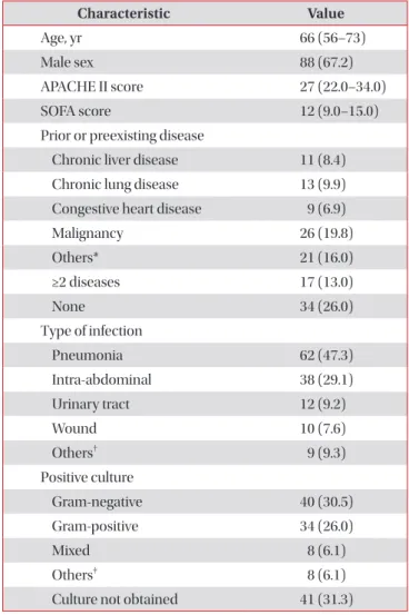
Table 1. Baseline characteristics of the patients (n=131) on day 1 of septic shock
Characteristic Value
Age, yr 66 (56–73)
Male sex 88 (67.2)
APACHE II score 27 (22.0–34.0)
SOFA score 12 (9.0–15.0)
Prior or preexisting disease
Chronic liver disease 11 (8.4) Chronic lung disease 13 (9.9) Congestive heart disease 9 (6.9)
Malignancy 26 (19.8)
Others* 21 (16.0)
≥2 diseases 17 (13.0)
None 34 (26.0)
Type of infection
Pneumonia 62 (47.3)
Intra-abdominal 38 (29.1)
Urinary tract 12 (9.2)
Wound 10 (7.6)
Others
†9 (9.3)
Positive culture
Gram-negative 40 (30.5)
Gram-positive 34 (26.0)
Mixed 8 (6.1)
Others
‡8 (6.1)
Culture not obtained 41 (31.3)
Values are presented as the median (interquartile range, 25%–75%) or number (%).
*Others: diabetes mellitus, neurologic disease, rheumatologic dis- ease.
†Others: primary bacteremia, leptospirosis, meningitis.
‡Oth- ers, anaerobe, intracellular bacteria, leptospira, fungus.
APACHE II: acute physiology, age, and chronic health evaluation II;
SOFA: sequential organ failure assessment.
value of the MBL level was calculated using bootstrapping, a nonparametric method that takes 1,000 samples of the data.
All data were analyzed using SPSS version 20.0 software (IBM Corp., Armonk, NY, USA).
Results
Baseline demographic, clinical, and microbiological data of all included patients (n=131) are summarized in Table 1.
The median age was 66 years. Eighty-eight men (67.2%) and 43 women (32.8%) were included. The most common cause of septic shock was pneumonia. The 28-day overall mortality rate was 35.9%.
Because we measured serum MBL levels on days 1 and 7 in 73 patients with septic shock, only 73 patients were enrolled in the final analysis. The median serum MBL level was 1.75 mg/mL (range, 0.84–2.65 mg/mL) on day 1 and 2.17 mg/mL (range, 1.0–3.66 mg/mL) on day 7. The serum MBL level on day 1 was negatively correlated with the SOFA score (r=–0.237, p=0.015).
We divided the patients with septic shock based on their serum MBL levels (<1.3 mg/mL or ≥1.3 mg/mL) on days 1 and 7. The group with a low MBL level (<1.3 mg/mL) on day 1
and increased MBL level (≥1.3 mg/mL) on day 7 had a good prognosis for 28-day survival (odds ratio, 1.96; 95% confidence interval, 1.10–2.87; p=0.087) (Table 2).
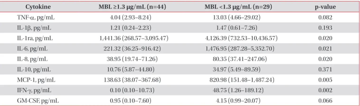
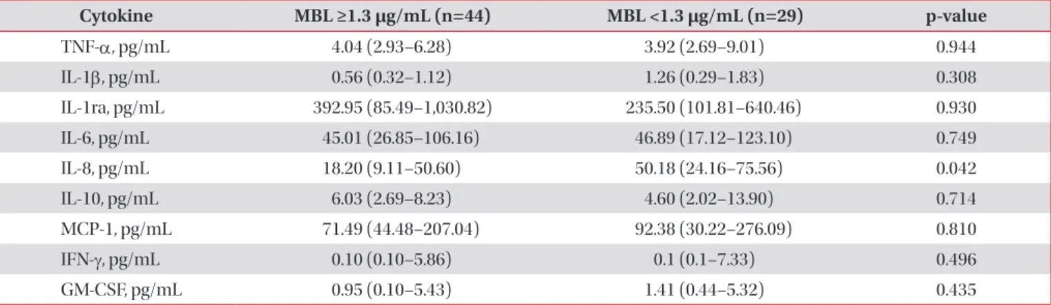
The group with low MBL levels showed a significant in- crease in MCP-1, IL-6, IL-8, IFN-γ, and IL-1ra levels compared with the group with high MBL levels (≥1.3 mg/mL) on day 1 (Table 3). Though the levels of the inflammatory and anti- inflammatory cytokines on day 7 was decreased, the group with low MBL (<1.3 mg/mL) showed a significant increase in IL-8 level compared with the group with higher MBL (Table 4).
Even though the initial MBL level was low, the group with increased MBL levels on day 7 showed a significant decrease in MCP-1, IL-1β, IL-6, IL-8, IFN-γ, and granulocyte macro- phage colony-stimulating factor levels (Table 5).
The levels of MCP-1 (r=–0.420, p=0.020) were negatively correlated with the MBL level on day 1 (Figure 1).
Discussion
We reported in a previous study that the initial MBL level (≥1.3 mg/mL) is an independent predictive factor of survival in septic shock
18. In this study, we observed that the increase of serum MBL level (≥ 1.3 mg/mL) on day 7 was a good prognos-
Table 3. Cytokine levels according to the initial MBL level (<1.3 μg/mL vs. ≥1.3 μg/mL) on day 1
Cytokine MBL ≥1.3 μ g/mL (n=44) MBL <1.3 μ g/mL (n=29) p-value
TNF-a, pg/mL 4.04 (2.93–8.24) 13.03 (4.66–29.02) 0.082
IL-1β, pg/mL 1.21 (0.24–2.23) 1.47 (0.61–7.26) 0.193
IL-1ra, pg/mL 1,441.36 (268.57–3,095.47) 4,126.39 (732.53–10,436.57) 0.020
IL-6, pg/mL 221.32 (36.25–916.42) 1,476.95 (287.28–5,352.70) 0.021
IL-8, pg/mL 38.95 (19.74–71.26) 80.35 (37.41–247.06) 0.020
IL-10, pg/mL 10.76 (5.87–44.80) 34.97 (5.49–89.59) 0.371
MCP-1, pg/mL 138.63 (38.07–367.68) 820.98 (151.48–1,487.24) 0.005
IFN-γ, pg/mL 0.10 (0.10–10.73) 48.75 (1.26–189.12) 0.002
GM-CSF, pg/mL 0.95 (0.10–7.60) 4.15 (0.99–20.07) 0.066
Values are presented as median (interquartile range, 25%–75%).
MBL: mannose-binding lectin; TNF-a: tumor necrosis factor a; IL: interleukin; IL-1ra: IL-1 receptor antagonist; MCP-1: monocyte chemoat- tractant protein 1; IFN-γ: interferon γ; GM-CSF: granulocyte-macrophage stimulating factor.
Table 2. Twenty-eight-day mortality according to serum MBL levels (<1.3 μg/mL or ≥1.3 μg/mL) on days 1 and 7
MBL level Nonsurvivor Survivor
<1.3 mg/mL on days 1 and 7 5/22 (22.7) 17/22 (77.3)
<1.3 mg/mL on day 1 and ≥1.3 mg/mL on day 7 0 (0) 7/7 (100)
≥1.3 mg/mL on days 1 and 7 1/4 (25.0) 3/4 (75.0)
≥1.3 mg/mL on day 1 and <1.3 mg/mL on day 7 14/40 (35.0) 26/40 (65.0) Values are presented as number (%).
MBL: mannose-binding lectin.
tic factor, even though initial MBL levels were low (<1.3 mg/
mL) in patients with septic shock.
Furthermore, to investigate the relationship of MBL levels with key pro-/anti-inflammatory cytokines in sepsis, we com- pared the cytokine profile and MBL levels in patients with septic shock. The group with high MBL levels showed signifi- cantly lower levels of pro- and anti-inflammatory cytokines such as IL-1ra, IL-6, IL-8, MCP-1, and IFN-γ compared to the group with low MBL levels (<1.3 mg/mL) on day 1. Comple- ment activation through MBL might contribute to pro- and anti-inflammatory responses in the early stage of sepsis. In ad- dition, if the immune response of pro-inflammatory cytokines, such as IL-1β, IL-6, IL-8, MCP-1, GM-CSF, and IFN-γ at initial was attenuated, MBL levels on day 7 could be restored. This subgroup showed good prognosis, even though the MBL level on day 1 was low.
Low levels of MBL result in defects in opsonization and phagocytosis
11,13. Although the relationship between cyto- kines and MBL is not completely clear, MBL, a component of the innate immune system, downregulates monocyte-medi- ated inflammation while enhancing phagocyte recruitment, thereby influencing the adaptive immune response
10,20-22. Inflammatory monocytes respond rapidly to microbial stimuli by secreting cytokines and antimicrobial factors; for example, they express the chemokine receptor CCR2 and traffic to sites of microbial infection in response to MCP-1 secretion
23. MCP- 1 is a potent chemoattractant of mononuclear cells and an im- portant immunomodulator in control of the balance between pro- and anti-inflammatory responses in sepsis
24. Although we did not investigate monocyte differentiation and function, our study shows a significant negative correlation between MBL and MCP-1 in patients with septic shock. Moreover, IL-8 Table 5. Cytokine levels according to the change of MBL level on day 1
Cytokine MBL levels <1.3 μ g/mL on days 1 and 7 MBL level <1.3 μ g/mL on day 1 and
MBL level ≥1.3 μ g/mL on day 7 p-value
TNF-a, pg/mL 19.5 (6.3–34.3) 5.5 (4.2–9.3) 0.217
IL-1β, pg/mL 2.3 (0.8–9.1) 0.4 (0.2–1.7) 0.036
IL-1ra, pg/mL 5,542.1 (2,327.8–10,597.9) 191.4 (143.3–15,135.5) 0.119
IL-6, pg/mL 2,607.3 (761.8–7,273.9) 119.8 (17.82–3,131.6) 0.042
IL-8, pg/mL 121.2 (66.0–379.5) 35.0 (11.9–67.6) 0.019
IL-10, pg/mL 45.3 (6.3–97.1) 10.0 (1.0–29.4) 0.066
MCP-1, pg/mL 992.3 (386.9–1,838.3) 34.9 (22.3–921.4) 0.042
IFN-γ, pg/mL 67.7 (20.6–369.3) 0.3 (0.1–12.9) 0.013
GM-CSF, pg/mL 8.1 (3.1–41.4) 0.6 (0.1–2.0) 0.014
Values are presented as median (interquartile range, 25%–75%).
MBL: mannose-binding lectin; TNF-a: tumor necrosis factor a; IL: interleukin; IL-1ra: IL-1 receptor antagonist; MCP-1: monocyte chemoat- tractant protein 1; IFN-γ: interferon γ; GM-CSF: granulocyte-macrophage stimulating factor.
Table 4. Cytokine levels according to the initial MBL level (<1.3 μg/mL vs. ≥1.3 μg/mL) on day 7
Cytokine MBL ≥1.3 μ g/mL (n=44) MBL <1.3 μ g/mL (n=29) p-value
TNF-a, pg/mL 4.04 (2.93–6.28) 3.92 (2.69–9.01) 0.944
IL-1β, pg/mL 0.56 (0.32–1.12) 1.26 (0.29–1.83) 0.308
IL-1ra, pg/mL 392.95 (85.49–1,030.82) 235.50 (101.81–640.46) 0.930
IL-6, pg/mL 45.01 (26.85–106.16) 46.89 (17.12–123.10) 0.749
IL-8, pg/mL 18.20 (9.11–50.60) 50.18 (24.16–75.56) 0.042
IL-10, pg/mL 6.03 (2.69–8.23) 4.60 (2.02–13.90) 0.714
MCP-1, pg/mL 71.49 (44.48–207.04) 92.38 (30.22–276.09) 0.810
IFN-γ, pg/mL 0.10 (0.10–5.86) 0.1 (0.1–7.33) 0.496
GM-CSF, pg/mL 0.95 (0.10–5.43) 1.41 (0.44–5.32) 0.435
Values are presented as median (interquartile range, 25%–75%).
MBL: mannose-binding lectin; TNF-a: tumor necrosis factor a; IL: interleukin; IL-1ra: IL-1 receptor antagonist; MCP-1: monocyte chemoat-
tractant protein 1; IFN-γ: interferon γ; GM-CSF: granulocyte-macrophage stimulating factor.
production is induced by bacteria, viruses, and other proin- flammatory cytokines (IL-1β and TNF-a) and IL-8 is identi- fied as a chemotactic factor for neutrophils
25. Activation of the complement system promotes the opsonization and killing of bacteria as well as the inflammatory response. In our study, these effects of MBL might suppress IL-8 level on days 1 and 7.
This study has limitations by the small sample size and the selection bias because we could not analyze all samples of the enrolled patients on day 7. Only patients who survived after 7 days were included to compare MBL levels on day 1 with day 7. We could not provide strong evidence supporting the rela- tionship between MBL levels and cytokine profile in vivo and in vitro, and we compared the results of the MBL levels and cytokine levels using the cut-off level of our previous study be- cause reference value has not been established.
In conclusion, our results indicate that the serial change in MBL levels may associate with the outcome of septic patients.
Moreover, we observed the possibility of a role of MBL func- tion in immune modulation. This result needs to be confirmed in a large cohort study and in vitro studies.
Authors' Contributions
Conceptualization: Huh JW, Koh Y. Acquisition of data:
Huh JW, Hong SB, Lim CM. Analysis of data: Song K, Yum JS.
Interpretation of data: Huh JW, Kim HJ. Writing - original draft preparation: Huh JW, Koh Y. Writing - review and editing: all authors. Approval of final manuscript: all authors.
Conflicts of Interest
No potential conflict of interest relevant to this article was reported.
References
1. Angus DC, Linde-Zwirble WT, Lidicker J, Clermont G, Carcillo J, Pinsky MR. Epidemiology of severe sepsis in the United States: analysis of incidence, outcome, and associated costs of care. Crit Care Med 2001;29:1303-10.
2. Moreno R, Afonso S, Fevereiro T. Incidence of sepsis in hospi- talized patients. Curr Infect Dis Rep 2006;8:346-50.
3. Annane D, Aegerter P, Jars-Guincestre MC, Guidet B; CUB- Réa Network. Current epidemiology of septic shock: the CUB- Rea Network. Am J Respir Crit Care Med 2003;168:165-72.
4. Kwiatkowski D. Genetic dissection of the molecular patho- genesis of severe infection. Intensive Care Med 2000;26 Suppl 1:S89-97.
5. Sorensen TI, Nielsen GG, Andersen PK, Teasdale TW. Genet- ic and environmental influences on premature death in adult adoptees. N Engl J Med 1988;318:727-32.
6. Hill AV, Allsopp CE, Kwiatkowski D, Anstey NM, Twumasi P, Rowe PA, et al. Common west African HLA antigens are associated with protection from severe malaria. Nature 1991;352:595-600.
7. Turner MW, Hamvas RM. Mannose-binding lectin: structure, function, genetics and disease associations. Rev Immuno- genet 2000;2:305-22.
8. Moine P, Abraham E. Immunomodulation and sepsis: impact of the pathogen. Shock 2004;22:297-308.
9. Martins PS, Brunialti MK, da Luz Fernandes M, Martos LS, Gomes NE, Rigato O, et al. Bacterial recognition and induced cell activation in sepsis. Endocr Metab Immune Disord Drug Targets 2006;6:183-91.
10. Garred P, Strom JJ, Quist L, Taaning E, Madsen HO. Associa- tion of mannose-binding lectin polymorphisms with sepsis and fatal outcome, in patients with systemic inflammatory response syndrome. J Infect Dis 2003;188:1394-403.
11. Koch A, Melbye M, Sorensen P, Homoe P, Madsen HO, Mol- bak K, et al. Acute respiratory tract infections and mannose- binding lectin insufficiency during early childhood. JAMA 2001;285:1316-21.
12. Fidler KJ, Wilson P, Davies JC, Turner MW, Peters MJ, Klein NJ. Increased incidence and severity of the systemic inflam- matory response syndrome in patients deficient in mannose- binding lectin. Intensive Care Med 2004;30:1438-45.
13. Kakkanaiah VN, Shen GQ, Ojo-Amaize EA, Peter JB. Asso- ciation of low concentrations of serum mannose-binding protein with recurrent infections in adults. Clin Diagn Lab Immunol 1998;5:319-21.
14. Summerfield JA, Ryder S, Sumiya M, Thursz M, Gorchein A, Figure 1. Correlation between the level of monocyte chemoattrac-
tant protein 1 (MCP-1) and serum mannose-binding lectin (MBL) levels at day 1. There was a significant negative correlation between the level of MCP-1 and MBL level.
2,500
2,000
1,500
1,000
500
0
MCP-1(pg/mL)
2 4 6
0 8
MBL ( g/mL)
r= 0.420, p=0.020