Received:December 5, 2014, Revised:January 31, 2015, Accepted:February 2, 2015
Corresponding to:Yeong Wook Song, Department of Internal Medicine, Seoul National University Hospital, 101 Daehak-ro, Jongno-gu, Seoul 110-744, Korea. E-mail:[email protected]
pISSN: 2093-940X, eISSN: 2233-4718
Copyright ⓒ 2015 by The Korean College of Rheumatology. All rights reserved.
This is a Free Access article, which permits unrestricted non-commerical use, distribution, and reproduction in any medium, provided the original work is properly cited.
Compound K, a Metabolite of Ginsenosides, Attenuates Collagen-induced Arthritis in Mice
Yun Jong Lee1,2, Kye Yong Song3, Eun Young Lee2,4, Heun Soo Kang5, Yeong Wook Song4,6
1Department of Internal Medicine, Seoul National University Bundang Hospital, Seongnam, 2Department of Internal Medicine, Seoul National University College of Medicine, 3Department of Dermatology and Histopathology, College of Medicine, Chung-Ang University, 4Department of Internal Medicine, Seoul National University Hospital, 5Metabolab Inc., Cancer Research Institute, Seoul National University College of Medicine, 6Department of Molecular Medicine and Biopharmaceutical Sciences, Graduate School of Convergence Science and Technology, Medical Research Center, Seoul National University, Seoul, Korea
Objective. Although several ginsenosides have been reported to have anti-arthritic activity, few in vivo studies of the anti-ar- thritic effects of compound K (CK), a major metabolite of ginsenosides, have been conducted. Therefore, we investigated the preventative and therapeutic effects of CK on collagen-induced arthritis (CIA). Methods. CK was administered to CIA mice pre- ventively and therapeutically and post-treatment bone microarchitectural characteristics, histopathological changes, and serum levels of anti-collagen antibodies, tumor necrosis factor-α, and interleukin (IL)-17 were investigated. We also examined cyto- kine production by type II collagen (CII)-stimulated splenocytes and mRNA expression of matrix metalloproteinases (MMPs), tissue inhibitors of metalloproteinase (TIMP)-1, receptor activator of nuclear factor-κB ligand (RANKL), and osteoprotegerin (OPG) in the joint tissues. Results. CK reduced the severity of CIA preventively and therapeutically (all p<0.05). Additionally, CK dose-dependently decreased histopathological signs of arthritis and improved microarchitectural characteristics (all p
<0.05) at 10 to 20 mg/kg/d in CIA mice. CK treatment significantly decreased the serum levels of anti-CII immunoglobulin G (p<0.01) and the secretion of interferon-γ and IL-2 from stimulated splenocytes (all p<0.05). Furthermore, MMP-3/TIMP-1 and RANKL/OPG ratios were suppressed in CK treated mice (all p<0.01). Conclusion. CK attenuated CIA via suppression of the humoral immune response and modulation of joint-destructive mediators. These results suggest that CK has therapeutic po- tential in rheumatoid arthritis (J Rheum Dis 2015;22:154-166)
Key Words. Panax, Ginsenoside M1, Experimental arthritis, Rheumatoid arthritis
INTRODUCTION
Rheumatoid arthritis (RA) is a chronic inflammatory disease of autoimmune origin, which is characterized by synovial inflammation in multiple joints and can lead to progressive joint destruction and functional disability.
The world-wide prevalence of RA has been estimated to be as high as 1% of the population and RA places a sig- nificant burden on public health and socio-economic re- sources [1]. Because RA is not curable with current thera- pies, long-term remission has been accepted as the ulti- mate goal in the treatment of RA [2]. However, despite
the introduction of new biologic anti-rheumatic drugs for the treatment of RA, remission rates remain suboptimal [3]. Additionally, RA is a heterogeneous disease in which responses to established anti-rheumatic drugs differ from patient to patient and therefore, new drugs may facilitate individualized treatment strategies [4].
Previous research has revealed that several ginseno- sides, the biologically active compounds from the roots of Panax ginseng C.A. Meyer, have anti-arthritic or chon- droprotective effects [5-14]. However, the bioavailability of ginsenosides and their metabolites after oral admin- istration was very low [15]. This finding suggests that the
attainable in vivo concentrations of ginsenosides may be below the levels that have demonstrated in vitro pharma- cological activity. On the other hand, compound K (CK), a major intestinal metabolite of ginsenosides, was effi- ciently absorbed from the gastrointestinal tract. We re- cently reported that CK suppressed receptor activator of nuclear factor-κB ligand (RANKL)-induced osteoclasto- genesis and tumor necrosis factor α (TNF-α) induced production of matrix metalloproteinases (MMPs) in RA fibroblast-like synoviocytes (RA-FLS) [16]. In addition, during the preparation of the manuscript, a Chinese group reported that CK could suppress the development of arthritis in rats with adjuvant-induced arthritis (AIA) [17-19]. But, they administered CK for only 15 days be- cause arthritis in AIA usually resolved spontaneously within 1 month. Also, AIA has been considered an in- adequate model for RA in several aspects [20]. Therefore, in the present study, we investigated the clinical effects of CK on collagen-induced arthritis (CIA), the most com- monly used RA model, in both preventive and therapeutic designs. And the mechanisms of its anti-rheumatic action were studied in therapeutic trials since the anti-rheu- matic therapy starts after the onset of clinical RA.
MATERIALS AND METHODS
Preparation and analyses of CK
A suspension of the root of Panax ginseng C.A. Meyer was mixed with 5% (v/v) Pectinex (Novozyme, Copenhagen, Denmark) for 3 days at pH 5 and 50oC. The pellets were extracted with ethanol for 48 h. The resulting ethanol ex- tract was then extracted with ethyl acetate (EtOAc). The combined EtOAc layers were evaporated to dryness in va- cuo and the residue was chromatographed on silica gel to isolate CK (eluted with CHCl3-MeOH-EtOAc-H2O=
2:1:4:1, lower phase). Further purification was performed by recrystallization from acetonitrile-water.
The absorbance of the purified CK in methanol was measured by high performance liquid chromatography (HPLC) at wavelength of 203 nm. The HPLC system in- cluded an HPLC pump (model 600 controller; Waters Corporation, Milford, MA, USA), a reverse-phase column (5-μm, 250×4.6 mm; Discovery C18, Supelco, Bellefonte, PA, USA), a dual absorbance detector (Waters Corpora- tion), and EmpowerTM 2 software (Waters Corporation).
Quantitative analysis for CK was obtained by comparison with standard curves and its purity was greater than 99%.
Additionally, qualitative analysis for CK was conducted
using an Agilent Technologies 6530 Q-Tof LC/MS spec- trometer (Agilent Technologies, Santa Clara, CA, USA).
The purified CK corresponded to the standard CK from ChromaDEXTM (Irvine, CA, USA). The 1H-NMR and
13C-NMR spectroscopy data (Varian Unity Inova AS 400 FT-NMR spectrometer; Varian Inc., Palo Alto, CA, USA) were virtually identical to those reported by Zhou et al [21]. Purified CK was dissolved in ethanol and then di- luted in 0.5% methylcellulose solution for oral administration.
Mice and CIA induction
Female 7-week-old DBA/1 OlaHsd mice (weight range 15 to 20 g) were purchased from Harlan (San Jose, CA, USA). The mice were housed in wire cages at 22±2oC and 55±15% humidity with a normal light/dark cycle. The animals were fed standard rodent chow without anti- biotics (Samyang Feed Co., Wonju, Korea) and allowed water ad libitum.
After a 7-day adaptation period, the mice were immu- nized intradermally at the base of the tail with 100 μg of bovine type II collagen (CII; Chondrex, Redmond, WA, USA) emulsified in 0.1 mL of Complete Freund’s Adjuvants (Chondrex). Twenty-one days later, mice were re-immunized with 100 μg of CII in 0.1 mL of Incomplete Freund’s Adjuvants (Chondrex). From day 22, the onset and progression of arthritis were monitored daily and clinical arthritis was graded in each paw as previously de- scribed [22]. The arthritis score for an individual mouse was calculated by summing the scores of each limb. These experiments were conducted according to the guidelines of the Institutional Animal Care and Use Committee in College of Medicine Seoul National University (IACUC;
No. 10-0124).
In vivo treatment of Compound K in CIA mice (Figure 1A)
To evaluate the preventive effect of CK on CIA develop- ment, immunized mice with no signs of arthritis were se- lected on day 22 and divided into four groups (n=9 per group). Treatment groups were given 100 μL of CK sol- ution orally once a day (5, 10, or 20 mg/kg/d) and the control group was given 100 μL of normal saline orally for 6 weeks. To examine the therapeutic effect of CK on CIA progression, immunized mice were maintained with- out any intervention for 2 weeks after re-immunization.
Mice with arthritis scores ≥6 were selected on day 35 and divided into four groups (n=9 per group). CK (5, 10, or
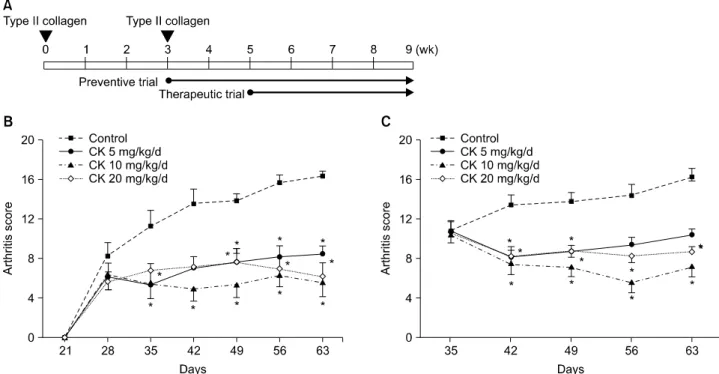
Figure 1. The effect of compound K (CK) on collagen-induced arthritis (CIA) disease activity. CK (0, 5, 10, or 20 mg/kg/d) was ad- ministered 1 day (preventive trial) or 14 days (therapeutic trial) after the boosting immunization of type II collagen (A). Preventive (B, n=9) and therapeutic (C, n=9) administration of CK significantly decreased arthritis scores along the disease course. However, the suppressive effects of CK were not dose-dependent in the dose range tested (5 to 20 mg/kg/d). Error bars represent the standard error of the mean. *p<0.05 by Kruskal-Wallace test with Dunn's multiple comparison test.
20 mg/kg/d) or vehicle was orally administered once a day for 4 weeks.
Measurement of serum anti-type II collagen anti- bodies
Whole blood was collected by cardiocentesis at the time of sacrifice, and serum was prepared and stored at −70oC until anlayzed. Serum levels of anti-CII immunoglobulin (IG) G2a and IgG2b antibodies were analyzed using com- mercially available enzyme-linked immunosorbent as- says (Chondrex). The levels of TNF-α and interleukin (IL)-17 in the serum were measured using a Luminex multiplexing platform (Milipore, Billerica, MA, USA).
Microscopic assessment
The knee joints and hind paws were removed at the time of sacrifice. The joints were fixed in 10% formalin solution for 24 h, decalcified in 15% ethylenediaminetetraacetic acid (EDTA, pH 7.5) for 1 week, and embedded in paraffin.
Tissue sections (20 μm thickness) were stained with hematoxylin and eosin and Masson-Trichrome. Histologic extent of knee arthritis was assessed semi-quantitatively by a pathologist (Song KY) blinded to the treatment as-
signment of specific samples. Histologic extent of arthritis was graded according to the method described by Wooley [23]: 0=normal synovium, 1= ynovial membrane hyper- trophy and cell infiltrates, 2=pannus and cartilage ero- sion, 3=major erosion of cartilage and subchondral bone, and 4=loss of joint integrity and ankylosis.
Quantitative real-time polymerase chain reaction
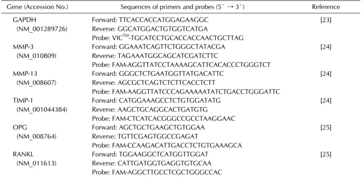
Total RNA was isolated from snap frozen joints by RNAzol (Invitrogen, Carlsbad, CA, USA). Frist-strand cDNA was synthesized using SuperScriptTM III Reverse Transcriptase (Invitrogen). Quantitative real-time poly- merase chain reaction (qRT-PCR) was performed using TaqMan probes for the genes of matrix metalloproteinase (MMP)-3 and MMP-13, tissue inhibitors of metallo- proteinase (TIMP)-1, RANKL, and osteoprotegerin (OPG).mRNA expression for these genes was normalized to glyceraldehyde-3-phosphate dehydrogenase (GAPDH) mRNA expression. The sequences of primers and probes combination used are listed in Table 1 [24-26].
Cytokine production from CII stimulated splenocytes
Spleens were harvested from CIA mice at sacrifice.Table 1. Primers and probes used for quantitative real-time polymerase chain reaction
Gene (Accession No.) Sequences of primers and probes (5` → 3`) Reference GAPDH
(NM_001289726)
Forward: TTCACCACCATGGAGAAGGC Reverse: GGCATGGACTGTGGTCATGA
Probe: VICTM-TGCATCCTGCACCACCAACTGCTTAG
[23]
MMP-3 (NM_010809)
Forward: GGAAATCAGTTCTGGGCTATACGA Reverse: TAGAAATGGCAGCATCGATCTTC
Probe: FAM-AGGTTATCCTAAAAGCATTCACACCCTGGGTCT
[24]
MMP-13 (NM_008607)
Forward: GGGCTCTGAATGGTTATGACATTC Reverse: AGCGCTCAGTCTCTTCACCTCTT
Probe: FAM-AAGGTTATCCCAGAAAAATATCTGACCTGGGATTC
[24]
TIMP-1
(NM_001044384)
Forward: CATGGAAAGCCTCTGTGGATATG Reverse: AAGCTGCAGGCACTGATGTG
Probe: FAM-CTCATCACGGGCCGCCTAAGGAAC
[24]
OPG
(NM_008764)
Forward: AGCTGCTGAAGCTGTGGAA Reverse: TGTTCGAGTGGCCGAGAT
Probe: FAM-CCAAGACATTGACCTCTGTGAAAGCA
[25]
RANKL (NM_011613)
Forward: TGGAAGGCTCATGGTTGGAT Reverse: CATTGATGGTGAGGTGTGCAA Probe: FAM-AGGCTTGCCTCGCTGGGCCAC
[25]
FAM: 6-carboxyfluorescein, GAPDH: glyceraldehyde 3-phosphate dehydrogenase, MMP: matrix metalloproteinase, OPG:
osteoprotegerin, RANKL: receptor activator of nuclear factor-κB ligand, TIMP: tissue inhibitors of metalloproteinase, VICTM:2'-chloro-7'-phenyl-1,4-dichloro-6-carboxyfluorescein.
Splenocytes (5×10⁵ cells/well) were isolated after red blood cell lysis and plated on CII-coated 96-well plate and cultured in RPMI medium with 10% fetal bovine serum (Hyclone, Logan, UT, USA). The levels of TNF-α, IL-2, interferon (IFN)-γ, and IL-4 in the supernatant were measured 48 h after stimulation using the Luminex assay (Milipore).
Microcomputed tomography analysis
After sacrifice, the hind paws of mice with CIA were scanned perpendicular to their longitudinal axis and their microarchitectural characteristics were analyzed at the an- kle and third metatarsal bone using SkyScan 1,076 device for micro-computed tomography (micro-CT) (Bruker AXS GmbH, Karlsruhe, Germany). Each joint was scanned at an isotropic voxel resolution of 35×35×35 μm. The scanning parameters were as follows: spatial resolution, 18 μm; tube voltage, 59 kV; tube current, 169 μA; filter, 1.0 mm; exposure, 1,770 ms; and rotation angle, 0.7o. The region of interest for metatarsal bone was selected as the 1.35 mm-long distal region of the third metatarsal bone (measured from 0.45 mm proximal to the third meta- tarsophalangeal joint) and that for ankle was as the tarsal joint area (from the proximal end of the third metatarsal bone to the posterior end of the talus). The micro-
architectural parameters were calculated as bone volume (BV)/total tissue volume (TV) ratio, bone surface area (BS)/BV ratio, cross-sectional thickness (Cs.th), and tra- becular thickness (Tb.th).
Statistical analysis
The data are presented as a mean±standard error.
Differences between the four experimental groups were analyzed by non-parametric Kruskal-Wallis test and all pairwise multiple comparison procedures (Dunn’s Method). The non-parametric Jonckheere’s trend test was used to analyze changes across different groups. A p-value less than 0.05 was considered statistically significant. Statistical calculations were done using SPSS Statistics ver. 17.0 (SPSS Inc., Chicago, IL, USA), except for the Dunn’s method which were performed using Prism 5.0 (GraphPad Software, San Diego, CA).
RESULTS
CK preventively and therapeutically reduced ar- thritis severity in CIA models
When CK was administered before the development of clinical arthritis, CK-treated groups showed significantly lower arthritis scores than the control group from day 49
Figure 2. Representative sections of joint tissues (hindfoot and knee joints) stained with hematoxylin-eosin (H&E) and Masson's Trichrome (MT) in therapeutic models (scale bar, 50 μm; A). Control group showed typical findings of collagen-induced arthritis (CIA); severe inflammatory cells infiltration, synovial hypertrophy, joint space narrowing, and bone and cartilage damage. These arthritic findings were decreased in mice therapeutically administered with compound K (CK). The histologic arthritis scores in knee joints significantly decreased with increasing dose of CK (B, n=6). Error bars represent the standard error of the mean. †p-val- ue was calculated by Jonckheere's trend test.
through the end of the treatment period (all p<0.05 by Kruskal-Wallis test with Dunn’s test; Figure 1B). Further- more, when CK was given at a dose of 10 to 20 mg/kg/d
after the development of clinical arthritis, CK groups sig- nificantly decreased arthritis scores throughout the study (all p<0.05; Figure 1C). However, the preventive or ther-
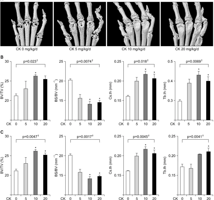
Figure 3. Microarchitectural analysis using micro-computed tomography (micro-CT) in therapeutic models (n=4 in each group, respectively). Representative micro-CT images of the hind paws (A) showed that the erosion in the metatarsophalangeal joints was decreased in compound K (CK)-treated groups. (B, C) Microarchitectural parameters included bone volume/total tissue volume (BV/TV), bone surface/bone volume (BS/BV), cross-sectional thickness (Cs.th), and trabecular thickness (Tb.th). Therapeutic CK ad- ministration significantly increased BV/TV, Cs.th, and Tb.th values and significantly decreased BS/BV values. Error bars represent the standard error of the mean. *p<0.05 by Kruskal-Wallace test with Dunn's multiple comparison test; †p-values were calcu- lated by Kruskal-Wallis test.
apeutic effect on CIA was not dose-dependent over the dose range tested.
In histologic examinations, inflammatory cell infiltra- tion, cartilage damage, and bone damage were decreased in CK-administered groups, when compared to the con- trol group for both knee and hind paw joints (Figure 2A).
The mean histologic scores in knee joints of CK-ad-
ministered mice tended to be reduced in comparison to those in control mice, in a significant dose-dependent manner: 2.83±0.17 in control group, 2.17±0.17 in 5 mg/kg CK group, 2.33±0.31 in 10 mg/kg CK group, and 1.83±0.31 in 20 mg/kg CK group (p=−0.036 by Jonckheere’s trend test; Figure 2B).
Figure 4. Serum levels of anti-type II collagen (CII) antibodies in therapeutic models. Therapeutical administration of compound K (CK) produced a significant reduction of anti-CII antibody IgG2a (A) and IgG2b (B, n=6 in each group). Error bars represent the standard error of the mean. *p<0.05 by Kruskal-Wallace test with Dunn's multiple comparison test; †p-values were calculated by Kruskal-Wallis test.
CK attenuated bone damage in CIA models
On reconstructed micro-CT images of the hind paws, the destruction in the metatarsophalangeal joints was re- duced in CK-treated groups (Figure 3A). In examining microarchitectural characteristics, it has been reported that BV, BV/TV, and Tb.th values decreased and BS/BV values increased along the course of CIA [27]. In mi- cro-CT analysis at the ankle (Figure 3B) and the third metatarsal bone (Figure 3C), BV/TV, Cs.th, and Tb.th val- ues significantly increased and BS/BV values were sig- nificantly decreased in in CK 10 mg/kg or 20 mg/kg group (all p<0.05 by Kruskal-Wallis test with Dunn’s test).These findings were consistent with the suppression of the clinical severity of arthritis.
CK suppressed the levels of anti-CII IgG in CIA models
We measured the serum levels of anti-CII IgG, which is considered a pathogenic mediator in CIA [28]. Anti-CII IgG2a levels in sera were reduced in all CK groups, espe- cially at a dose of 10 to 20 mg/kg (p<0.01 by Kruskal- Wallis test; Figure 4A). Serum levels of anti-CII IgG2b tended to decrease in CK 5 mg/kg and 10 mg/kg groups and were significantly lower in the CK 20 mg/kg group than controls (p<0.01; Figure 4B).
CK decreased IFN-γ and IL-2 secretion from CII-stimulated splenocytes
To investigate the change of cytokine production by CK
treatment, we analyzed cytokine levels in conditioned media from isolated splenocytes after stimulation with bovine CII. The concentrations of TNF-α were not sig- nificantly changed by CK treatment (Figure 5A). However, the levels of IL-2 and IFN-γ from the CII-stimulated splenocytes tended to decrease in the CK 5 mg/kg and 10 mg/kg groups and were significantly lower in the CK 20 mg/kg group than in control mice (both p<0.05 by Kruskal-Wallis test; Figure 5B and 5C). The CK 20 mg/kg group showed a tendency for an increase in the levels of IL-4 from the CII-stimulated splenocytes (Figure 5D).
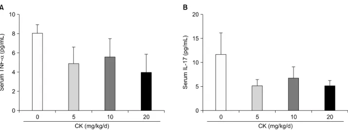
The serum concentrations of TNF-α and IL-17 were not significantly changed by CK treatment even though their levels tended to be lower in the CK-administered groups (Figure 6).
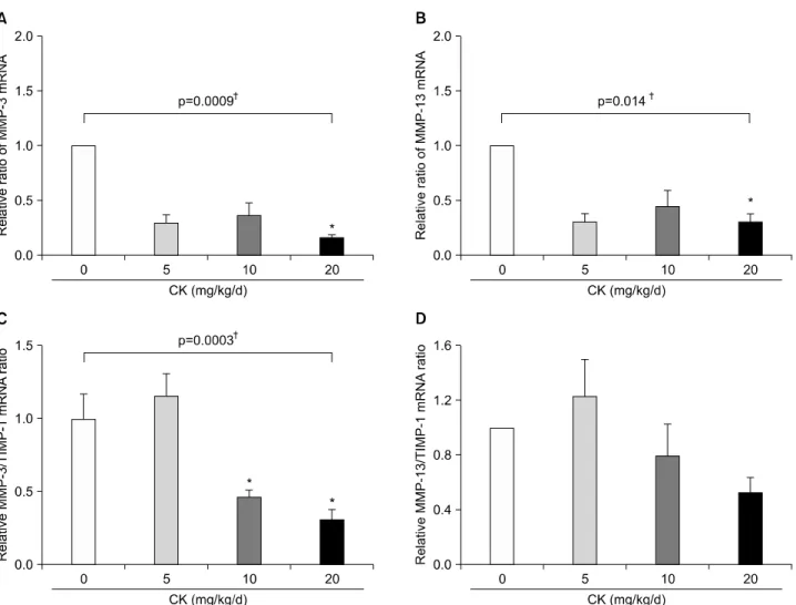
The expression of MMP-3 and RANKL in the ar- thritic joints was reduced by CK
Because the serum levels of TNF-α and IL-17, which play a major role in inflammatory bone destruction, were not affected by CK treatment, we investigated the ex- pression of effector molecules involved in bone and carti- lage damage including MMPs and RANKL, directly in joint tissue. In qRT-PCR analyses, the levels of MMP-3 and MMP-13 mRNA were significantly suppressed by CK treatment (both p<0.05 by Kruskal-Wallis test; Figure 7A and 7B). Furthermore, MMP-3/TIMP-1 mRNA ratios were significantly reduced in CK 10 mg/kg and 20 mg/kg groups compared to the control group (p<0.01; Figure
Figure 5. Cytokine production from the type II collagen stimulated splenocytes in therapeutic models. Splenocytes were isolated at the time of sacrifice after 4 weeks of compound K (CK) treatment and were stimulated with bovine CII for 48 h (n=6 in each group). When tumor necrosis factor (TNF)-α (A), interleukin (IL)-2 (B), interferon (IFN)-γ (C), and IL-4 (D) levels were measured in the culture media, IL-2 and IFN-γ were dose-dependently decreased in CK-treated groups. However, no significant effects on TNF-α or IL-4 production were observed. Error bars represent the standard error of the mean. *p<0.05 by Kruskal-Wallace test with Dunn's multiple comparison test; †p-values were calculated by Kruskal-Wallis test.
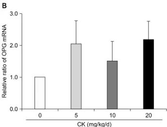
7C). MMP-13/TIMP-1 mRNA ratios tended to be de- creased in a dose-dependent manner even though the ef- fects did not reach statistical significance. In addition, RANKL mRNA levels were reduced (p<0.05; Figure 8A) and OPG mRNA levels were augmented (Figure 8B) by CK treatment. As a result, RANKL/OPG ratios, an in- dicator of osteoclastogenesis, were significantly de- creased by CK treatment (p<0.01; Figure 8C).
DISCUSSION
A wide range of pharmacological activities of ginseng has been attributed to ginsenosides, a class of steroid-like compounds. Approximately 40 ginsenosides have been
identified to date. Among them, Ro, Rb1, Rg1, and CK have been studied in animal arthritis models [5,9,17- 19,29] and all but one of the ginsenosides, Ro, were shown to have anti-arthritic effects. However, the bio- availability after oral administration was low, 3.29% for Rg1 and 0.64% for Rb1 [30].
Although each ginsenoside may have different pharma- cological effects, poor oral bioavailability can be a major limitation to their therapeutic potential. Additionally, the intestinal metabolism of ginsenosides was found to be de- pendent on an individual's intestinal microflora making oral absorption of Rg1 and Rb1 inconsistent [31]. Howev- er, CK, a major metabolite of ginsenosides, was readily ab- sorbed from the gastrointestinal tract independent of the
Figure 6. Serum levels of tumor necrosis factor (TNF)-α and interleukin (IL)-17 in therapeutic models (n=6 in each group).
Therapeutic compound K (CK) administration did not significantly affect TNF-α (A) and IL-17 (B) levels. Error bars represent the standard error of the mean.
parent compounds [32]. Based on these findings, the an- ti-arthritic effects of CK need to be established when eval- uating the therapeutic effects of ginsenosides.
The present study showed that CK attenuated disease activity in CIA mice, both preventively and therapeu- tically. In previous studies on the effect of ginsenosides on CIA, ginsenosides administration was initiated 18 to 26 days after the first immunization [5,6,8,9]. Consistent with these studies, our data showed that CK was prophy- lactically effective in suppressing progression of arthritis in the CIA model. In human RA, anti-rheumatic drugs are introduced after clinical disease is already established.
Therefore, our therapeutic model might be closer to a real world scenario than the preventive model. Thus, we fo- cused on the therapeutic action of CK in CIA through its administration in the mice that presented arthritis scores
≥6 on day 35 (the therapeutic model). A significant an- ti-arthritic effect of CK was clearly observed in the ther- apeutic model, as shown in Figure 1C. In the study of Chinese researchers [17-19], CK was administered for only 15 days because the arthritis activity spontaneously fell down and rarely lasts longer than 4 weeks in AIA.
However, in our models, control mice had persistent dis- ease activity during the therapeutical administration of CK for 4 weeks (Figure 1C).
The range of CK dosage used in the previous animal studies varied from 1.25 mg/kg to 160 mg/kg with the most frequently used dose being 10 mg/kg. Since Gao et al. [33] reported that the treatment of CK ≥20 mg/kg for 90 days led to hepatotoxicity in Beagle dogs, we decided to use 5 to 20 mg/kg CK in our studies. We did not find
dose-dependent effects in CIA mice in this dose range in either the prophylactic or the therapeutic models. In pre- vious studies with CIA, researchers generally used a sin- gle concentration of ginsenosides or ginseng extracts, ex- cept for Kim et al. [5] They administered ginsenoside Rb1 at two doses but found significant effects only at the high- er dose. In addition, Wu et al. observed anti-inflammatory action of CK at 40 to 160 mg/kg in a dose-independent manner in rats with AIA [17-19]. Therefore, prior to stud- ies in humans, further investigation is needed to establish the adequate dose of CK.
Our study showed a significant decrement in serum IgG2a and IgG2b anti-CII autoantibodies levels in CIA mice after administration of CK. Because both isotypes of anti-CII are known to be arthritogenic [34], the reduction of the IgG2 levels could be an anti-arthritic mechanism of CK. The CII-specific T-cell response was found to be pre- dominately due to Th1 cells producing IL-2 and IFN-γ [35]. The decrement in serum levels of tissue damaging CII-specific IgG2 by CK was consistent with the sup- pression of IFN-γ and IL-2 secretion from CII-stimu- lated splenocytes by CK [36]. Even though we did not di- rectly evaluate the action of CK on B cells, Wu et al. [17]
reported that CK inhibited B cell proliferation at the dose of 40 mg/kg or more and decreased the total levels of IgG2a at the dose of 160 mg/kg in rats with AIA. However, these changes could not directly explain the therapeutic effects in their AIA rats, since there has been no docu- mented role for B cells in AIA rats whereas B cells play a crucial role in the development of CIA.
Many cytokines have been implicated in the onset and
Figure 7. Effects of compound K (CK) on matrix metalloproteinase (MMP)-3 and MMP-13 mRNA expression in the hind feet of ther- apeutic models. When MMP-3 and MMP-13 mRNA levels in the hind foot tissue were analyzed using quantitative real-time poly- merase chain reaction (n=6 in each group), therapeutic administration of CK significantly decreased expression of MMP-3 (A) and MMP-13 (B). The suppressive effect of CK on MMP-3 expression remained significant after adjustment for tissue inhibitors of metal- loproteinase (TIMP)-1 mRNA (C, n=4 in each group). MMP-13/TIMP-1 ratios (D, n=4 in each group) tended to decrease in a dose dependent manner. Error bars represent the standard error of the mean. *p<0.05 by Kruskal-Wallace test with Dunn's multiple comparison test; †p-values were calculated by Kruskal-Wallis test.
progression of CIA. Among them, TNF-α and IL-17 are considered important cytokines mediating a variety of processes in RA and CIA [37]. Overexpression of TNF-α and IL-17 resulted in synovitis and joint destruction and their deficiency markedly suppressed the development of CIA [38,39]. However, in our model, CK did not affect the production of TNF-α and IL-17 in CII-stimulated sple- nocytes or their circulating levels in mice with CIA. This finding was in agreement with the observation of Wu et al. [17]. They found that 5 to 20 mg/kg of CK did not de- crease the levels of TNF-α and IL-17 from peritoneal macrophages of AIA rats. Therefore, the anti-arthritic ef- fects of CK might be mediated via a suppression of patho- genic auto-antibody production through Th1 cytokine
modulation, not inhibition of pro-inflammatory TNF-α and IL-17 production.
On the other hand, CK was reported to inhibit TNF-α induced cellular responses in RA-FLS, HUVECs, and as- troglial cells [16,40,41]. In RA and CIA, bone and carti- lage destruction is considered to be mediated by TNF-α and IL-17, because they can induce the expression of ma- trix-degrading enzymes and osteoclast differentiation.
We previously reported that CK significantly inhibited MMP-1 and MMP-3 production from RA-FLS and RANKL-mediated osteoclastogenesis in vitro [16]. In the present study, MMP-3 and MMP-13 mRNA levels were decreased in joint tissues after CK administration.
Additionally, RANKL/OPG ratios were significantly low-
Figure 8. Effects of compound K (CK) on receptor activator of nuclear factor-κB ligand (RANKL) and osteoprotegerin (OPG) mRNA expression in the hind feet of therapeutic models. When RANKL and OPG mRNA levels in the hind foot tissue were analyzed using quantitative real-time poly- merase chain reaction (n=6 in each group), therapeutic CK administration significantly decreased RANKL mRNA ex- pression (A) and tended to increase OPG mRNA (B).
Consequently, the ratios of RANKL/OPG were significantly reduced in CK administered mice (C). Error bars represent the standard error of the mean. *p<0.05 by Kruskal-Wallace test with Dunn's multiple comparison test; †p-values were calculated by Kruskal-Wallis test.
er in CK-treated mice than in control mice. Because this shift of the RANKL/OPG ratio can lead to a reduction of bone resorption, these results were congruent with mi- croarchitectural changes in CK-treated CIA mice. Wu et al. [17] also found that CK decreased the expression lev- els of RANKL and up-regulated those of OPG in RA-FLS.
Therefore, suppression of bone and cartilage degradation could be explained by the reduction of matrix-degrading enzymes levels and RANKL/OPG ratios.
CONCLUSION
The results of our study demonstrate that a natural gin- senoside CK exhibits anti-arthritic effects in CIA mice by suppression of Th1 cell-mediated humoral responses and joint-destructive effector molecules such as MMPs and RANKL.
ACKNOWLEDGMENTS
This study was supported by a grant of the Korea Health technology R&D Project, Ministry of Health & Welfare, Republic of Korea (A090933).
CONFLICT OF INTEREST
No potential conflict of interest relevant to this article was reported.
REFERENCES
1. Furneri G, Mantovani LG, Belisari A, Mosca M, Cristiani M, Bellelli S, et al. Systematic literature review on econom- ic implications and pharmacoeconomic issues of rheuma- toid arthritis. Clin Exp Rheumatol 2012;30(4 Suppl 73):S72-84.
2. Smolen JS, Aletaha D, Bijlsma JW, Breedveld FC, Boumpas D, Burmester G, et al; T2T Expert Committee. Treating rheumatoid arthritis to target: recommendations of an in-
ternational task force. Ann Rheum Dis 2010;69:631-7.
3. Jung YO, Kim HA. Recent paradigm shifts in the diagnosis and treatment of rheumatoid arthritis. Korean J Intern Med 2012;27:378-87.
4. Moreland L. Unmet needs in rheumatoid arthritis. Arthri- tis Res Ther 2005;7 Suppl 3:S2-8.
5. Kim HA, Kim S, Chang SH, Hwang HJ, Choi YN. Anti-ar- thritic effect of ginsenoside Rb1 on collagen induced ar- thritis in mice. Int Immunopharmacol 2007;7:1286-91.
6. Chang SH, Choi Y, Park JA, Jung DS, Shin J, Yang JH, et al.
Anti-inflammatory effects of BT-201, an n-butanol extract of Panax notoginseng, observed in vitro and in a colla- gen-induced arthritis model. Clin Nutr 2007;26:785-91.
7. Shin JS, Park N, Ra J, Kim Y, Shin M, Hong M, et al. Panax ginseng C.A. Meyer modulates the levels of MMP3 in S12 murine articular cartilage cell line. J Ethnopharmacol 2009;124:397-403.
8. Kim KR, Chung TY, Shin H, Son SH, Park KK, Choi JH, et al. Red ginseng saponin extract attenuates murine colla- gen-induced arthritis by reducing pro-inflammatory re- sponses and matrix metalloproteinase-3 expression. Biol Pharm Bull 2010;33:604-10.
9. Du J, Cheng B, Zhu X, Ling C. Ginsenoside Rg1, a novel glucocorticoid receptor agonist of plant origin, maintains glucocorticoid efficacy with reduced side effects. J Immunol 2011;187:942-50.
10. Na JY, Kim S, Song K, Lim KH, Shin GW, Kim JH, et al.
Anti-apoptotic activity of ginsenoside Rb1 in hydrogen peroxide-treated chondrocytes: stabilization of mitochon- dria and the inhibition of caspase-3. J Ginseng Res 2012;
36:242-7.
11. Kim S, Na JY, Song KB, Choi DS, Kim JH, Kwon YB, et al.
Protective effect of ginsenoside Rb1 on hydrogen pero- xide-induced oxidative stress in rat articular chondrocytes.
J Ginseng Res 2012;36:161-8.
12. Cheng W, Wu D, Zuo Q, Wang Z, Fan W. Ginsenoside Rb1 prevents interleukin-1 beta induced inflammation and apoptosis in human articular chondrocytes. Int Orthop 2013;37:2065-70.
13. So MW, Lee EJ, Lee HS, Koo BS, Kim YG, Lee CK, et al.
Protective effects of ginsenoside Rg3 on human osteo- arthritic chondrocytes. Mod Rheumatol 2013;23:104-11.
14. Lee JH, Lim H, Shehzad O, Kim YS, Kim HP. Ginsenosides from Korean red ginseng inhibit matrix metallopro- teinase-13 expression in articular chondrocytes and pre- vent cartilage degradation. Eur J Pharmacol 2014;724:
145-51.
15. Yu K, Chen F, Li C. Absorption, disposition, and pharma- cokinetics of saponins from Chinese medicinal herbs:
what do we know and what do we need to know more?
Curr Drug Metab 2012;13:577-98.
16. Choi YS, Kang EH, Lee EY, Gong HS, Kang HS, Shin K, et al. Joint-protective effects of compound K, a major ginse- noside metabolite, in rheumatoid arthritis: in vitro evidence. Rheumatol Int 2013;33:1981-90.
17. Wu H, Chen J, Wang Q, Jia X, Song S, Yuan P, et al.
Ginsenoside metabolite compound K attenuates in- flammatory responses of adjuvant-induced arthritis rats.
Immunopharmacol Immunotoxicol 2014;36:124-9.
18. Liu KK, Wang QT, Yang SM, Chen JY, Wu HX, Wei W.
Ginsenoside compound K suppresses the abnormal activa-
tion of T lymphocytes in mice with collagen-induced arthritis. Acta Pharmacol Sin 2014;35:599-612.
19. Chen J, Wu H, Wang Q, Chang Y, Liu K, Song S, et al.
Ginsenoside metabolite compound K alleviates adjuvant- induced arthritis by suppressing T cell activation. Inflam- mation 2014;37:1608-15.
20. Kim HO, Lee SI. Experimental animal models for rheuma- toid arthritis: methods and applications. J Rheum Dis 2012;19:189-95.
21. Zhou W, Feng MQ, Li JY, Zhou P. Studies on the prepara- tion, crystal structure and bioactivity of ginsenoside com- pound K. J Asian Nat Prod Res 2006;8:519-27.
22. Brand DD, Latham KA, Rosloniec EF. Collagen-induced arthritis. Nat Protoc 2007;2:1269-75.
23. Wooley PH. Collagen-induced arthritis in the mouse.
Methods Enzymol 1988;162:361-73.
24. Xiao J, Shimada M, Liu W, Hu D, Matsumori A. Anti-in- flammatory effects of eplerenone on viral myocarditis. Eur J Heart Fail 2009;11:349-53.
25. Wells JE, Rice TK, Nuttall RK, Edwards DR, Zekki H, Rivest S, et al. An adverse role for matrix metallopro- teinase 12 after spinal cord injury in mice. J Neurosci 2003;23:10107-15.
26. Palmqvist P, Lundberg P, Persson E, Johansson A, Lundgren I, Lie A, et al. Inhibition of hormone and cyto- kine-stimulated osteoclastogenesis and bone resorption by interleukin-4 and interleukin-13 is associated with in- creased osteoprotegerin and decreased RANKL and RANK in a STAT6-dependent pathway. J Biol Chem 2006;281:
2414-29.
27. Lee JH, Chun KJ, Kim HS, Kim SH, Lee KY, Kim DJ, et al.
Changes in microarchitectural characteristics at the tibial epiphysis induced by collagen-induced rheumatoid arthri- tis over time. Clin Interv Aging 2012;7:373-82.
28. Nandakumar KS, Bäcklund J, Vestberg M, Holmdahl R.
Collagen type II (CII)-specific antibodies induce arthritis in the absence of T or B cells but the arthritis progression is enhanced by CII-reactive T cells. Arthritis Res Ther 2004;6:R544-50.
29. Matsuda H, Samukawa K, Kubo M. Anti-inflammatory ac- tivity of ginsenoside Ro. Planta Med 1990;56:19-23.
30. Leung KW, Wong AS. Pharmacology of ginsenosides: a lit- erature review. Chin Med 2010;5:20.
31. Kim KA, Jung IH, Park SH, Ahn YT, Huh CS, Kim DH.
Comparative analysis of the gut microbiota in people with different levels of ginsenoside Rb1 degradation to com- pound K. PLoS One 2013;8:e62409.
32. Kim HK. Pharmacokinetics of ginsenoside Rb1 and its me- tabolite compound K after oral administration of Korean Red Ginseng extract. J Ginseng Res 2013;37:451-6.
33. Gao YL, Liu ZF, Li CM, Shen JY, Yin HX, Li GS. Subchronic toxicity studies with ginsenoside compound K delivered to dogs via intravenous administration. Food Chem Toxicol 2011;49:1857-62.
34. Rowley MJ, Nandakumar KS, Holmdahl R. The role of col- lagen antibodies in mediating arthritis. Mod Rheumatol 2008;18:429-41.
35. Germann T, Bongartz M, Dlugonska H, Hess H, Schmitt E, Kolbe L, et al. Interleukin-12 profoundly up-regulates the synthesis of antigen-specific complement-fixing IgG2a, IgG2b and IgG3 antibody subclasses in vivo. Eur J Immu-
nol 1995;25:823-9.
36. Terato K, Hasty KA, Reife RA, Cremer MA, Kang AH, Stuart JM. Induction of arthritis with monoclonal anti- bodies to collagen. J Immunol 1992;148:2103-8.
37. Billiau A, Matthys P. Collagen-induced arthritis and re- lated animal models: how much of their pathogenesis is auto-immune, how much is auto-inflammatory? Cytokine Growth Factor Rev 2011;22:339-44.
38. Nakae S, Nambu A, Sudo K, Iwakura Y. Suppression of im- mune induction of collagen-induced arthritis in IL-17- de- ficient mice. J Immunol 2003;171:6173-7.
39. Mori L, Iselin S, De Libero G, Lesslauer W. Attenuation of
collagen-induced arthritis in 55-kDa TNF receptor type 1 (TNFR1)-IgG1-treated and TNFR1-deficient mice. J Immunol 1996;157:3178-82.
40. Lee ES, Choi JS, Kim MS, You HJ, Ji GE, Kang YH.
Ginsenoside metabolite compound K differentially antag- onizing tumor necrosis factor-α-induced monocyte- en- dothelial trafficking. Chem Biol Interact 2011;194:13-22.
41. Choi K, Kim M, Ryu J, Choi C. Ginsenosides compound K and Rh(2) inhibit tumor necrosis factor-alpha-induced ac- tivation of the NF-kappaB and JNK pathways in human as- troglial cells. Neurosci Lett 2007;421:37-41.