Korean J Endocr Surg 2016;16:100-106
https://doi.org/10.16956/kaes.2016.16.4.100
Endocrine Surgery
원격전이가 동반된 미세 침윤형 여포암 환자의 임상병리학적 양상
1서울대학교 의과대학 서울대학교병원 외과, 2서울대학교 의과대학 암연구소, 3서울대학교암병원 갑상선센터 외과,
4서울대학교병원운영 서울특별시보라매병원 외과, 5서울대학교 의과대학 분당서울대학교병원 외과
주영욱
1ㆍ김수진
1,2,3ㆍ채영준
2,4ㆍ이진욱
1,2,3ㆍ성찬용
1,2,3ㆍ김종규
1,2,3ㆍ유형원
2,5ㆍ최준영
2,5ㆍ이규언
1,2,3Clinicopathologic Features in Minimally Invasive Follicular Thyroid Cancer Patients with Distant Metastasis
Purpose: Although minimally invasive follicular thyroid carcinoma (MIFTC) is considered a thyroid tumor with low malignant potential, some MIFTC can spread, metastasize, and eventually lead to death. This study investigates the risk factors for distant metastasis in MIFTC patients.
Methods: Between 1981 and 2014, the records of 365 consecutive patients who underwent thyroidectomy for MIFTC at Seoul National University Hospital were reviewed. Univariate and multivariate analyses were performed to identify risk factors associated with distant metastasis.
Results: Of 351 patients, 10 (2.9%) presented with distant metastasis. Of these, two (0.6%) were found at the time of thyroidectomy, while eight (2.3%) were detected at later exams, over a median of 7.3 years (range, 0.2∼30.8). In univariate analysis, lymph node metastasis (P<0.001) was significantly associated with distant metastasis. In multivariate analysis, lymph node metastasis (P<0.001) and locoregional recurrence (P=0.008) were significantly associated with distant metastasis.
Conclusion: Distant metastasis in MIFTC patients were associated with high-risk clinicopathologic features and more aggressive clinical courses. Further study will be needed to ascertain these results with long-term surveillance.
Key Words: Minimally invasive follicular thyroid cancer, Distant metastasis, Clinicopathologic features
중심 단어: 미세 침윤형 갑상선 여포암, 원격전이, 임상병리학적 양상
Young Wook Ju1, Su-jin Kim1,2,3, Young Jun Chai2,4, Jin Wook Yi1,2,3, Chan-Yong Seong1,2,3, Jong-Kyu Kim1,2,3, Hyeong Won Yu2,5, June Young Choi2,5, Kyu Eun Lee1,2,3
1Department of Surgery, Seoul National University Hospital, Seoul National University College of Medicine, 2Cancer Research Institute, Seoul National University College of Medicine, 3Division of Surgery, Thyroid Center, Seoul National University Cancer Hospital,
4Department of Surgery, Seoul Metropolitan Government-Seoul National University Boramae Medical Center, Seoul, 5Department of Surgery, Seoul National University Bundang Hospital, Seoul National University College of Medicine, Seongnam, Korea
Received September 25, 2016, Revised October 4, 2016, Accepted October 14, 2016 Correspondence: Su-jin Kim Department of Surgery, Seoul National University Hospital, Seoul National University College of Medicine, 101 Daehak-ro, Jongno-gu, Seoul 03080, Korea Tel: +82-2-2072-7208 Fax: +82-2-766-3975 E-mail: [email protected]
Copyright © 2016 Korean Association of Thyroid and Endocrine Surgeons; KATES. All Rights Reserved.
cc This is an Open Access article distributed under the terms of the Creative Commons Attribution Non-Commercial License (http://creativecommons.org/licenses/by-nc/4.0) which permits unrestricted non-commercial use, distribution, and reproduction in any medium, provided the original work is properly cited.
서 론
갑상선 여포암은 갑상선암 중 상대적으로 드문 형태지만 요오 드 섭취 정도에 따라 빈도 차이를 보여 4∼39%라고 보고되고 있 다.(1) 일반적으로 요오드 섭취가 많으면 여포암의 빈도가 줄어 드는 것으로 알려져 있고 우리나라의 경우 전체 갑상선암 중 5∼
15% 정도로 보고되고 있다.(2) 여성에 호발하며 발병시 평균 연 령이 50세 정도로 알려져 있다. 보통 단일 결절로 나타나나 10%
정도는 다발성 결절과 공존하기도 한다. 갑상선 여포암은 유두암 과 같은 고분화 갑상선암(well-differentiated thyroid cancer) 으로 분류되고 있지만 여포암은 유두암보다 고령에서 발생하고 림프절 전이는 흔하지 않으나, 원격전이는 종종 보이는 등 다른 임상병리학적 특징을 보이고 있다.(3)
갑상선 여포암은 일반적으로 침윤 정도에 따라 미세 침윤형과 광범위 침윤형으로 분류한다. 미세 침윤형은 육안적으로 피막침 윤이 있고 현미경적으로 종양이 피막이나 피막 주위 갑상선 실질
Characteristics Number (%) Age at diagnosis (y)
Mean Range
<45 y
≥45 y
45.4 15∼79 158 (45.0) 193 (55.0) Gender
Male Female
67 (19.1) 284 (80.9) Size of tumor
≤4 cm
>4 cm
275 (78.3) 76 (21.7)
Multifocality 37 (10.5)
Capsular invasion Lymphatic invasion Vascular invasion
344 (98.0) 9 (2.6) 33 (9.4) Positive resection margins 3 (0.9) Lymph node metastasis
Central node metastasis Lateral node metastasis
Central & lateral node metastasis Unknown
Locoregional recurrence
6 (1.7) 1 (0.3) 1 (0.3) 3 (0.9) 1 (0.3) 2 (0.6) Distant metastasis
- At presentation - During follow-up Site of distant metastasis
- Lung - Bone - Lung & bone - Lung & mediastinum
10 (2.9) 2 (0.6) 8 (2.3)
4 (1.1) 1 (0.3) 4 (1.1) 1 (0.3) TNM stage
I II III IV
212 (60.4) 89 (25.4) 44 (12.5) 6 (1.7) Final extent of thyroidectomy
- Less than total thyroidectomy - Total thyroidectomy
245 (69.8) 106 (30.2) Radioactive iodine therapy
External radiotherapy
146 (41.6) 1 (0.3) TNM = Tumor-node-metastasis staging system.
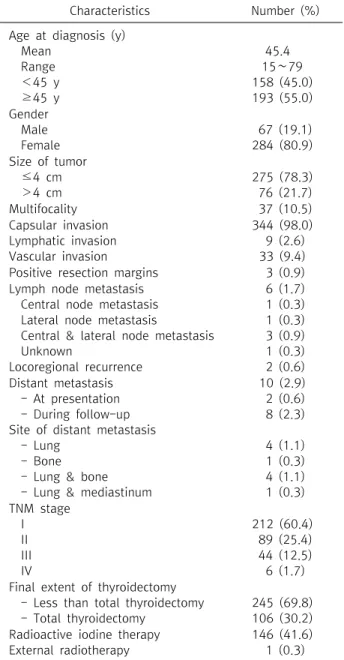
Table 1. Clinicopathologic characteristics of the study popula- tion (n=351)
내 혈관을 침범하고, 광범위 침윤형은 큰 혈관과 피막에 광범위 한 침윤을 보이며 갑상선 실질 조직으로의 침범을 보인다.
여포암은 갑상선 상피에서 발생하는 악성종양으로 진단은 피 막이나, 혈관 또는 림프관을 침윤한 것이 증명되어야 하므로 유 두암과 달리 수술 전 미세침 흡인 세포검사(fine needle aspira- tion cytology)나 수술 중 동결절편 검사만으로 정확한 진단이 거의 불가능하다.(4) 여포암, 여포 선종, 그리고 여포성 변이 유두 암(follicular variant papillary carcinoma) 등이 수술 전 미세침 흡인 세포검사 결과 여포성 종양(follicular neoplasm)으로 진단 될 수 있으나 여포암의 수술 전 진단은 원격전이가 발견되지 않으 면 불가능하다.
암의 원격전이에 대한 분석은 암의 병태생리학적 특성 파악 및 적합한 치료를 위해서 매우 중요하다. 본 저자들은 미세 침윤 형 여포암에서 원격전이와 임상병리학적 특징, 치료 방법, 환자 의 예후와의 연관성을 알아보고자 본 연구를 시행하였다.
방 법
1980년 8월부터 2014년 2월까지 갑상선암으로 서울대학교 병원에서 수술적 치료를 받은 11,998명의 환자 중 미세 침윤형 여포암으로 진단된 365명의 환자들을 대상으로 후향적으로 의 무기록을 조사하였다. 수술 후 추적관찰 기간 2개월 미만의 환자 14명은 본 연구에서 제외하였다. 여포암의 진단은 최종 병리결 과 조직학적으로 피막이나 혈관에 암세포의 침윤이 있거나, 원 격전이가 확인된 경우로 하였다. 각 환자에 대해 성별, 나이, 종 양의 크기, 다발성, 피막 침윤, 림프관 침윤, 혈관 침윤, 절단면 침 범, 림프절 전이, TNM 병기, 수술범위, 진단 당시 전이 여부, 원 격전이 여부 및 장소, 방사성 요오드 치료, 국소재발, 생존 여부 등을 조사하였다.
수술적 치료는 미세침 흡인 세포검사, 수술 소견, 수술 중 동결 절편 검사에서 악성이 강력히 의심이 되거나 종양의 크기가 큰 경우, 혈관 침범이 의심되는 경우, 다발성인 경우, 환자가 전절제 술을 희망하는 경우 등에 한하여 처음부터 갑상선 전절제를 시행 하였다. 처음 수술 시 일엽절제술이나 아전절제술을 시행한 환 자중 최종 조직 검사 결과상 여포암으로 확인된 경우 고위험군에 서 완결절제술(completion thyroidectomy)을 권하여 시행하 였고, 그 이외의 경우에는 추적관찰만 시행하였다.
수술 후에는 갑상선 자극 호르몬(TSH) 억제 목적으로 갑상선 호르몬제(levothyroxine)를 복용하였다. 갑상선 전절제술을 시 행 후 위험요소가 있는 경우 잔여 조직 소멸 및 치료 목적으로 방 사성 요오드 치료를 시행하였다. 재발 및 전이 여부 검사를 위해 임상적 진찰, 혈액검사, 흉부 및 뼈 단순 X선 촬영(X-ray), 경부
초음파, 컴퓨터 단층촬영(CT), 양전자 단층촬영(PET) 등을 시행 하였다. 국소재발은 수술 당시 발견되지 않았던 암 조직이 수술 후 경과관찰 기간 동안 잔존 갑상선이나 주변 림프절에서 발견되 는 것으로 정의 하였으며, 림프절 재발이 의심되는 경우 미세침 흡인 세포검사를 시행 하였다. 원격전이는 진단 당시 또는 수술 후 경과관찰 기간 중 암 조직이 갑상선과 주변 림프절을 제외한 부위에서 발견되는 것으로 정의 하였다.
모든 통계적 분석에는 SPSS 22.0 (SPSS Inc, Chicago, IL)을 사용하였다. 원격전이에 영향을 미치는 인자는 단변량 분석의 경우 교차분석(χ2 test) 시행 시 Fisher의 정확한 검정(Fisher’s
Case Age*/
Sex
Operation date
Follow-up (month)
Extent of
thyroidectomy pTNM Size†
(cm) LN‡ RAI§
(mCi) EBRT Location∥
Time of diagnosis of distant metastasis
Status
1 32/F 1981-08-18 2.3 TT T3N1M1 6.0 Yes 150 No Lung 1981-10-27 Unavailabe
2 31/F 1989-02-09 331.2 TT T3N0M1 6.1 No 30 No Bone 2014-03-28 Alive/PD
3 64/M 1992-08-17 288.5 Lobectomy
& subtotal
T2N0M1 3.5 No 1,560 No Lung 2002-05-24 Alive/NED
4 22/F 2001-10-12 168.4 TT
& MRND, Rt.
T1N1M1 1.0 Yes 840 No Lung 2005-02-05 Alive/NED
5 67/M 2003-05-23 156.9 TT T1N0M1 1.2 No 550 No Lung, bone 2012-06-25 Alive/NED
6 44/F 2004-01-15 147.4 TT T2N0M1 2.2 No 910 No Lung,
mediastinum
2005-03-04 Alive/PD
7 46/F 2006-01-05 116.8 Lobectomy
& subtotal
T1N0M1 1.3 No 60 No Lung 2006-06-10 Alive/NED
8 64/F 2006-04-27 123.6 TT T3N0M1 4.2 No 1,290 No Lung, bone 2008-10-31
(lung) 2009-11-02
(bone)
Alive/PD
9 40/F 2007-09-04 105.8 TT T2N0M1 3.6 No 1,210 No Lung, bone 2007-08-30
(at diagnosis) 2011-06-17
(bone)
Alive/PD
10 58/M 2008-12-16 58.8 TT T2N1M1 3.2 Yes 1,000 Yes Lung, bone 2008-10-22
(at diagnosis)
Alive/PD
pTNM = Pathological tumor-node-metastasis staging system; EBRT = External beam radiotherapy; TT = Total thyroidectomy; MRND
= Modified radical neck dissection; PD = Progressive disease; NED = No evidence of disease.
*Age: Age at diagnosis; †Size: Tumor size; ‡LN: Lymph node metastasis; §RAI: Total accumulative dose of radioactive I-131 ablation;
∥Location: Location of distant metastasis.
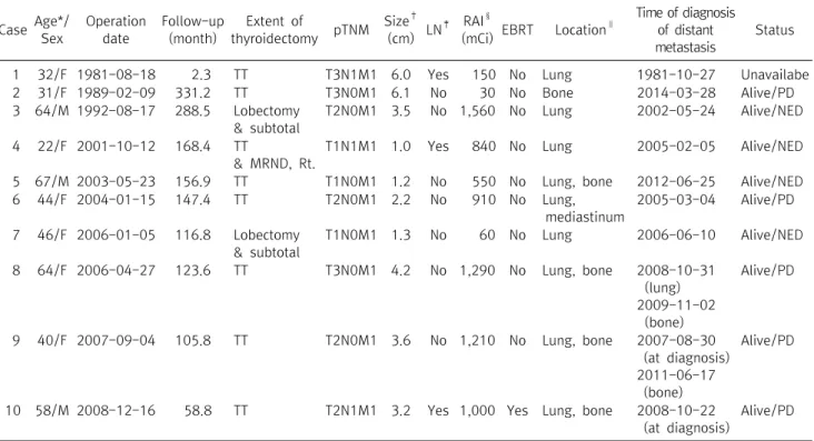
Table 2. Characteristics of the 10 patients who had distant metastasis
exact test)을 함께 시행하였고, 다변량 분석의 경우 로지스틱 회 귀분석(Logistic regression)을 시행하였다. P값이 0.05 미만인 경우 통계학적으로 의미 있는 것으로 판정하였다.
결 과
본 연구의 대상 환자 351명 중 남자는 67명(19.1%), 여자는 284명(80.9%)이었고, 남녀 비는 남:여=1:4.2였으며 진단 당 시 평균연령은 45.4세(15∼79세)였다. 종양의 크기가 4 cm 이 하인 경우가 275예(78.3%), 4 cm 초과인 경우가 76예(21.7%) 였다. 다발성을 보이는 경우가 37예(10.5%)였으며, 병리조직학 적으로 피막 침윤이 344예(98.0%), 림프관 침윤이 9예(2.6%), 혈관 침윤이 33예(9.4%)에서 있었다. 수술 후 최종 조직검사에 서 절단면 침범 소견을 보인 경우는 3예(0.9%)였고, 이중 완결절 제술은 1예에서 시행하였다. 6예(1.7%)에서 림프절 전이를 보 였는데 각각 중앙 경부 림프절 침범 1예(0.3%), 측경부 림프절 침 범 1예(0.3%), 중앙 및 측경부 림프절 침범 3예(0.9%) 였으며 나 머지 1예에서는 기록상 정확한 위치를 확인할 수 없었다. 수술 후 88개월(2∼369개월)의 추적 기간 중 국소재발은 2예에서 있 었는데, 원격전이가 없었던 341명 중 국소재발이 1예(0.3%), 원 격전이가 있었던 10명 중 국소재발이 1예(10%)에서 확인되었
다. 원격전이는 총 10예(2.9%)에서 확인되었다. 진단 당시 원격 전이가 확인된 경우는 2예(0.6%)에서 있었는데 골 전이 1예와 폐 전이 1예가 있었다. 추적 기간 중 전이가 있었던 8예(2.3%)의 경우 전이부위는 폐 4예, 뼈 1예, 폐와 뼈에서 발견된 경우가 4예, 폐와 종격에 발견된 경우가 1예 있었다. TNM 병기상 T stage에 서 T1 212예(60.4%), T2 89예(25.4%), T3 44예(12.5%), T4 6 예(1.7%)였다. 수술 방법은 아전절제술 50예(14.2%), 일엽절제 술 195예(55.6%)였으며, 완결절제술을 시행한 15예(4.3%)를 포함하여 최종 전절제술 상태의 환자는 총 106예(30.2%)였다.
완결절제술(completion thyroidectomy)은 최종 조직 검사 결 과에서 여포암으로 확인된 고위험군 중 잔여 갑상선 조직을 제거 하기 위해 시행하였으며, 총 15예(4.3%)에서 시행하였다. 방사 성 요오드 치료를 시행한 경우는 총 146예(41.6%)였는데, 진단 당시 전이가 있었던 환자는(2예) 방사성 요오드 치료 전 갑상선 전절제술을 시행하였다. 수술전 골 전이가 확인된 1예(0.3%)에 서는 방사선 치료를 시행하였다(Table 1).
수술 후 국소재발 기간은 평균 150개월(109∼191개월), 원 격전이의 진단까지는 평균 78개월(2∼306개월)이었다. 생존은 처음 수술일로부터 사망 또는 마지막 추적 관찰일 까지로 정의하 였고, 전체 환자 중 미세 침윤형 여포암으로 사망한 경우는 없었 다(Table 2).
Risk factors Patients (n) Distant metastasis n (%)
No distant metastasis n (%)
Fisher’s exact test (P value) Age at diagnosis (y)
<45 y
≥45 y
158 193
5 (50.0) 5 (50.0)
153 (44.9) 188 (55.1)
0.759
Gender Male Female
67 284
3 (30.0) 7 (70.0)
64 (18.8) 277 (81.2)
0.410
Size of tumor
≤4 cm
>4 cm
275 76
7 (70.0) 3 (30.0)
268 (78.6) 73 (21.4)
0.456
Multifocality Unifocal Multifocal
314 37
8 (80.0) 2 (20.0)
306 (89.7) 35 (10.3)
0.285
Capsular invasion Lymphatic invasion Vascular invasion
344 9 33
10 (100) 1 (10.0) 1 (10.0)
334 (95.2) 8 (2.3) 32 (9.4)
1.000 0.231 1.000
Positive resection margins 3 0 (0) 3 (0.9) 1.000
Lymph node metastasis Central node
Lateral node
Central & lateral node Unknown
6 1 1 3 1
3 (30.0) 0 1 (10.0) 1 (10.0) 1 (10.0)
3 (0.9) 1 (0.3) 0 2 (0.3) 0
<0.001
Locoregional recurrence 2 1 (10.0) 1 (0.3) 0.056
MIFTC = Minimally invasive follicular thyroid carcinoma.
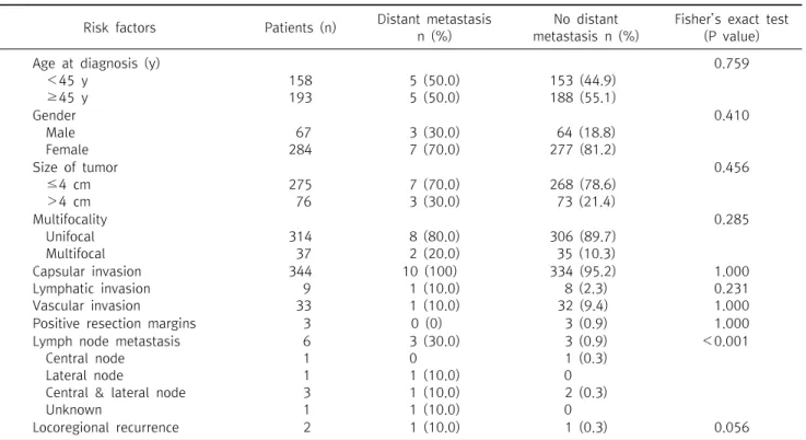
Table 3. Association between distant metastasis and clinicopathologic features in 351 patients with MIFTC
Risk factors Univariate Multivariate*
Hazard ratio (95% CI) P Hazard ratio (95% CI) P
Age at diagnosis (y)
<45 y
≥45 y
Ref.
0.81 (0.23∼2.86) 0.759
Gender Male Female
Ref.
0.54 (0.14∼2.14) 0.410
Size of tumor
≤4 cm
>4 cm
Ref.
1.57 (0.40∼6.24) 0.456
Multifocality Unifocal Multifocal
Ref.
2.19 (0.45∼10.70) 0.285 Capsular invasion
Lymphatic invasion Vascular invasion
0.98 (0.97∼1.00) 4.63 (0.52∼41.0) 1.07 (0.13∼8.74)
1.000 0.231 1.000 Positive resection margins 0.99 (0.98∼1.00) 1.000
Lymph node metastasis 48.29 (8.25∼282.54) <0.001 51.32 (8.29∼317.57) <0.001
Locoregional recurrence 37.79 0.056 57.42 (2.87∼1149.92) 0.008
MIFTC = Minimally invasive follicular thyroid carcinoma; CI = Confidence interval.
*Multivariate: adjusted for age at diagnosis (y), gender.
Table 4. Univariate and multivariate analysis for distant metastasis in 351 patients with MIFTC
방사선 치료를 시행한 1예는 58세 남자로 좌측 대퇴골 골절로 사지구제술(Limb salvage surgery with prosthesis)을 시행하 는 한편 양측 대퇴골 및 골반 부위에 대하여 고식적 방사선 치료 를 시행하였다. 이후 여포암의 골 전이로 진단되어 갑상선 전절
제술을 시행하는 한편 방사성 요오드 치료와 고식적 방사선 치료 를 시행하였으나 이후 대퇴골, 골반, 흉추, 오른쪽 어깨뼈에 대한 다발성 골 전이로 진행하였다. 사망한 경우가 없어서 통계학적 으로 생존곡선을 구할 수는 없었으며, 사망에 대한 예후인자는
분석할 수 없었다.
원격전이 소견을 보인 환자의 위험인자에 대한 단변량 분석 (Fisher’s exact test)에서 림프절 전이 (P<0.001)가 유의한 위 험인자였다(Table 3, 4). 다변량 분석(Logistic regression)에서 는 림프절 전이(P<0.001) 및 국소재발 여부(P=0.008)가 유의 한 독립적인 예후인자였다(Table 4).
고 찰
갑상선 여포암은 조직학적 특징에 따라 미세 침윤형 여포암과 광범위 침윤형으로 분류된다.(5) 세계보건기구(WHO)는 미세 침윤형 갑상선암을 육안적으로 피막에 쌓여있는 단일성 종양에 혈관침윤이 동반되고, 피막의 전층을 침윤하기도 하는 양상으로 정의한다. 광범위 침윤형 여포암은 광범위한 혈관침윤 및 피막 침윤이 동반되어 주위 갑상선 실질로의 침범을 동반하게 된 다.(6) 원격전이는 미세 침윤형에서는 드물며 광범위 침윤형에 서 더 많은 것으로 보고되고 있다.(7) 미세 침윤형 여포암은 재발 율이 낮고 전이가 적어 장기적인 예후는 매우 좋은 것으로 알려 져 있는데, 연구에 따라 0에서 22%까지 보고되고 있다.(8,9)
미세 침윤형 여포암의 수술적 치료방침에 대해 과거 일부에서 는 유두암에 비해 나쁜 예후 때문에 갑상선 전절제술과 방사선 요오드 치료를 해야 한다고 주장하기도 하였다.(10,11) Sugino 등 (12)의 연구에 의하면 미세 침윤형 여포암 환자에서 완결절제술을 통 해 통계적으로 유의한 결과를 도출할 수는 없었으나, 완결절제술을 시행한 환자들에서 사망이 없었음을 주목할 만한 결과라고 하였다.
유럽 내분비외과 학회(The European Society of Endocrine Surgeons)에서는 최근 미세 침윤형 여포암 환자의 치료방침에 대하여 “45세 이상, 40 mm 이상의 종양, 혈관 침윤이나 림프절 양성, 원격전이가 있는 환자에서는 갑상선 전절제술을 시행하여 야 한다”는 데에 동의하였다.(13)
Goffredo 등(14)은 Surveillance, Epidemiology, and End Results (SEER) 자료를 토대로 1,200명의 미세 침윤형 여포암 환자를 분석하였을 때, 2명만이 미세 침윤형 여포암로 사망하였 고 이는 미국의 전체 인구 사망률과 견주어도 비슷한 정도임을 확인하였는데, 갑상선 전절제술이나 방사성 요오드 치료가 예후 의 개선과 무관하다는 결론을 내렸다. 그러나 여포암의 원격전 이와 관련된 Ban 등(6)의 연구에 따르면, 10명의 전이성 여포암 환자중 4명이 미세 침윤형 여포암으로 확인되었고, 병변의 크기 가 작고 혈관 침윤 소견이 없어도 원격전이의 양상은 비슷하다고 하였으며, 이전 연구에서 전이성 여포암 환자의 질병 관련 10년 생존율이 50%까지 감소할 수 있음을 언급하였다. 본 연구에서 원 격전이가 확인된 10명의 환자들 중 5명은 T2 이하의 크기에 림프
절 전이가 없는 상태다. 이는 미세 침윤형 여포암이 경우에 따라 뚜렷한 원격전이 성향을 지닐 수 있음을 시사하는 결과로, 양성 종양과 같은 양상을 보인다는 기존의 연구와 다른 부분이다.
몇몇 연구들에서 미세 침윤형 여포암의 경과는 양호하여 일부 환자들에서는 일엽절제술 만으로도 충분하다는 결과를 보고하 였던 바 있다.(1,14-18) 그리고 다른 연구에서는 미세 침윤형 여 포암 환자들의 예후가 항상 좋은 것은 아니기 때문에, 특히 원격 전이의 위험인자가 있는 환자군에서는 갑상선 전절제술과 방사 성 요오드 치료가 필요하다는 주장도 있다.(6,13,19-21) 갑상선 전절제술과 방사성 요오드 치료가 원격전이를 줄이는지 여부는 아직 명확치 않으나 원격전이를 조기에 발견하는데 도움이 되는 것은 틀림이 없다.(22-24) 원격전이의 조기발견과 발견 이후의 방 사성 요오드 치료는 생존률을 개선하는 것으로 알려져 있 다.(25-27) 본 연구에서 원격전이가 확인된 10명 중 최종 전절제 상태로 확인된 환자는 총 8명이었으며, 방사성 요오드 치료는 모 든 환자에서 시행하였다. 추적 관찰 기간 중 사망한 경우는 없었다.
본 연구에서 미세침 흡인 세포검사, 수술 소견, 수술 중 동결 절편 검사에서 악성이 강력히 의심이 되고 종양의 크기가 4 cm 이상이거나 혈관 침범이 의심되는 경우, 다발성인 경우에는 처 음부터 갑상선 전절제술을 시행하였다. 일엽절제술이나 아전절 제술을 시행한 경우 최종 조직 검사 결과상 크기가 4 cm 이상이 거나, 혈관 침범이 있을 경우 완결절제술을 시행하였고, 그 이외 의 경우에는 일엽 절제술을 시행하였다.
다른 연구들에 의하면 미세 침윤형 여포암의 재발률은 18.2%
(5∼42.8%), 사망률은 13.9% (2∼42.8%) 정도이며, 미세 침윤 형 중 피막 침범만 보였던 경우 7%에서, 피막 침범과 상관없이 혈 관 침범을 보였던 경우는 17%에서 재발한다는 보고가 있 다.(28-30) 위 연구들의 평균 추적관찰 기간은 약 10년 정도로 비교적 길었던 반면, 본 연구는 평균 7.3년으로 추적 기간이 짧아 서 재발률이 낮게 나타난 것으로 생각된다.
본 연구의 제한점으로는 상대적으로 짧은 추적기간에 의한 낮 은 재발률로 인해 원격전이가 확인된 환자 수가 적어, 소규모 표본 에 의한 오차 가능성이 존재한다는 것이다. 또한 사망자가 없어 생 존과 관련된 위험인자에 대한 분석을 할 수 없었다. 이는 본 연구의 한계점으로 향후 추가적인 추적관찰 및 연구가 필요한 부분이다.
결 론
일반적으로 미세 침윤형 여포암 환자의 예후는 양호하나, 본 연구를 통해 미세 침윤형 여포암 환자에서 원격전이에 의한 임상 경과의 악화를 확인할 수 있었다. 본 연구에서 림프절 전이 및 국 소재발이 미세 침윤형 여포암 환자에서의 원격전이와 유의한 관
계가 있었으나, 원격전이가 있는 환자수가 적어서 이러한 연구 결과를 검증하기 위하여 향후 대규모의 장기간 경과관찰을 가지 는 연구가 필요할 것으로 생각된다.
REFERENCES
1. Thompson LD, Wieneke JA, Paal E, Frommelt RA, Adair CF, Heffess CS. A clinicopathologic study of minimally invasive fol- licular carcinoma of the thyroid gland with a review of the English literature. Cancer 2001;91:505-24.
2. Hong EK, Lee JD. A national study on biopsy-confirmed thyroid diseases among Koreans: an analysis of 7758 cases. J Korean Med Sci 1990;5:1-12.
3. Kim WW, Hur SM, Kim SH, Lee SK, Kim S, Oh YL, et al.
Prognostic factors and treatment in follicular thyroid carcinoma. J Korean Surg Soc 2010;78:149-56.
4. Hirokawa M, Carney JA, Goellner JR, DeLellis RA, Heffess CS, Katoh R, et al. Observer variation of encapsulated follicular le- sions of the thyroid gland. Am J Surg Pathol 2002;26:1508-14.
5. Baloch ZW, LiVolsi VA. Our approach to follicular-patterned le- sions of the thyroid. J Clin Pathol 2007;60:244-50.
6. Ban EJ, Andrabi A, Grodski S, Yeung M, McLean C, Serpell J.
Follicular thyroid cancer: minimally invasive tumours can give rise to metastases. ANZ J Surg 2012;82:136-9.
7. Lo CY, Chan WF, Lam KY, Wan KY. Follicular thyroid carcino- ma: the role of histology and staging systems in predicting survival. Ann Surg 2005;242:708-15.
8. Ghossein R. Update to the College of American Pathologists re- porting on thyroid carcinomas. Head Neck Pathol 2009;3:86- 93.
9. Sugino K, Ito K, Nagahama M, Kitagawa W, Shibuya H, Ohkuwa K, et al. Prognosis and prognostic factors for distant metastases and tumor mortality in follicular thyroid carcinoma. Thyroid 2011;21:751-7.
10. Loh KC, Greenspan FS, Gee L, Miller TR, Yeo PP. Pathological tumor-node-metastasis (pTNM) staging for papillary and fol- licular thyroid carcinomas: a retrospective analysis of 700 patients. J Clin Endocrinol Metab 1997;82:3553-62.
11. Cobin RH, Gharib H, Bergman DA, Clark OH, Cooper DS, Daniels GH, et al. AACE/AAES medical/surgical guidelines for clinical practice: management of thyroid carcinoma. American Association of Clinical Endocrinologists. American College of Endocrinology. Endocr Pract 2001;7:202-20.
12. Sugino K, Kameyama K, Nagahama M, Kitagawa W, Shibuya H, Ohkuwa K, et al. Does completion thyroidectomy improve the outcome of patients with minimally invasive follicular carcino- ma of the thyroid? Ann Surg Oncol 2014;21:2981-6.
13. Dionigi G, Kraimps JL, Schmid KW, Hermann M, Sheu- Grabellus SY, De Wailly P, et al. Minimally invasive follicular thyroid cancer (MIFTC)--a consensus report of the European Society of Endocrine Surgeons (ESES). Langenbecks Arch Surg 2014;399:165-84.
14. Goffredo P, Cheung K, Roman SA, Sosa JA. Can minimally in- vasive follicular thyroid cancer be approached as a benign le- sion?: a population-level analysis of survival among 1,200
patients. Ann Surg Oncol 2013;20:767-72.
15. Collini P, Sampietro G, Rosai J, Pilotti S. Minimally invasive (encapsulated) follicular carcinoma of the thyroid gland is the low-risk counterpart of widely invasive follicular carcinoma but not of insular carcinoma. Virchows Arch 2003;442:71-6.
16. Shaha AR. Invited commentary: Minimally invasive follicular thyroid carcinoma. Surgery 2001;130:119-20.
17. Huang CC, Hsueh C, Liu FH, Chao TC, Lin JD. Diagnostic and therapeutic strategies for minimally and widely invasive fol- licular thyroid carcinomas. Surg Oncol 2011;20:1-6.
18. van Heerden JA, Hay ID, Goellner JR, Salomao D, Ebersold JR, Bergstralh EJ, et al. Follicular thyroid carcinoma with capsular invasion alone: a nonthreatening malignancy. Surgery 1992;
112:1130-6; discussion 1136-8.
19. Delbridge L, Parkyn R, Philips J, Barraclough B, Robinson B.
Minimally invasive follicular thyroid carcinoma: completion thyroidectomy or not? ANZ J Surg 2002;72:844-5.
20. Ito Y, Hirokawa M, Masuoka H, Yabuta T, Kihara M, Higashiyama T, et al. Prognostic factors of minimally invasive follicular thyroid carcinoma: extensive vascular invasion sig- nificantly affects patient prognosis. Endocr J 2013;60:637-42.
21. Sugino K, Kameyama K, Ito K, Nagahama M, Kitagawa W, Shibuya H, et al. Outcomes and prognostic factors of 251 pa- tients with minimally invasive follicular thyroid carcinoma.
Thyroid 2012;22:798-804.
22. American Thyroid Association (ATA) Guidelines Taskforce on Thyroid Nodules and Differentiated Thyroid Cancer, Cooper DS, Doherty GM, Haugen BR, Kloos RT, Lee SL, et al. Revised American Thyroid Association management guidelines for pa- tients with thyroid nodules and differentiated thyroid cancer.
Thyroid 2009;19:1167-214.
23. Mazzaferri EL, Robbins RJ, Spencer CA, Braverman LE, Pacini F, Wartofsky L, et al. A consensus report of the role of serum thyro- globulin as a monitoring method for low-risk patients with papillary thyroid carcinoma. J Clin Endocrinol Metab 2003;88:
1433-41.
24. Schlumberger M, Berg G, Cohen O, Duntas L, Jamar F, Jarzab B, et al. Follow-up of low-risk patients with differentiated thyroid carcinoma: a European perspective. Eur J Endocrinol 2004;150:
105-12.
25. Casara D, Rubello D, Saladini G, Masarotto G, Favero A, Girelli ME, et al. Different features of pulmonary metastases in differ- entiated thyroid cancer: natural history and multivariate stat- istical analysis of prognostic variables. J Nucl Med 1993;34:
1626-31.
26. Durante C, Haddy N, Baudin E, Leboulleux S, Hartl D, Travagli JP, et al. Long-term outcome of 444 patients with distant meta- stases from papillary and follicular thyroid carcinoma: benefits and limits of radioiodine therapy. J Clin Endocrinol Metab 2006;91:2892-9.
27. Schlumberger M, Arcangioli O, Piekarski JD, Tubiana M, Parmentier C. Detection and treatment of lung metastases of differentiated thyroid carcinoma in patients with normal chest X-rays. J Nucl Med 1988;29:1790-4.
28. Emerick GT, Duh QY, Siperstein AE, Burrow GN, Clark OH.
Diagnosis, treatment, and outcome of follicular thyroid
carcinoma. Cancer 1993;72:3287-95.
29. D'Avanzo A, Treseler P, Ituarte PH, Wong M, Streja L, Greenspan FS, et al. Follicular thyroid carcinoma: histology and prognosis.
Cancer 2004;100:1123-9.
30. Lang W, Choritz H, Hundeshagen H. Risk factors in follicular thyroid carcinomas. A retrospective follow-up study covering a 14-year period with emphasis on morphological findings. Am J Surg Pathol 1986;10:246-55.