ISSN 2234-3806 • eISSN 2234-3814
http://dx.doi.org/10.3343/alm.2015.35.2.187
Bone Marrow Flow Cytometry in Staging of Patients With B-cell Non-Hodgkin Lymphoma
Borahm Kim, M.D., Seung-Tae Lee, M.D., Hee-Jin Kim, M.D., and Sun Hee Kim, M.D.
Department of Laboratory Medicine and Genetics, Samsung Medical Center, Sungkyunkwan University School of Medicine, Seoul, Korea Background: Bone marrow biopsies are routinely performed for staging patients with B-
cell non-Hodgkin lymphoma (NHL). In addition to histomorphological studies, ancillary tools may be needed for accurate diagnosis. We investigated the clinical utility of multipa- rameter flow cytometric examination of bone marrow aspirates.
Methods: A total of 248 bone marrow specimens from 232 patients diagnosed with B-cell NHL were examined. Monoclonal antibodies directed against CD19, CD20, CD10 (or CD5), and κ and λ immunoglobulins were used. Multi-stage sequential gating was per- formed to select specific cells of interest, and the results were compared with bone mar- row histology.
Results: The concordance rate between histomorphology and flow cytometry was 91.5%
(n=227). Eight cases (3.2%) were detected by flow cytometry alone and were missed by histomorphology analysis, and 6 of these 8 cases showed minimal bone marrow involve- ment (0.09-2.2%). The diagnosis in these cases included large cell lymphoma (n =3), mantle cell lymphoma (n=3), and mucosa-associated lymphoid tissue (MALT) lymphoma (n=2). Thirteen cases were histopathologically positive and immunophenotypically nega- tive, and the diagnoses in these cases included diffuse large cell lymphoma (n=7), T-cell/
histiocyte-rich large B-cell lymphoma (n=2), anaplastic lymphoma kinase (ALK)-positive large B-cell lymphoma (n=1), follicular lymphoma (n=1), MALT lymphoma (n=1), and unclassifiable lymphoma (n=1).
Conclusions: Multi-color flow cytometry can be a useful method for assessing bone mar- row in staging NHL and also plays a complementary role, especially in detecting small numbers of lymphoma cells.
Key Words: Bone marrow, Immunophenotyping, Flow cytometry, Non-Hodgkin lymphoma
Received: May 12, 2014 Revision received: July 31, 2014 Accepted: December 1, 2014 Corresponding author: Sun Hee Kim Department of Laboratory Medicine and Genetics, Samsung Medical Center, 81 Ilwon-ro, Gangnam-gu, Seoul 135-710, Korea
Tel: +82-2-3410-2704 Fax: +82-2-3410-2719 E-mail: [email protected]
© The Korean Society for Laboratory Medicine This is an Open Access article distributed under the terms of the Creative Commons Attribution Non-Commercial License (http://creativecom- mons.org/licenses/by-nc/3.0) which permits unrestricted non-commercial use, distribution, and reproduction in any medium, provided the original work is properly cited.
INTRODUCTION
Complete and accurate staging of non-Hodgkin lymphoma (NHL) is essential in determining the extent of disease, which may af- fect both the prognosis and treatment strategies [1-5]. Although there has been a continual growth in the number of ancillary tools that can be used in the laboratory to evaluate malignant lymphoma over the last decade [6-9], evaluation of bone marrow involvement of malignant lymphoma is still an important aspect, and bilateral trephine biopsies have been regarded as the stan-
dard method [10].
The utility of flow cytometric analysis in the routine staging of NHL has been evaluated by several previous studies [11-20];
however, sufficient data and standardization of protocols are lacking. Moreover, with the advance of technology, the useful- ness of a multi-color strategy for diagnosis has been increasingly recognized, but has not been fully evaluated. For this reason, we evaluated the roles of six-color multiparameter flow cytometric analysis of bone marrow aspirate in the staging of B-cell NHL in the Korean patient population.
METHODS
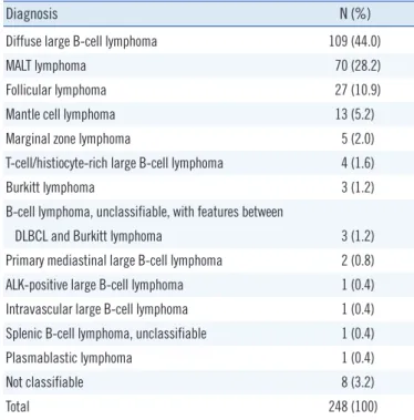
1. Study populationWe collected 248 bone marrow specimens from 232 patients (137 men and 95 women) who were diagnosed as having B-cell malig- nancy between December 2012 and July 2013 at our center: 198 at diagnosis and 50 during the course of the disease. Regarding diagnoses, diffuse large B-cell lymphoma was most common (44.0%), followed by mucosa-associated lymphoid tissue (MALT) lymphoma (28.2%) and follicular lymphoma (10.9%) (Table 1).
2. Bone marrow aspirate and biopsy
Wright-Giemsa–stained slides of bilateral bone marrow aspirate smears were examined for atypical lymphoid/lymphoma cells in- dependent of immunophenotypic studies. Of 248 bone marrow aspirate specimens, five had no cellular component and were excluded from the study. Bilateral bone marrow trephine biop- sies were obtained and tissue samples were fixed in 10% neu- tral-buffered formalin, decalcified, and paraffin-embedded. He- matoxylin and eosin (H&E) staining and CD3 and CD20 immu- nostaining were performed to determine lymphoma involvement.
3. Flow cytometry
Flow cytometric immunophenotyping of an EDTA anticoagulated
bone marrow aspirate specimen was performed in each case. A standard bone marrow assay with erythrocyte cell lysis was used for all bone marrow aspirate specimens. Aspirate specimens from one side or a mixture of both right and left sides were used depending on the quality and amount of specimens. The num- ber of cells analyzed was between 50,000 and 250,000, and the instrumental sensitivity was 0.1%.
Samples were analyzed with a two-step protocol; screening was done first with six-color multiparameter flow cytometry to detect the abnormal lymphoid cell population followed by sec- ond-line, detailed immunophenotyping for specific characteriza- tion of lymphoma cells. For the first step, an analysis with six markers for B-cell lymphoma was performed with monoclonal antibodies directed against CD19, CD20, CD10, and κ and λ immunoglobulins (Igs). In the case of mantle cell lymphoma, a monoclonal antibody against CD5 was used instead of CD10.
These antibodies were combined as κ/λ-fluorescein isothiocya- nate/lambda-phycoerythrin (PE), CD19-peridinin chlorophyll (PerCP), CD10-allophycocyanin (APC), CD20-PE-cyanine7 (Cy7), and CD45-APC-Cy7. In the case of mantle cell lymphoma, CD5- PerCP and CD19-APC were used. Antibodies were supplied by Becton Dickinson immunocytometry systems (Becton Dickin- son, San Jose, CA, USA) except for CD5, which was supplied by Beckman Coulter (Beckman Coulter, Miami, FL, USA). Because lymphomas associated with these cases were classified on the basis of lymph node or tissue biopsies, the first bone marrow im- munophenotypic analyses were solely centered on involvement of malignant lymphoma, i.e., the presence of a monoclonal B-cell population. Cutoffs of κ-to-λ ratios have been pre-established as 0.25:1 and 4:1 in our laboratory with normal bone marrow spec- imens.
The second-line flow cytometric analysis was performed with positive cases in the initial clonality test. This step is aimed at evaluating the detailed immunophenotype of lymphoma cells. A selected panel of antibodies was used and combined as the fol- lowing: FMC (developed at Flinders Medical Centre) 7/CD23/
CD10/CD45, CD5/CD20/CD22/CD45, CD38/CD3/CD56/CD45, and terminal deoxynucleotidyltransferase (TdT). Both forward/
side light scatter and CD45/side light scatter were used in each case as primary gating methodologies. Further gating was per- formed as necessary on either lymphoid subpopulations based on cell size or backgating in the event of CD19+ B-cell staining.
4. Data comparison
The results of flow cytometry were compared with that of bone marrow biopsy, in terms of concordance rate.
Table 1. Patient distribution in accordance with histological diagnosis
Diagnosis N (%)
Diffuse large B-cell lymphoma 109 (44.0)
MALT lymphoma 70 (28.2)
Follicular lymphoma 27 (10.9)
Mantle cell lymphoma 13 (5.2)
Marginal zone lymphoma 5 (2.0)
T-cell/histiocyte-rich large B-cell lymphoma 4 (1.6)
Burkitt lymphoma 3 (1.2)
B-cell lymphoma, unclassifiable, with features between
DLBCL and Burkitt lymphoma 3 (1.2)
Primary mediastinal large B-cell lymphoma 2 (0.8)
ALK-positive large B-cell lymphoma 1 (0.4)
Intravascular large B-cell lymphoma 1 (0.4)
Splenic B-cell lymphoma, unclassifiable 1 (0.4)
Plasmablastic lymphoma 1 (0.4)
Not classifiable 8 (3.2)
Total 248 (100)
Abbreviations: DLBCL, diffuse large B-cell lymphoma; MALT, mucosa-asso- ciated lymphoid tissue; ALK, anaplastic lymphoma kinase.
RESULTS
Approximately 24.6% of bone marrow evaluations showed evi- dence of NHL; bone marrow trephine biopsies showed histo- logic involvement of lymphoma cells in 50 cases (20.2%) and flow cytometric analysis of bone marrow aspirate showed the presence of lymphoma cells in 45 cases (18.1%).
1. Flow cytometric immunophenotype
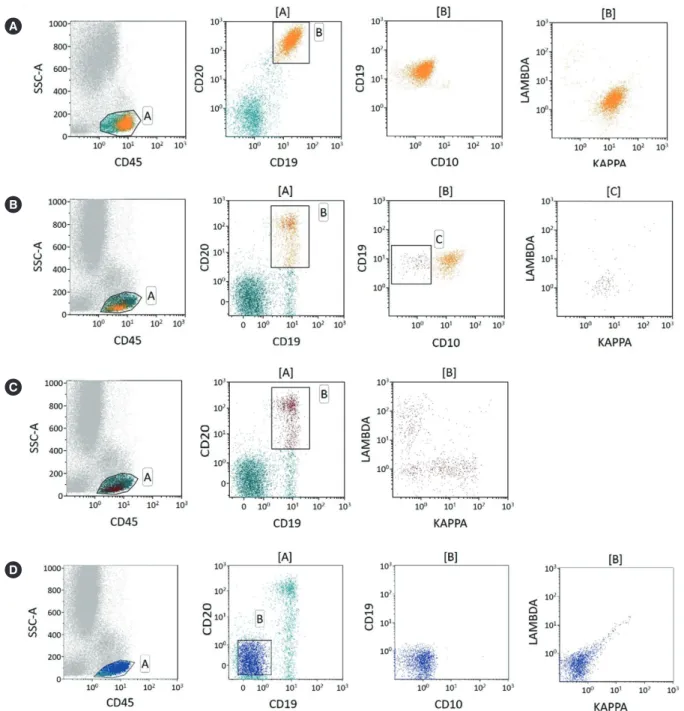
Forty-five cases (18.1%) showed bone marrow involvement by NHL by flow cytometry, with monoclonal B-cell populations rang- ing from 0.09% to 60.00% (median 5.12%) of total cell popula- tions. Among the positive cases, 19 (42.2%) showed minimal bone marrow involvement (malignant cells less than 5%; Fig. 1).
The 203 cases with a negative flow cytometric study typically
Fig. 1. Examples of κ-to-λ ratios for patients with B-cell non-Hodgkin lymphoma using different gating strategies. (A) Plots showing unequiv- ocal light chain restriction. (B) Increased sensitivity using multi-color antibodies. Gating on CD19-positive and CD20-positive and CD10-neg- ative cells could identify very small amounts of clonal B-cells (κ-to-λ ratio=11.5) (C) whereas gating without CD10 masks the clonal B-cell population (κ-to-λ ratio=1.412). (D) Plots showing negative controls.
A
B
C
D
showed a mixture of polyclonal B cells and a small population of CD19+/Ig- cells representing normal CD19+ precursor B cells.
In these cases, the κ-to-λ ratios of polyclonal B cells ranged from 0.98 to 1.85, and the median was 1.35, which was concordant with previously established normal ranges.
2. Discrepancies between flow cytometry and histopathology The concordance rate between flow cytometry and trephine bi- opsy histopathology was 91.5%; both were negative in 190 cases (76.6%) and both were positive in 37 cases (14.9%). Dis- crepant results were found in 21 cases (8.5%) (Table 2). There were eight cases with histomorphologically negative and immu- nophenotypically positive results, and the diagnosis in these cases included diffuse large B-cell lymphoma (n =3), mantle cell lymphoma (n =3), and MALT lymphoma (n =2). Of those eight cases, six showed minimal bone marrow involvement (0.09-2.2%). Thirteen cases were histopathologically positive and immunophenotypically negative, and the diagnoses in these cases included diffuse large cell lymphoma (n=7), T-cell/histio- cyte-rich large B-cell lymphoma (n =2), anaplastic lymphoma kinase (ALK)-positive large B-cell lymphoma (n =1), follicular lymphoma (n=1), MALT lymphoma (n=1), and unclassifiable lymphoma (n=1).
3. Bone marrow aspirate and biopsy
Morpholgic examination and analysis of immunostaining of both bone marrow biopsy and aspirate showed 91.8% concordance rate; both were negative in 190 cases (78.2%) and both were positive in 33 cases (13.6%) (Table 3).
DISCUSSION
Complete and accurate staging of malignant lymphoma is es- sential in providing adequate management and predicting out- comes. As a part of lymphoma staging, the evaluation of bone marrow involvement is an important process, and morphological assessment of bone marrow biopsy specimens remains the standard method. Recently, flow cytometric analysis of bone marrow aspirate has been performed as a routine procedure.
Although flow cytometry provides more objective results with higher sensitivity, previous studies did not support a significant benefit beyond morphological examination alone [18-20]. How- ever, the low detection rate by flow cytometry in those studies may be partly related to the use of less sensitive and less spe- cific single- or dual-color analyses, which were the standard at that time, compared with the four- or six-color multiparameter analysis technology that is currently used. With the advanced Table 2. B-cell NHL involvement of bone marrow by flow cytometry (F) and core biopsy (B)
Diagnosis F+B+ F+B- F-B+ F-B-
Diffuse large B-cell lymphoma (N=109) 9 (8.3) 3 (2.8) 7 (6.4) 90 (82.6)
MALT lymphoma (N=70) 2 (2.9) 2 (2.9) 1 (1.4) 65 (92.9)
Follicular lymphoma (N=27) 9 (33.3) 1 (3.7) 17 (63.0)
Mantle cell lymphoma (N=13) 5 (38.5) 3 (23.1) 5 (38.5)
Marginal zone lymphoma (N=5) 3 (60.0) 2 (40.0)
T-cell/histiocyte-rich large B-cell lymphoma (N=4) 1 (25.0) 2 (50.0) 1 (25.0)
Burkitt lymphoma (N=3) 1 (33.3) 2 (66.7)
B-cell lymphoma, unclassifiable, with features between
DLBCL and Burkitt lymphoma (N=3) 3 (100)
Primary mediastinal large B-cell lymphoma (N=2) 2 (100)
ALK-positive large B-cell lymphoma (N=1) 1 (100)
Intravascular large B-cell lymphoma (N=1) 1 (100)
Splenic B-cell lymphoma, unclassifiable (N=1) 1 (100)
Plasmablastic lymphoma (N=1) 1 (100)
Not classifiable (N=8) 5 (62.5) 1 (12.5) 2 (25.0)
Total (N=248) 37 (14.9) 8 (3.2) 13 (5.2) 190 (76.6)
Data is presented as number (percentage).
Abbreviations: NHL, non-Hodgkin lymphoma; DLBCL, diffuse large B-cell lymphoma; MALT, mucosa-associated lymphoid tissue; ALK, anaplastic lymphoma kinase.
technology currently available, six-color or eight-color multipa- rameter analysis can improve the sensitivity and specificity [21, 22]. At this point, evaluation of the utility of flow cytometric anal- ysis on bone marrow using multiparameter analysis could be helpful. We performed both morphological examination of bone marrow biopsy specimens and flow cytometric analysis of bone marrow aspirates concurrently, and compared the results.
The immunophenotyping in this study was performed with six-color multiparameter flow cytometry, which has advantages compared to examination with only CD19, κ, and λ light chains.
Including CD10 enabled increased detection sensitivity by ex- cluding normal hematogones, and the inclusion of both CD19 and CD20 was additionally helpful for a more specific selection of B-cell populations according to differentiation status. More- over, the specificity may be improved by using CD10 in follicular lymphoma and by using CD5 in mantle cell lymphoma. This will result in an improved sensitivity by lowering the possibility of masking small portions of malignant cells by the presence of many polyclonal cells.
The flow cytometric analysis is based on the clonality test.
Detection of immunoglobulin light chain-restriction expression represents the most reliable evidence for the presence of malig-
nant cells. In this study, κ-to-λ ratio ranged from 0.98 to 1.85 and the median was 1.35, confirming the normal range previ- ously determined. The second-line, detailed immunophenotyp- ing to detect abnormal immunophenotypes of malignant B-cell populations may increase the sensitivity when the role of screening immunophenotyping is limited, as in cases of malig- nant B cells with no light chain expression. Detailed immuno- phenotyping can also provide complementary information, which may be helpful in the accurate subclassification of NHL [23, 24]. This is very useful in cases without easily accessible lymph nodes or in cases with no definite lymphadenopathy.
There were 21 discrepant cases between the results of the bone marrow biopsy and flow cytometric analysis. Among those, eight cases were reported, in which lymphoma cells were not detected by morphological examination. The discrepancy could be explained in part by a low sensitivity of morphological exami- nation in detecting minimal bone marrow involvement. Indeed, six of eight cases showed minimal bone marrow involvement of malignant lymphoma. In 13 cases, however, the involvement of the lymphoma was detected only in the biopsy specimen. This discrepancy may be attributed to differences in the specimens analyzed. For example, when the disease does not diffusely in- Table 3. B-cell NHL involvement by bone marrow aspirate (A) and core biopsy (B)
Diagnosis A+B+ A+B- A-B+ A-B-
Diffuse large B-cell lymphoma (N=106) 9 (8.5) 5 (4.7) 4 (3.8) 88 (8.30)
MALT lymphoma (N=70) 2 (2.9) 1 (1.4) 1 (1.4) 66 (94.3)
Follicular lymphoma (N=27) 7 (25.9) 3 (11.1) 17 (63.0)
Mantle cell lymphoma (N=13) 4 (30.8) 2 (15.4) 1 (7.7) 6 (46.2)
T-cell/histiocyte-rich large B-cell lymphoma (N=2) 1 (50.0) 1 (50.0)
Marginal zone lymphoma (N=5) 2 (40.0) 1 (20.0) 2 (40.0)
Burkitt lymphoma (N=3) 1 (33.3) 2 (66.7)
B-cell lymphoma, unclassifiable, with features intermediate between
DLBCL and Burkitt lymphoma (N=3) 3 (100)
Primary mediastinal large B-cell lymphoma (N=2) 2 (100)
ALK-positive large B-cell lymphoma (N=1) 1 (100)
Intravascular large B-cell lymphoma (N=1) 1 (100)
Splenic B-cell lymphoma, unclassifiable (N=1) 1 (100)
Plasmablastic lymphoma (N=1) 1 (100)
Not classifiable (N=8) 5 (62.5) 1 (12.5) 2 (25.0)
Total (N=243*) 33 (13.6) 8 (3.3) 12 (4.9) 190 (78.2)
Data is presented as number (percentage).
*Among 248 specimens, five bone marrow aspirate specimens were inadequate in quality and excluded from the study (3 with diffuse large B-cell lympho- ma and 2 with T-cell/histiocyte-rich large B-cell lymphoma).
Abbreviations: See Table 2.
volve the bone marrow and the lymphoid infiltration foci are small, the bone marrow may produce aspirate samples free of disease while the biopsy may be positive for lymphoma cells.
Evidence for this is the improved sensitivity of bilateral bone marrow biopsy over unilateral biopsy. The same principle can be applied in the cases, in which lymphoma involvement was not detectable on bone marrow aspirate despite positive results on bone marrow biopsies. The presence of these cases con- firms previously published results, which concluded that there is no substitute for a bone marrow biopsy [13, 14].
Additionally, collection of bone marrow aspirate is a simple and minimally invasive procedure that is considered an ancillary tool in the staging of NHL. The utility of collecting bone marrow aspirate has been advocated by some groups [25, 26], while others have not supported the utility of this procedure [27]. To evaluate the usefulness of this specimen, we simultaneously compared bone marrow aspirate and biopsy samples. When as- pirate was compared with biopsy specimens by morphological examination and immunostaining, the concordance rate was 91.8% with 20 discrepant cases. On morphological examination of aspirate specimens, it was difficult to confirm the involvement of NHL when a minimal number of lymphoma cells were present or the specimen was diluted with peripheral blood. The utility of flow cytometric examination could be maximized when the cyto- morphological findings are equivocal.
We conclude that multi-color flow cytometry and examination of bone marrow aspirate are useful methods for assessing bone marrow in NHL staging. Although they are not a substitute for bone marrow biopsy, they do have a complementary role in de- tecting a small portion of lymphoma cells in a small subset of pa- tients. Particularly, multi-color flow cytometric analysis can in- crease the sensitivity of this method, enabling more specific gat- ing of abnormal cell populations. It may also be useful in cases where the morphological evaluation of the bone marrow biopsy is inconclusive or unavailable. Since the clinical significance of such a minimal bone marrow involvement is not fully estab- lished, further clinical studies are needed to elucidate the effect of minimal bone marrow involvement on the clinical course and prognosis of patients.
Authors’ Disclosures of Potential Conflicts of Interest
No potential conflicts of interest relevant to this article were re- ported.
REFERENCES
1. A predictive model for aggressive non-Hodgkin’s lymphoma. The Inter- national Non-Hodgkin’s Lymphoma Prognostic Factors Project. N Engl J Med 1993;329:987-94.
2. Bastion Y and Coiffier B. Is the International Prognostic Index for Ag- gressive Lymphoma patients useful for follicular lymphoma patients? J Clin Oncol 1994;12:1340-2.
3. Coiffier B, Gisselbrecht C, Vose JM, Tilly H, Herbrecht R, Bosly A, et al.
Prognostic factors in aggressive malignant lymphomas: description and validation of a prognostic index that could identify patients requiring a more intensive therapy. The Groupe d’Etudes des Lymphomes Agres- sifs. J Clin Oncol 1991;9:211-9.
4. Munck JN, Dhermain F, Koscielny S, Girinsky T, Carde P, Bosq J, et al.
Alternating chemotherapy and radiotherapy for limited-stage intermedi- ate and high-grade non-Hodgkin’s lymphomas: long-term results for 96 patients with tumors > 5 cm. Ann Oncol 1996;7:925-31.
5. Shipp MA, Neuberg D, Janicek M, Canellos GP, Shulman LN. High- dose CHOP as initial therapy for patients with poor-prognosis aggressive non-Hodgkin’s lymphoma: a dose-finding pilot study. J Clin Oncol 1995;13:2916-23.
6. Coad JE, Olson DJ, Christensen DR, Lander TA, Chibbar R, McGlennen RC, et al. Correlation of PCR-detected clonal gene rearrangements with bone marrow morphology in patients with B-lineage lymphomas. Am J Surg Pathol 1997;21:1047-56.
7. Murphy TD, Crisan D, Farkas DH. Use of molecular diagnostic methods for lymphoma staging in bilateral bone marrow biopsies. Mol Diagn 1997;2:177-82.
8. Nedomova R, Papajik T, Prochazka V, Indrak K, Jarosova M. Cytogenet- ics and molecular cytogenetics in diffuse large B-cell lymphoma (DLB- CL). Biomed Pap Med Fac Univ Palacky Olomouc Czech Repub 2013;
157:239-47.
9. Reichard KK and Robinett S. Detection of genetic translocations in lym- phoma using fluorescence in situ hybridization. Methods Mol Biol 2013;
999:189-202.
10. Juneja SK, Wolf MM, Cooper IA. Value of bilateral bone marrow biopsy specimens in non-Hodgkin’s lymphoma. J Clin Pathol 1990;43:630-2.
11. Liu W, Medeiros LJ, Lin P, Romaguera JE, Wang SA, Jorgensen JL.
Usefulness of flow cytometrici mmunophenotyping for bone marrow staging in patients with mantle cell lymphoma after therapy. Am J Clin Pathol 2012;137:634-40.
12. Merli M, Arcaini L, Boveri E, Rattotti S, Picone C, Passamonti F, et al.
Assessment of bone marrow involvement in non-Hodgkin’s lymphomas:
comparison between histology and flow cytometry. Eur J Haematol 2010;85:405-15.
13. Duggan PR, Easton D, Luider J, Auer IA. Bone marrow staging of pa- tients with non-Hodgkin lymphoma by flow cytometry: correlation with morphology. Cancer 2000;88:894-9.
14. Hanson CA, Kurtin PJ, Katzmann JA, Hoyer JD, Li CY, Hodnefield JM, et al. Immunophenotypic analysis of peripheral blood and bone marrow in the staging of B-cell malignant lymphoma. Blood 1999;94:3889-96.
15. Morice WG, Kurtin PJ, Hodnefield JM, Shanafelt TD, Hoyer JD, Rem- stein ED, et al. Predictive value of blood and bone marrow flow cytome- try in B-cell lymphoma classification: comparative analysis of flow cy- tometry and tissue biopsy in 252 patients. Mayo Clin Proc 2008;83:
776-85.
16. Palacio C, Acebedo G, Navarrete M, Ruiz-Marcellán C, Sanchez C, Blan- co A, et al. Flow cytometry in the bone marrow evaluation of follicular and diffuse large B-cell lymphomas. Haematologica 2001;86:934-40.
17. Perea G, Altés A, Bellido M, Aventín A, Bordes R, Ayats R, et al. Clinical utility of bone marrow flow cytometry in B-cell non-Hodgkin lymphomas (B-NHL). Histopathology 2004;45:268-74.
18. Dunphy CH. Combining morphology and flow cytometric immunophe- notyping to evaluate bone marrow specimens for B-cell malignant neo- plasms. Am J Clin Pathol 1998;109:625-30.
19. Iancu D, Hao S, Lin P, Anderson SK, Jorgensen JL, McLaughlin P, et al.
Follicular lymphoma in staging bone marrow specimens: correlation of histologic findings with the results of flow cytometry immunophenotypic analysis. Arch Pathol Lab Med 2007;131:282-7.
20. Naughton MJ, Hess JL, Zutter MM, Bartlett NL. Bone marrow staging in patients with non-Hodgkin’s lymphoma: is flow cytometry a useful test?
Cancer 1998;82:1154-9.
21. Carulli G and Marini A. Diagnosis and classification of B-cell non-Hodg- kin lymphomas. The role of multiparameter flow cytometry. Clin Ter 2012;
163:47-57.
22. Stacchini A, Aliberti S, Demurtas A, Benevolo G, Godio L. Ten antibod- ies, six colors, twelve parameters: a multiparameter flow cytometric ap-
proach to evaluate leptomeningeal disease in B-cell non-Hodgkin’s lym- phomas. Cytometry B Clin Cytom 2012;82:139-44.
23. Fineberg S, Marsh E, Alfonso F, Espiritu E, Gottesman SR, Amorosi E, et al. Immunophenotypic evaluation of the bone marrow in non-Hodg- kin’s lymphoma. Hum Pathol 1993;24:636-42.
24. Jennings CD and Foon KA. Recent advances in flow cytometry: applica- tion to the diagnosis of hematologic malignancy. Blood 1997;90:2863-92.
25. Barekman CL, Fair KP, Cotelingam JD. Comparative utility of diagnostic bone-marrow components: a 10-year study. Am J Hematol 1997;56:37- 41.
26. Lee SH, Erber WN, Porwit A, Tomonaga M, Peterson LC; International Council for Standardization In Hematology. ICSH guidelines for the standardization of bone marrow specimens and reports. Int J Lab He- matol 2008;30:349-64.
27. Winfield DA and Polacarz SV. Bone marrow histology. 3: Value of bone marrow core biopsy in acute leukaemia, myelodysplastic syndromes, and chronic myeloid leukaemia. J Clin Pathol 1992;45:855-9.