ISSN 2234-3806 • eISSN 2234-3814
434 www.annlabmed.org https://doi.org/10.3343/alm.2017.37.5.434 Ann Lab Med 2017;37:434-437
https://doi.org/10.3343/alm.2017.37.5.434
Brief Communication
Clinical Microbiology
Identification of viridans streptococci With Matrix- Assisted Laser Desorption & Ionization Time-of-flight Mass Spectrometry by an In-house Method and a Commercially Available System
Catalina-Suzana Stingu, M.D.1, Klaus Eschrich, Ph.D.2, Juliane Thiel, M.D.1, Toralf Borgmann, M.D.1, Reiner Schaumann, M.D.1, and Arne C. Rodloff, M.D.1
Institute for Medical Microbiology and Epidemiology of Infectious Diseases1, University Hospital, University of Leipzig; Rudolf-Schoenheimer-Institute for Biochemistry2, University of Leipzig, Leipzig, Germany
Two matrix-assisted laser desorption ionization time-of-flight mass spectrometry (MALDI- TOF-MS)-based methods were compared for their ability to identify viridans streptococci.
One approach employed a reference database and software developed in-house. All in- house measurements were performed using an Autoflex II Instrument (Bruker Daltonics GmbH, Germany). The other system, a VITEK-MS (BioMérieux, France) was operated on the commercially available V2.0 Knowledge Base for Clinical Use database. Clinical iso- lates of viridans streptococci (n=184) were examined. Discrepant results were resolved by 16S rDNA sequencing. Species-level identification percentages were compared by a chi- square test. The in-house method correctly identified 179 (97%) and 175 (95%) isolates to the group and species level respectively. In comparison, the VITEK-MS system correctly identified 145 (79%) isolates to the group and species level. The difference between the two methods was statistically significant at both group and species levels. Using the Auto- flex II instrument combined with an extraction method instead of whole cell analysis re- sulted in more reliable viridans streptococci identification. Our results suggest that com- bining extraction with powerful analysis software and the careful choice of well-identified strains included into the database was useful for identifying viridans streptococci species.
Key Words: Viridans streptococci, MALDI-TOF MS, In-house database
Received: September 23, 2016 Revision received: January 12, 2017 Accepted: May 15, 2017
Corresponding author: Catalina-Suzana Stingu
Institute for Medical Microbiology and Epidemiology of Infectious Diseases, University of Leipzig, Liebigstr. 21, Leipzig 04103, Germany
Tel: +49-3419715242 Fax: +49-3419715209
E-mail: CatalinaSuzana.Stingu@medizin.
uni-leipzig.de
© Korean Society for Laboratory Medicine This is an Open Access article distributed under the terms of the Creative Commons Attribution Non-Commercial License (http://creativecom- mons.org/licenses/by-nc/4.0) which permits unrestricted non-commercial use, distribution, and reproduction in any medium, provided the original work is properly cited.
Identification of viridans streptococci at the species level is diffi- cult because of the high degree of genotypic and phenotypic similarity of some species. Unfortunately, phenotypic test sys- tems that are widely used in clinical laboratory, like Rapid ID32 Strep (bioMérieux, Lyon, France) and VITEK 2 (bioMérieux), do not always accurately identify some species in this heterogeneous bacterial group [1-3]. Matrix-assisted laser desorption ionization time-of-flight mass spectrometry (MALDI-TOF-MS)-based meth- odologies seem to have great potential for reliably identifying viridans streptococci in clinical laboratories [4, 5].
In this study, two MALDI-TOF-MS-based approaches were compared for their ability to identify viridans streptococci iso- lates.
Clinical strains of viridans streptococci (n=184) were used in this study. All isolates were previously biochemically identified as streptococci by using the Rapid ID32 Strep System (bioMérieux) and then stored at –80°C in CRYOBANK BLUE tubes from Mast Diagnostica (Reinfeld, Germany). For analysis, the strains were cultured on Columbia Blood Agar (Thermo Fisher Scientific, Ox- oid Microbiology Products, Hampshire, UK) at 36–37°C with 5%
1 / 1 CROSSMARK_logo_3_Test
2017-03-16 https://crossmark-cdn.crossref.org/widget/v2.0/logos/CROSSMARK_Color_square.svg
Stingu C-S, et al.
Identification of viridans streptococci with MALDI-TOF MS
https://doi.org/10.3343/alm.2017.37.5.434 www.annlabmed.org 435
CO2 for 18–24 hr.
A reference database comprising a well-characterized spec- trum of reference and clinical strains of Streptococcus mitis, S.
oralis, S. pneumoniae, S. intermedius, S. salivarius, S. sangui- nis, S. parasanguinis, S. gordonii, S. constellatus, S. anginosus, S. mutans, and S. sobrinus was developed in-house (Table 1).
For this, individual colonies of streptococcal isolates were recul- tured in brain-heart infusion broth overnight and prepared by using the extraction methods described by Friedrichs et al [4].
Mass spectra were acquired by using an Autoflex II MALDI-TOF mass spectrometer (Bruker Daltonics GmbH, Bremen, Germany) with a nitrogen laser (337 nm) operating in positive linear mode (delay 150 nsec, voltage 20 kV, mass range 2–20 kDa) using FLEXCONTROL software version 2.4 (Bruker Daltonics). The spec- tra were externally calibrated by using the standard calibrant mixture, Protein Calibration Standard I (Bruker Daltonics). Auto- mated peak extraction from data files of the reading was performed after transferring in FLEXANALYSIS software version 2.4 (Bruker
Daltonics). To identify bacterial species, similarity analysis be- tween peak lists was performed by using a hierarchical cluster- ing procedure in MatLab software (Version 7.10.0.499; The Math- Works Inc., Natick, MA, USA), as described by Friedrichs et al [4].
VITEK MS (bioMérieux) was operated on the V2.0 Knowledge Base for clinical use and employed for identification a direct col- ony method. Colonies were picked from Columbia blood agar plates and spotted onto the polymeric target slides (bioMérieux) in duplicate. After air drying, 1 µL matrix solution (α-cyano-4- hydroxycinnamic acid, VITEK MS CHCA) was added. The target slides were then loaded into the mass spectrometer (VITEK MS).
The spectra were compared to the VITEK MS V2.0 Knowledge Base for clinical use. Escherichia coli ATCC 8739 was used to calibrate and control the method according to the manufactur- er’s instructions. Successful identification was considered only at 99.9% probability. For the unidentified or misidentified strains, we repeated the protocol once. Since the V2.0 Knowledge Base for clinical use cannot differentiate between S. mitis and S. ora-
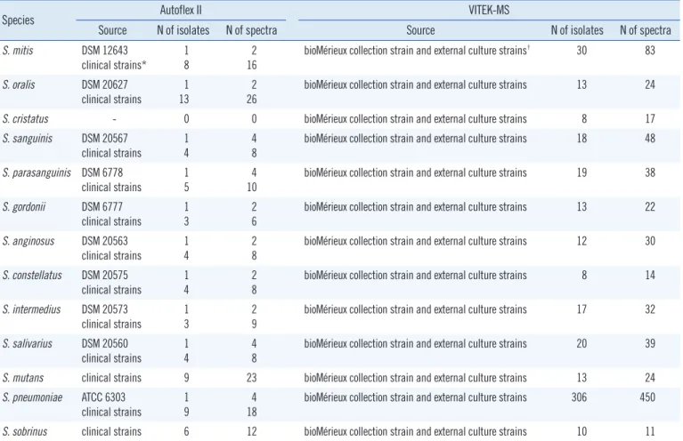
Table 1. Details of the in-house database used by Autoflex II and V2.0 Knowledge Base used with VITEK-MS
Species Autoflex II VITEK-MS
Source N of isolates N of spectra Source N of isolates N of spectra
S. mitis DSM 12643
clinical strains*
1 8
2 16
bioMérieux collection strain and external culture strains† 30 83 S. oralis DSM 20627
clinical strains 1
13 2
26 bioMérieux collection strain and external culture strains 13 24
S. cristatus - 0 0 bioMérieux collection strain and external culture strains 8 17
S. sanguinis DSM 20567 clinical strains
1 4
4 8
bioMérieux collection strain and external culture strains 18 48 S. parasanguinis DSM 6778
clinical strains 1
5 4
10 bioMérieux collection strain and external culture strains 19 38 S. gordonii DSM 6777
clinical strains
1 3
2 6
bioMérieux collection strain and external culture strains 13 22 S. anginosus DSM 20563
clinical strains 1
4 2
8 bioMérieux collection strain and external culture strains 12 30 S. constellatus DSM 20575
clinical strains
1 4
2 8
bioMérieux collection strain and external culture strains 8 14 S. intermedius DSM 20573
clinical strains 1
3 2
9 bioMérieux collection strain and external culture strains 17 32 S. salivarius DSM 20560
clinical strains
1 4
4 8
bioMérieux collection strain and external culture strains 20 39
S. mutans clinical strains 9 23 bioMérieux collection strain and external culture strains 13 24
S. pneumoniae ATCC 6303
clinical strains 1
9 4
18 bioMérieux collection strain and external culture strains 306 450
S. sobrinus clinical strains 6 12 bioMérieux collection strain and external culture strains 10 11
*All clinical strains included in the Autoflex II Database were identified by using 16S rRNA gene sequencing; †ll strains were identified by using Vitek 2, Rap- id ID 32 Strep, or 16S rRNA sequencing.
Stingu C-S, et al.
Identification of viridans streptococci with MALDI-TOF MS
436 www.annlabmed.org https://doi.org/10.3343/alm.2017.37.5.434 lis, S. mitis/S. oralis was considered as a match for species iden-
tification.
Discrepant results between the two systems (including the unidentified strains) were resolved by 16S rRNA gene sequenc- ing. MALDI-TOF MS results discordant with 16S rRNA gene se- quencing were considered incorrect.
Chi-square test was used to compare species-level identifica- tion percentages.
The Autoflex II-based method correctly identified 179 (97%) isolates to the group level and 175 (95%) isolates to the species level. Five strains were incorrectly identified to the group level.
Two strains belonging to the S. sanguinis group were misidenti- fied as members of the S. mitis group, and three members of the S. mitis group were misidentified as members of the S. san- guinis group. In comparison, VITEK-MS correctly identified 145 (79%) isolates to the group and species levels (Table 2). Eight strains were misidentified, and 31 remained repeatedly uniden- tified. Four strains belonging to the S. sanguinis group were mis- identified as members of the S. mitis group, and 4 strains be- longing to the S. mitis group were misidentified as members of the S. sanguinis group. The 31 unidentified strains were as fol- lows: 17 members of the S. mitis group, 12 members of the S.
sanguinis group, and two members of the S. anginosus group.
The difference between the two methods was statistically signifi- cant (P <0.05) for both group and species levels.
The taxonomic changes over the past several years, poor ca-
pacity of Rapid ID 32 Strep and VITEK 2 systems to differentiate them, and their genetic and proteomic homology have made the identification of viridans streptococci challenging in clinical labo- ratories [2, 6].
In this study, two MALDI-TOF-MS-based methods were com- pared for their ability to identify viridans streptococci. Three dif- ferent groups and eight different species were represented in this study: S. mitis group (n=105), S. anginosus group (n=9), and S. sanguinis group (n=70).
The Autoflex II-based in-house identification method showed superior performance to the commercial VITEK-MS system at both group and species levels. Its database did not include Strep- tococcus cristatus, so consequently all 3 S. cristatus strains were misidentified as S. oralis. The VITEK-MS system, which includes S. cristatus in its database, correctly identified two of these three strains, with only one strain being misidentified as S. parasan- guinis.
Our in-house method misidentified three strains belonging to the S. mitis group as S. gordonii (n=2) and S. sanguinis (n=1).
Furthermore, two strains belonging to the S. sanguinis group were misidentified as members of the S. mitis group. One mis- identification at the species level occurred within the S. sangui- nis group: an S. parasanguinis strain was misidentified as S. gor- donii.
VITEK-MS failed repeatedly to identify 31 strains (17%), 17 (16%) of them belonging to the S. mitis group, 12 (17%) strains from the S. sanguinis group, and two (22%) strains as members of the S. anginosus group. Whenever VITEK-MS correctly identi- fied a strain at the group level, the species identification was also correct. Four different strains of the S. sanguinis group (two strains of S. sanguinis, one strain each of S. gordonii and S. parasan- guinis) were misidentified as S. mitis/oralis. Additional four strains belonging to the S. mitis group were misidentified as members of the S. sanguinis group (two S. gordonii strains and two S. para- sanguinis strains).
Both systems misidentified strains belonging to all groups;
hence, we cannot conclude that one specific group of strepto- cocci proved to be more difficult to differentiate than another.
While the Autoflex II-based method identified all strains, cor- rectly or not, VITEK-MS failed to identify 17% of strains. The un- identified strains were members of all tested groups.
Another study compared the performance of two commercially available systems: VITEK-MS (bioMérieux) and MALDI Biotyper (Bruker Daltonics) using 54 strains and showed that both sys- tems performed better than the commercially available biochem- ical methods [7]. Overall, the MALDI Biotyper and VITEK-MS sys- Table 2. Comparison of identification results for 184 strains of viri-
dans streptococci obtained with the Autoflex II-based method or with the VITEK-MS-based method
Species N of
isolates
N of isolates (% correct) identified by:
Autoflex II VITEK-MS Group
level Species
level Group
level Species level
S. mitis group 105 102 (97) 84 (80)
S. mitis/oralis 102 99 (97) 82 (80)
S. cristatus 3 0 2 (66.6)
S. sanguinis group 70 68 (97) 54 (77)
S. sanguinis 41 40 (97.5) 33 (80)
S. parasanguinis 6 5 (83) 5 (83)
S. gordonii 23 22 (96) 16 (69.5)
S. anginosus group 9 9 (100) 7 (77.7)
S. anginosus 4 4 (100) 3 (75)
S. constellatus 5 5 (100) 4 (80)
Total 184 179 (97) 175 (95) 145 (79) 145 (79)
Stingu C-S, et al.
Identification of viridans streptococci with MALDI-TOF MS
https://doi.org/10.3343/alm.2017.37.5.434 www.annlabmed.org 437
tems gave a correct species-level identification in 94% and 69%
of strains, respectively. In our study using more strains (n=184), we observed better performance of VITEK-MS, with 75% of strains being correctly identified. Rychert et al [8] and Dubois et al [9]
reported an even better performance of VITEK-MS with 82% of strains being correctly identified from 218 streptococcal isolates and 90.8% being correctly identified from 335 viridans strepto- cocci strains. It is not straightforward to compare these results with the performance of the Autoflex II-based method used in this study, because it is not a commercial system but uses cell extracts instead of whole cells and in-house developed software and database. When compared with the results by Friedrichs et al [4] that used the same system, our results were similar.
A combination of cell extraction, spectra acquisition using the Autoflex II, in-house developed software, and our own database seemed to more reliably identify viridans streptococci strains than the VITEK-MS system. The combination of an extraction method with a powerful analysis software and a database con- taining carefully chosen, well-identified strains can provide a useful tool for identifying viridans streptococci species.
Authors’ Disclosures of Potential Conflicts of Interest
No potential conflicts of interest relevant to this article were re- ported.
Acknowledgments
The authors appreciate the laboratory work of Angela Pöschel, Annett Hennig-Rolle, and Helga Stache.
REFERENCES
1. Teles C, Smith A, Ramage G, Lang S. Identification of clinically relevant viridans group streptococci by phenotypic and genotypic analysis. Eur J Clin Microbiol Infect Dis 2011;30:243-50.
2. Gavin P J, Warren JR, Obias AA, Collins SM, Peterson LR. Evaluation of the Vitek 2 system for rapid identification of clinical isolates of gram-neg- ative bacilli and members of the family Streptococcaceae. Eur J Clin Mi- crobiol Infect Dis 2002;21:869-74.
3. Ikryanikova LN, Lapin KN, Malakhova MV, Filimonova AV, Ilina EN, Dubo- vickaya VA, et al. Misidentification of alpha-haemolytic streptococci by routine tests in clinical practice. Infect Genet Evol 2011;11:1709-15.
4. Friedrichs C, Rodloff AC, Chhatwal GS, Schellenberger W, Eschrich K.
Rapid identification of viridans streptococci by mass spectrometric dis- crimination. J Clin Microbiol 2007;45:2392-7.
5. Moon HW, Lee SH, Chung HS, Lee M, Lee K. Performance of the Vitek MS matrix-assisted laser desorption ionization time-of-flight mass spec- trometry system for identification of gram-positive cocci routinely isolated in clinical microbiology laboratories. J Med Microbiol 2013;62:1301-6.
6. Davies AP, Reid M, Hadfield SJ, Johnston S, Mikhail J, Harris LG, et al.
Identification of clinical isolates of α-hemolytic streptococci by 16S rRNA gene sequencing, matrix-assisted laser desorption ionization-time of flight mass spectrometry using MALDI Biotyper, and conventional phenotypic methods: a comparison. J Clin Microbiol 2012; 50:4087-90.
7. Kärpänoja P, Harju I, Rantakokko-Jalava K, Haanperä M, Sarkkinen H.
Evaluation of two matrix-assisted laser desorption ionization-time of flight mass spectrometry systems for identification of viridans group strepto- cocci. Eur J Clin Microbiol Infect Dis 2014; 33:779-88.
8. Rychert J, Burnham CA, Bythrow M, Garner OB, Ginocchio CC, Jen- neman R, et al. Multicenter evaluation of the VITEK MS matrix-assisted laser desorption ionization-time of flight mass spectrometry system for identification of Gram-positive aerobic bacteria. J Clin Microbiol 2013;
51:2225-31.
9. Dubois D, Segonds C, Prere MF, Marty N, Oswald E. Identification of clinical Streptococcus pneumoniae isolates among other alpha and non- hemolytic streptococci by use of the Vitek MS matrix-assisted laser de- sorption ionization-time of flight mass spectrometry system. J Clin Mi- crobiol 2013;51:1861-7.