ISSN 2234-3806 • eISSN 2234-3814
Ann Lab Med 2021;41:409-413
https://doi.org/10.3343/alm.2021.41.4.409
Phospholipase C Beta 2 Protein Overexpression Is a Favorable Prognostic Indicator in Newly Diagnosed Normal Karyotype Acute Myeloid Leukemia
Mi Suk Park , Ph.D.1,*, Young Eun Lee , M.S.2,*, Hye Ran Kim , Ph.D.3, Jong Hee Shin , M.D., Ph.D.4, Hyun Wook Cho , Ph.D.5, Jun Hyung Lee , M.D., Ph.D.4, and Myung Geun Shin , M.D., Ph.D.2,4
1Department of Medical Laboratory Science, Gimhae College, Gimhae, Korea; 2Brain Korea 21 Plus Program, Chonnam National University Medical School and Chonnam National University Hwasun Hospital, Hwasun, Korea; 3College of Korean Medicine, Dongshin University, Naju, Korea; 4Department of Laboratory Medicine, Chonnam National University Medical School and Chonnam National University Hwasun Hospital, Hwasun, Korea; 5Department of Biology, Sunchon National University, Sunchon, Korea
Phospholipase C beta 2 (PLC-β2) regulates various essential functions in cell signaling, differentiation, growth, and mobility. We investigated the clinical implications of PLC-β2 protein expression in newly diagnosed normal karyotype acute myeloid leukemia (NK- AML). The PLC-β2 expression status in bone marrow tissues obtained from 101 patients with NK-AML was determined using semiquantitative immunohistochemistry (IHC). IHC results were compared with those for known prognostic markers. Using a cutoff score for positivity of 7.0, the PLC-β2 overexpression group showed superior overall survival (OS) (72.6% vs. 26.5%; P =0.016) and low hazard ratio (HR) (0.453; P =0.019) compared with the PLC-β2 low-expression group. The PLC-β2 overexpression group showed no sig- nificant gain in event-free survival (50.6% vs. 43.0%, P =0.465) and HR (0.735; P =0.464).
Among the known prognostic markers, only FLT3-ITD positivity was associated with a sig- nificantly low OS and high HR. In conclusion, PLC-β2 overexpression was associated with favorable OS in NK-AML patients. Our results suggest that PLC-β2 expression assessment using IHC allows prognosis prediction in NK-AML.
Key Words: Phospholipase C beta 2 protein, normal karyotype acute myeloid leukemia, prognosis
Received: August 9, 2020
Revision received: September 16, 2020 Accepted: January 14, 2021
Corresponding author:
Myung Geun Shin, M.D., Ph.D.
Department of Laboratory Medicine, Chonnam National University Medical School and Chonnam National University Hwasun Hospital, 322 Seoyang-ro, Hwasun-eup, Hwasun-gun, Jeollanam-do 58128, Korea
Tel: +82-61-379-7950 Fax: +82-61-379-7984 E-mail: [email protected] Co-corresponding author:
Jun Hyung Lee, M.D., Ph.D.
Department of Laboratory Medicine, Chonnam National University Medical School and Chonnam National University Hwasun Hospital, 322 Seoyang-ro, Hwasun-eup, Hwasun-gun, Jeollanam-do 58128, Korea
Tel: +82-61-379-7955 Fax: +82-61-379-7984 E-mail: [email protected]
* These authors contributed equally to this study.
© Korean Society for Laboratory Medicine This is an Open Access article distributed under the terms of the Creative Commons Attribution Non-Commercial License (https://creativecom- mons.org/licenses/by-nc/4.0) which permits unrestricted non-commercial use, distribution, and reproduction in any medium, provided the original work is properly cited.
2017-03-16 https://crossmark-cdn.crossref.org/widget/v2.0/logos/CROSSMARK_Color_square.svg
410 www.annlabmed.org https://doi.org/10.3343/alm.2021.41.4.409 Normal karyotype acute myeloid leukemia (NK-AML) is caused
by a variety of genetic abnormalities that result in various clinical outcomes [1-3]. New molecular markers are required to develop a more accurate risk stratification system for evaluating and aid- ing the treatment of NK-AML.
Phospholipase C (PLC) isozymes are widely distributed in mammals and play essential roles in cell growth, signaling, and the development of pathological conditions, such as cancer [4- 6]. Although associations between various cancers and PLC have been described [7-17], there has been no study relating NK-AML with PLC-β2 expression. Therefore, we investigated the clinicopathological implications of PLC-β2 expressionin NK-AML patients.
Immunohistochemistry (IHC) was employed to assess PLC-β2 expression in formalin-fixed, paraffin-embedded (FFPE) bone marrow (BM) sections. PLC-β2 expression was assessed in 101 patients newly diagnosed as having NK-AML from November 2010 to September 2016 at Chonnam National University Hwa- sun Hospital, Hwasun, Korea. This study was approved by the Institutional Review Board of Chonnam National University Hwa- sun Hospital (IRB No. 2009-35), which waived the need for in- formed consent.
FFPE BM sections of 3 μm thickness were deparaffinized in
xylene at 65°C for 10 minutes, rehydrated with ethanol, and rinsed with phosphate buffered saline. Thereafter, the slides were inserted to a Benchmark GX automatic stainer (Ventana Medical Systems, Oro Valley, AZ, USA). Rabbit polyclonal anti- human PLC-β2 antibody (clone# ab176012; Abcam, Cambridge, MA, USA) was applied at a 1:50 dilution. Monoclonal rabbit an- tihuman IgG1–4 antibody (clone# EPR4421, Abcam) at a 1:500 dilution was used as a negative isotype control. PLC-β2-immuno- stained slides were scored as follows: a brown granular cytoplas- mic staining was considered positive, and positivity was scored on a scale of 0–8 as the sum of a diffuseness score (0–5) and an intensity score (0–3) (Supplemental Data Table S1).
The final positivity score was determined as a median of scores by three independent pathology experts (Supplemental Data Fig.
S1). Overall survival (OS) and event-free survival (EFS) were an- alyzed using each positivity score as a cutoff and a median fol- low-up duration of 17.8 months. After determining the optimal cutoff value in the survival analysis, the IHC results were com- pared with those collected from the medical records for the fol- lowing prognostic markers: NPM1 variants and FLT3-ITD, BAALC, and WT1. OS was estimated using the Kaplan–Meier method.
Unadjusted hazard ratios (HRs) were calculated from a univari- ate Cox proportional hazards model. P <0.05 was considered
Table 1. Patient characteristics according to PLC-β2 protein expression
Patient characteristics Total
(N=101) PLC-β2 low expression
(N=48) PLC-β2 overexpression
(N=53) P
Age (yr) 57 [49;66] 56 [48;65] 59 [50;68] 0.496
Sex 0.813
Male 57 (56.4%) 26 (54.2%) 31 (58.5%)
Female 44 (43.6%) 22 (45.8%) 22 (41.5%)
PLC-β2 IHC score 7 [6;7] 6 [4;6] 7 [7;8] 0.000
Overall survival (month) 12.0 [3.0;26.0] 22.8 [6.0;36.5] 14.0 [2.0;26.0] 0.016
Event-free survival (month) 10.0 [3.0;21.0] 10.0 [3.5;16.0] 10.0 [2.0;24.0] 0.943
PB WBC count (106/L) 16,300 [3,800;59,290] 12,150 [4,310;45,515] 17,200 [3,330;84,200] 0.357
Blast % of PB 50 [10;80] 50 [10;80] 50 [10;80] 0.913
Blast % of BM 70 [50;80] 70 [50;80] 80 [60;90] 0.180
Complete remission 1.000
Achieved 44 (44.9%) 21 (44.7%) 23 (45.1%)
Failed 54 (55.1%) 26 (55.3%) 28 (54.9%)
Stem cell transplantation 1.000
None 63 (62.4%) 30 (62.5%) 33 (62.3%)
Transplanted 38 (37.6%) 18 (37.5%) 20 (37.7%)
Data are presented as median [interquartile ranges] or number (percentage).
Abbreviations: PLC, phospholipase C; IHC, immunohistochemistry; PB, peripheral blood; WBC, white blood cell; BM, bone marrow.
significant.
Median patient age at diagnosis was 57 years (23–83 years).
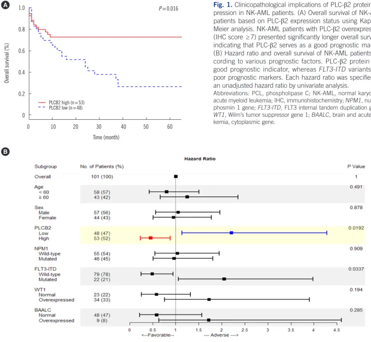
The male:female ratio was 57:44. Considering a positivity score of 7.0 as a cutoff, the PLC-β2 overexpression group showed higher OS (72.6% vs. 26.5%; P =0.016) and lower HR (0.453;
P =0.019) than the PLC-β2 low expression group. In EFS analy- sis, the PLC-β2 overexpression group showed no significant sur- vival gain (50.6% vs. 43.0%, P =0.465) and HR change (0.7357;
P =0.464) (Table 1 and Fig. 1).
There were no significant differences in age, sex, white blood cell counts in peripheral blood (PB), blast percentage in PB and
BM, complete remission rate, and enforcement of stem cell trans- plant between the PLC-β2 overexpression and low expression groups (Table 1). Among the known prognostic markers, only FLT3-ITD positivity was significantly associated with low OS (29.1%
vs. negative group 52.7%; P =0.032) and high HR (2.052; P = 0.034) (Fig. 1) [19].
NPM1-positive patients had a slightly, albeit not significantly, higher survival rate (P =0.892) and lower HR 0.96 (P =0.909).
However, patients with WT1 and BAALC expression, which are poor prognostic factors, exhibited a slightly, albeit not significantly, lower survival rate (WT1 P =0.189, BAALC P =0.280) and higher
Fig. 1. Clinicopathological implications of PLC-β2 protein ex- pression in NK-AML patients. (A) Overall survival of NK-AML patients based on PLC-β2 expression status using Kaplan–
Meier analysis. NK-AML patients with PLC-β2 overexpression (IHC score ≥7) presented significantly longer overall survival, indicating that PLC-β2 serves as a good prognostic marker.
(B) Hazard ratio and overall survival of NK-AML patients ac- cording to various prognostic factors. PLC-β2 protein is a good prognostic indicator, whereas FLT3-ITD variants are poor prognostic markers. Each hazard ratio was specified as an unadjusted hazard ratio by univariate analysis.
Abbreviations: PCL, phospholipase C; NK-AML, normal karyotype acute myeloid leukemia; IHC, immunohistochemistry; NPM1, nucleo- phosmin 1 gene; FLT3-ITD, FLT3 internal tandem duplication gene;
WT1, Wilm’s tumor suppressor gene 1; BAALC, brain and acute leu- kemia, cytoplasmic gene.
B 1.0
0.8
0.6
0.4
0.2
0
0 10 20 30 40 50 60 Time (month)
Overall survival (%)
P =0.016 A
PLCB2 high (n=53) PLCB2 low (n=48)
412 www.annlabmed.org https://doi.org/10.3343/alm.2021.41.4.409 HR (WT1 P =0.194, BAALC P =0.285) (Fig. 1) [1].
PLC-β2 expression decreased significantly in NK-AML patients, and the OS was significantly higher in the PLC-β2 overexpres- sion group than in the PLC-β2 low-expression group (P =0.016).
Given that the PLC-β2 pathway is involved in the regulation of platelet abundance, apoptosis, cell migration, and the cell cycle, these results suggest that PLC-β2 expression can be a potential prognostic factor.
AML cells produce anti-apoptotic factors in the cellular micro- environment that aid the survival of malignant cells, whereas the apoptosis of normal stem cells inhibits their survival [18]. Induc- ing cellular apoptosis is one of the main functions of PLC-β2; it is presumed that apoptosis via PLC-β2 expression promotes an anti-apoptotic pathway-inducing microenvironment, leading to a poor prognosis in AML [1].
In addition, PLC-β2 plays an essential role in cell migration mediated via intracellular and extracellular factors. PLC-β2 is an intracellular enzyme that mediates signal transduction from the C5a receptor to the C5 cut section (C5a) of the complement system, promoting granulocyte degranulation [13]. A study on PLC-β2-knockout mice has revealed that PLC-β2-mediated pro- tease degranulation and release are related to the normal migra- tion of the intracellular enzyme from the BM to the PB [13]. A decrease in PLC-β2 expression is thought to result from a di- minished ability of the BM to release hematopoietic cells into the PB, resulting in a decreased cell survival rate.
The direct effects of PLC-β2 on biological functions (cell pro- liferation and apoptosis) may indicate that PLC-β2 expression can serve a useful indicator of normal neutrophil function in AML patients, and might, therefore, indicate a good prognosis in AML. Further, PLC release from blood cells during inflamma- tion has been described in several pathological conditions; thus, PLC-β2 overexpression, which reflects a defense response in AML patients, may serve as an indicator of survival in these pa- tients. Finally, PLC-β2 overexpression in NK-AML patients is presumed to be an essential indicator of the status of neutrophil cell cycle regulation. However, further study is needed to fully elucidate the functions and mechanisms of PLC-β2 in AML.
In conclusion, in addition to current prognostic indicators, PLC- β2 overexpression is an independent favorable prognostic indi- cator of NK-AML, as it is associated with high survival and low risk rates in NK-AML patients. Our results suggest that the PLC-β2 expressionassessed by IHC allows prognosis prediction in NK- AML.
ACKNOWLEDGEMENTS
None.
AUTHOR CONTRIBUTIONS
Shin MG designed the study. Park MS, Lee JH, and Lee YE en- rolled patients, collected clinical laboratory data, and performed laboratory measurements. Shin MG, Lee JH, and Cho HW drafted the manuscript. Shin MG, Lee JH, Lee YE, Park MS, and Kim HR analyzed the data and revised the manuscript. Shin JH pro- vided valuable comments and recommendations. All the au- thors revised and accepted the final version of the manuscript.
CONFLICTS OF INTEREST
No potential conflicts of interest relevant to this article were re- ported.
RESEARCH FUNDING
This research was supported by the National Research Founda- tion of Korea (NRF) grant funded by Ministry of Science and ICT (MSIT) (No. 2019R1F1A1059859) and the Ministry of Edu- cation (No. 2018R1D1 A1B07040984).
ORCID
Mi Suk Park https://orcid.org/0000-0001-8678-5379 Young Eun Lee https://orcid.org/0000-0003-2700-3087 Hye Ran Kim https://orcid.org/0000-0002-0658-3345 Jong Hee Shin https://orcid.org/0000-0001-9217-8428 Hyun Wook Cho https://orcid.org/0000-0002-7280-7773 Jun Hyung Lee https://orcid.org/0000-0002-8682-3694 Myung Geun Shin https://orcid.org/0000-0002-0372-9185
REFERENCES
1. Marcucci G, Mrózek K, Bloomfield CD. Molecular heterogeneity and prognostic biomarkers in adults with acute myeloid leukemia and nor- mal cytogenetics. Curr Opin Hematol 2005;12:68-75.
2. Marcucci G, Baldus CD, Ruppert AS, Radmacher MD, Mrózek K, Whit- man SP, et al. Overexpression of the ETS-related gene, ERG, predicts a worse outcome in acute myeloid leukemia with normal karyotype: a Cancer and Leukemia Group B study. J Clin Oncol 2005;23:9234-42.
3. Mrózek K, Döhner H, Bloomfield CD. Influence of new molecular prog- nostic markers in patients with karyotypically normal acute myeloid leu- kemia: recent advances. Curr Opin Hematol 2007;14:106-14.
4. Park D, Jhon DY, Kriz R, Knopf J, Rhee SG. Cloning, sequencing, ex- pression, and Gq-independent activation of phospholipase C-β2. J Biol Chem 1992;267:16048-55.
5. Park SH, Ryu SH, Suh PG, Kim H. Assignment of human PLCB2 en- coding PLCβ2 to human chromosome 15q15 by fluorescence in situ hybridization. Cytogenet Cell Genet 1998;83:48-9.
6. Yang YR, Follo MY, Cocco L, Suh PG. The physiological roles of primary phospholipase C. Adv Biol Regul 2013;53:232-41.
7. Zhang T, Song X, Liao X, Wang X, Zhu G, Yang C, et al. Distinct prog- nostic values of phospholipase C beta family members for non-small cell lung carcinoma. BioMed Res Int 2019;2019:4256524.
8. Xiang Q, He X, Mu J, Mu H, Zhou D, Tang J, et al. The phosphoinosit- ide hydrolase phospholipase C delta1 inhibits epithelial-mesenchymal transition and is silenced in colorectal cancer. J Cell Physiol 2019;234:
13906-16.
9. Bae J, Kumazoe M, Takeuchi C, Hidaka S, Fujimura Y, Tachibana H.
Epigallocatechin-3-O-gallate induces acid sphingomyelinase activation through activation of phospholipase C. Biochem Biophys Res Commun 2019;520:186-91.
10. Mercurio L, Cecchetti S, Ricci A, Pacella A, Cigliana G, Bozzuto G, et al.
Phosphatidylcholine-specific phospholipase C inhibition down-regulates CXCR4 expression and interferes with proliferation, invasion, and gly- colysis in glioma cells. PloS One 2017;12:e0176108.
11. Cai S, Sun PH, Resaul J, Shi L, Jiang A, Satherley LK, et al. Expression of phospholipase C isozymes in human breast cancer and their clinical significance. Oncol Rep 2017;37:1707-15.
12. Lu G, Chang JT, Liu Z, Chen Y, Li M, Zhu JJ. Phospholipase C beta 1: a candidate signature gene for proneural subtype high-grade glioma. Mol Neurobiol 2016;53:6511-25.
13. Adamiak M, Poniewierska-Baran A, Borkowska S, Schneider G, Abdel- baset-Ismail A, Suszynska M, et al. Evidence that a lipolytic enzyme—
hematopoietic-specific phospholipase C-β2—promotes mobilization of hematopoietic stem cells by decreasing their lipid raft-mediated bone marrow retention and increasing the promobilizing effects of granulo- cytes. Leukemia 2016;30:919-28.
14. Luo XP. Phospholipase C ε-1 inhibits p53 expression in lung cancer.
Cell Biochem Funct 2014;32:294-8.
15. Li Y, An J, Huang S, Liao H, Weng Y, Cai S, et al. PLCE1 suppresses p53 expression in esophageal cancer cells. Cancer Invest 2014;32:236-40.
16. Cui XB, Peng H, Li S, Li TT, Liu CX, Zhang SM, et al. Prognostic value of PLCE1 expression in upper gastrointestinal cancer: a systematic re- view and meta-analysis. Asian Pac J Cancer Prev 2014;15:9661-6.
17. Chen J, Wang W, Zhang T, Ji J, Qian Q, Lu L, et al. Differential expres- sion of phospholipase C epsilon 1 is associated with chronic atrophic gastritis and gastric cancer. PloS One 2012;7:e47563.
18. Milojkovic D, Devereux S, Westwood NB, Mufti GJ, Thomas NSB, Bug- gins AGS. Antiapoptotic microenvironment of acute myeloid leukemia. J Immunol 2004;173:6745-52.
19. Kim B, Kim S, Lee ST, Min YH, Choi JR, Kim B, et al. FLT3 internal tan- dem duplication in patients with acute myeloid leukemia is readily de- tectable in a single next-generation sequencing assay using the Pindel algorithm. Ann Lab Med 2019;39:327-9.
www.annlabmed.org https://doi.org/10.3343/alm.2021.41.4.409 Supplemental Data Table S1. IHC scoring system for PLC-β2 ex-
pression in BM sections obtained from NK-AML patients at diagnosis
IS Score
None 0
Weak 1
Intermediate 2
Strong 3
DS Score
None 0
<10% 1
10%–30% 2
30%–50% 3
50%–70% 4
≥70% 5
Total score (IS+DS) 0–8
Abbreviations: IHC, immunohistochemistry; PLC, phospholipase C; BM, bone marrow; NK-AML, normal karyotype acute myeloid leukemia; IS, in- tensity; DS: diffuseness.
Supplemental Data Fig. S1. PLC-β2 expression measured by IHC. PLC-β2 expression was scored in BM sections from NK-AML patients as the sum of IHC DS (0–5) and IS (0–3). (A) Grade 0 (DS 0+IS 0), (B) grade 4 (3+1), (C) grade 6 (4+2), (D) grade 8 (5+3) PLC-β2 ex- pression.
Abbreviations: PLC, phospholipase C; IHC, immunohistochemistry; BM, bone marrow; NK-AML, normal karyotype acute myeloid leukemia; DS, diffuseness score; IS, intensity score.
A
C
B
D