with BBF complicated by an abscess which occurred after RFA. He was treated by placement of external drainage catheter into the liver abscess and percutaneous transhepatic biliary drainage (PTBD) into the right intrahepatic duct.
After 6 weeks, a complete obliteration of the BBF was confirmed by a repeated follow-up of computed tomography scan and cholangiography through PTBD. (Korean J Hepatobiliary Pancreat Surg 2012;16:110-114)
Key Words: Biliary fistula; Cholangiocarcinoma; Catheter ablation; Bronchial fistula
Received: July 2, 2012; Revised: July 30, 2012; Accepted: August 2, 2012 Corresponding author: Dong Eun Park
Department of Surgery, Wonkwang University Hospital, Wonkwang University School of Medicine, 344-1, Shinyong-dong, Iksan 570-711, Korea Tel: +82-63-859-1475, Fax: +82-63-855-2386, E-mail: [email protected]
Copyright Ⓒ 2012 by The Korean Association of Hepato-Biliary-Pancreatic Surgery Korean Journal of Hepato-Biliary-Pancreatic Surgery ∙ pISSN: 1738-6349
INTRODUCTION
Bronchobiliary fistula (BBF) is a very rare condition defined as an abnormal communication between the bili- ary tree and bronchial tree. It was first reported by Peakcock in 1850.1 The causes of acquired BBF are as follows: consequence of local infection such as hydatid or liver abscess, trauma, obstruction of biliary tract and neoplasm. In primary or metastatic liver tumor, the use of radiofrequency ablation (RFA) has been on the rise as an alternative or adjuvant treatment of surgical resection.
Thus incidence of complications associated with RFA has also been increasing. Although BBF is an extremely rare condition as a complication of RFA, the number of re- ported cases of BBF has increasing in recent years. Here, we report a case regarding management of BBF with a review of literature.
CASE
A 56 year-old man was diagnosed with metastatic chol-
angiocarcinoma in the liver. Four years ago, he had under- gone radical resection of the common bile duct and hep- atico-jejunostomy due to Bismuth type I perihilar cholan- giocarcinoma. During follow up after laparotomy, he had been diagnosed with stricture of hepaticojejunostomy without jaundice. Three metastatic lesions in the liver were found in the segments 5, 6, and 8 (each 2.9 cm, 1.6 cm, and 3.3 cm in size). Preferentially, radiofrequency ablation (RFA) was performed under ultrasonographic guidance in lesions of the segments 5 and 6.
After the first RFA, he complained of severe pain in the right upper quadrant of the abdomen. The pain gradu- ally improved over five days with the administration of analgesics. He refused additional RFA procedure of the remained metastatic lesion of the segment 8. So, com- bined chemotherapy of gemcitabine and cisplatin as a therapeutic strategy for the remained lesion proceeded over the following 18 weeks.
A follow-up computed tomography (CT) scan obtained after end of the chemotherapy showed partial response of metastatic cholangiocarcinoma in the segment 8 without
Fig. 1. A computed tomography scan obtained two weeks af- ter radiofrequency ablation shows that the previously ablated area has become hypodense with area formation, which was identified as a liver abscess (arrow).
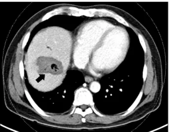
Fig. 2. A follow-up computed tomography shows liver ab- scess with inserted pigtail catheter in the previous radio- frequency ablated site (black circle) and consolidation with air formation in the adjacent diaphragm, which was diagnosed as a lung abscess (white circle).
recurrence of previous RFA site (segment 5 and 6).
However, the patient’s renal function was worsened by the chemotherapy and the patient refused further chemo- therapy because of deteriorated general condition. So, RFA of the remained metastatic lesion of segment 8 was performed. A follow-up CT scan performed immediately after RFA showed complete ablation of the metastatic cholangiocarcinoma without direct evidence of dia- phragmatic injury except for a small amount of reactive pleural effusion in right lung.
The patient developed a lower grade fever of less than 38.0oC during the 3 days after RFA. One week later, he revisited the hospital with intermittent fever and chill. A follow-up CT scan showed a liver abscess in the previous RFA site. Ultrasonography-guided percutaneous catheter drainage (PCD) of the liver abscess with a 10.2-Fr pigtail catheter was inserted, and antibiotics were administrated for three weeks (Fig. 1). The patient showed new symp- toms with complaints of productive cough with green-yel- low sputum accompanied with more than 38.9oC fever with chills. The coughing worsened to the point of chok- ing when the patient lay down and improved with sitting upright. In chest auscultation, breathing sounds decreased in the right chest with crackles. The laboratory test results revealed leucocyte 15.54×10³/µl (normal range: 4-10×103/ µl), alkaline phosphatase of 718 IU/L (normal range:
104-338 IU/L), gamma-glutamyl transpeptidase (GGT) of 216 IU/L (normal range: 8-61 IU/L), blood urea nitrogen
(BUN) of 54.4 mg/dl (normal range: 8-20 mg/dl), serum creatinine of 3.74 mg/dl (normal range: 0.5-1.3 mg/dl), and C-reactive protein of 214 mg/L (normal range: 0-5 mg/L). Serum albumin, bilirubin, prothrombin time, and activated partial thromboplastin time were within normal limits. Plain chest radiograph demonstrated the presence of ill-defined consolidation and infiltrative lesion with pleural effusion in the right lower lung field. In sputum culture, E. coli were isolated and an antibiotic was admin- istrated as appropriate to the susceptibility result.
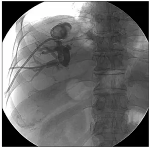
However, the coughing became worse and the amount of green-yellow sputum also increased. Follow-up abdomen and chest CT scans showed a persistent liver abscess (4 cm in size) at previous RFA site and abscess of the right lower lung (Fig. 2). Liver abscess in liver dome and lung abscess in right lower lung filed were attached to the diaphragm. Ultrasonography-guided PCD of the liver ab- scess with a 10.2-Fr pigtail catheter was reinserted for catheter change. Contrast material injection showed the presence of cavities of the lung and the bronchial tree communicating with the biliary tree, which was consistent with BBF (Fig. 3). E. coli were isolated from a culture of the drainage material. Following the catheter insertion, antibiotics therapy was continued. Although the amount of sputum was reduced after the insertion of the drainage catheter, bilioptysis and coughing were continued.
We performed percutaneous transhepatic biliary drain-
Fig. 3. Contrast material was injected through the percuta- neous abscess drainage catheter. This contrast material showed in the abscess cavity in liver (black arrow), bronchial tree (thin white arrow), and biliary tree (wide white arrow), confirming the presence of bronchobiliary fistula.
Fig. 4. Although the amount of sputum decreased after the insertion of the drainage catheter, bilioptysis and coughing continued. We performed percutaneous transhepatic biliary drainage (PTBD) in the dilated right hepatic duct to reduce the intrahepatic biliary tree pressure which was increased due to the hepaticojejunostomy site stricture.
Fig. 5. One month later, the follow-up coronary computed to- mography image shows improvement of liver abscess and no consolidation of lower lung field.
Fig. 6. After the percurtaneous drainage catheter was re- moved, contrast material was injected through PTBD catheter.
Cholangiography shows no communication between the bron- chial system and biliary tree.
age (PTBD) in the dilated right hepatic duct to reduce in- trahepatic pressure of biliary tree which was increased be- cause of stricture of the hepatico-jejunostomy site (Fig. 4).
After PTBD, coughing and bilioptysis were markedly improved. Follow-up abdominal CT scans obtained four weeks after the catheter insertion showed a markedly re- duced abscess cavity of the liver dome and improvement of the lung abscess in the right lower lobe. Thereafter, the
amount of percutaneous drainage decreased gradually.
When the amount of drainage reached less than 5 ml per day, the abscess cavity of the liver dome and right-side lung did not show in the follow up CT scan (Fig. 5). After PCD catheter at the liver dome was removed, repeated cholangiography showed no communication between the intrahepatic biliary tree and bronchial tree (Fig. 6).
DISCUSSION
Bronchobiliary fistula is defined as an abnormal inter- connection between the biliary tree in the liver and the bronchial tree. In the past, local infection such as hydatid or amebic abscess in the liver has been considered as a classic cause of BBF. According to recently published re- ports, the main cause of BBT is now recognized to be primary or metastatic tumor.2 The pathogenesis of BBF-associated tumor may involve iatrogenic damage, di- aphragm invasion, and intrahepatic or extrahepatic biliary obstruction. Among these, bile duct obstruction is known as the most common cause, most often due to lithiasis, tumor, hydatid cyst, or postoperative stricture.1,3 Our pa- tient showed mild dilatation of the bile duct to the post- operative stricture on the CT scan and experienced inter- mittent cholangitis. Before occurrence of BBF, he had a liver abscess in previous RFA site for metastatic cholan- giocarcinoma. In this circumstance, we had to consider for the possibility of occurrence of liver abscess or BBF when RFA had been performed at this site.
Pathogenesis of RFA-related BBF resulted from rupture of a growing biloma through a diaphragmatic defect caused by thermal injury. For this reason, the size and lo- cation of the tumor is an important risk factor for the de- velopment of BBF. In our patient, the tumor was located in the hepatic dome adjacent to the right diaphragm and was 5.5 cm on in size at the time of recurrence. The tu- mor was reduced to 3 cm after the combined chemo- therapy of gemcitabine and cisplatin for 3 months.
The most pathognomonic finding of BBF is bilioptysis defined productive cough of bile stained sputum.
Bilioptysis accounted for 67% of cases.2 Sometimes, it is diagnosed as acute pneumonitis with cough-producing greenish sputum.4 Other symptoms and signs include fe- ver, jaundice, abdominal pain, and chest pain. Early diag- nosis of BBF is very important because morbidity and mortality could be increasing by chemical pneumonitis, pneumonia or necrotizing bronchitis.5 Diagnosis can be confirmed by imaging procedures such as hepatobiliary scan, percutaneous transhepatic cholangiogram (PTC), en- doscopic retrograde cholangiograpy (ERCP), and mag- netic resonance cholangio-pancreatography (MRCP). Our patient was diagnosed by fistulogram through percuta- neous abscess drainage.
BBF requires a well-planned management strategy de- pending on the cause of each BBF. However, until now, there has been a consensus on the optimal treatment of BBF. Transitional therapeutic modality of BBF had been established as follows: surgical relief of biliary ob- struction, subphrenic drainage, excision of the fistulous tract and appropriate supportive measures. However, as more studies have been conducted regarding BBF, the therapeutic method of BBF has shifted from surgical man- agement to non-surgical approach including endoscopic management and percutaneous approach. Recently, in sev- eral cases, endoscopic interventions such as endoscopic nasogastric bile drainage (ENBD) with sphincterotomy of Oddi’ sphincter and PTBD were effective in resolving dis- tal biliary obstruction. Therefore, in these patients who have a restricted life span with malignant disease, con- servative treatment with non-surgical interventions is more accepted than the surgical approach.6,7 In the past years, direct embolization of fistula tract under broncho- scopic guidance was introduced as a new therapeutic method.8,9 However, these methods should be proven by more clinical cases.
If non-surgical managements of BBF fail, surgical cor- rection should be considered as the definitive therapeutic modality. Surgical procedures vary depending on the cause of BBF. The following procedures of literature were performed in BBF: drainage of the right subphrenic or hepatic abscess, closure of fistula, resection of hydatid cyst or tumor, biliary drainage using T-tube, and bilioen- teric anastomosis (e.g., Roux-en-Y hepaticojejunostomy).
In conclusion, although RFA is a safe and effective mo- dality for the treatment of primary or metastatic hepatic tumor, the possibility of BBF should be considered if the patient complains of bilioptysis after RFA. Because the optimal management of BBF is controversial, individuali- zed and multidisciplinary approach of BBF treatment should be applied depending on the cause of BBF.
ACKNOWLEDGEMENTS
This paper was supported by Wonkwang University in 2011.