J Korean Surg Soc 2012;83:36-42
http://dx.doi.org/10.4174/jkss.2012.83.1.36
ORIGINAL ARTICLE
Journal of the Korean Surgical Society
JKSS
pISSN 2233-7903ㆍeISSN 2093-0488
Received January 27, 2012, Revised April 30, 2012, Accepted May 13, 2012 Correspondence to: Ho-Seong Han
Department of Surgery, Seoul National University Bundang Hospital, Seoul National University College of Medicine, 82 Gumi-ro 173beon-gil, Bundang-gu, Seongnam 463-707, Korea
Tel: +82-31-787-7091, Fax: +82-31-787-4055, E-mail: [email protected]
cc Journal of the Korean Surgical Society is an Open Access Journal. All articles are distributed under the terms of the Creative Commons Attribution Non-Commercial License (http://creativecommons.org/licenses/by-nc/3.0/) which permits unrestricted non-commercial use, distribution, and reproduction in any medium, provided the original work is properly cited.
Safety and efficacy of laparoscopic radiofrequency ablation for hepatic malignancies
Seung Duk Lee, Ho-Seong Han, Jai Young Cho, Yoo-Seok Yoon, Dae Wook Hwang, Kyuwhan Jung, Chang Jin Yoon
1, Yujin Kwon, Ji Hoon Kim
Departments of Surgery and 1Radiology, Seoul National University Bundang Hospital, Seoul National University College of Medicine, Seoul, Korea
Purpose: Radiofrequency ablation (RFA) is an accepted treatment option for primary and metastatic liver tumors. As percu- taneous RFA has some limitations, laparoscopic RFA (LRFA) has been used as a therapeutic alternative for the treatment of hepatic malignancies. Methods: Between March 2006 and September 2009, thirty patients with hepatic malignancies that were contraindicated for resection or percutaneous RFA underwent LRFA. Indications for this procedure were hep- atocellular carcinoma (HCC, 21 patients), metastatic liver tumor (8 patients) and intrahepatic cholangiocarcinoma (1 patient).
Results: Among the 30 patients who underwent LRFA, 5 patients underwent concomitant laparoscopic liver resection.
Intraoperative laparoscopic ultrasound detected new malignant lesions in 4 patients (13.3%). A total of 46 lesions were ab- lated by LRFA. There was no postoperative mortality. The three-year overall survival rate was 83.7% for the HCC group and 64.3% for the metastatic group. Conclusion: LRFA for hepatic malignancies proved to be a safe and effective treatment. Also, this procedure is indicated for lesions that are not amenable to percutaneous RFA or liver resection.
Key Words: Hepatocellular carcinoma, Liver neoplasms, Laparoscopy, Radiofrequency ablation
INTRODUCTION
Hepatic resection is the most effective treatment for pa- tients with primary or metastatic hepatic malignancies [1].
Liver resection is usually limited by the poor functional re- serve of the liver in cirrhotic patients, multifocal bilateral lesions and tumor proximity to major vascular or biliary structures [2]. Recently, percutaneous radiofrequency ablation (RFA) has gained widespread acceptance as treat- ment for primary and metastatic liver cancers, especially
when liver resection is not well-indicated [3-5]. However, this procedure has limitations when the lesion is located close to a visceral organ (e.g., the gall bladder, stomach, and colon) or is located on the liver surface or the dia- phragm [2]. For these patients, laparoscopic RFA (LRFA) can offer a better and effective treatment as it provides for a safer approach to liver lesions difficult or impossible to treat with a percutaneous approach [6-10].
The aim of this study was to evaluate the safety and effi- cacy of LRFA for primary and metastatic hepatic
malignances. Perioperative morbidity, mortality, overall cumulative survival and recurrence rates were retro- spectively evaluated.
METHODS
Patients and methods
Between March 2006 and September 2009, 30 consec- utive patients with hepatic malignancies underwent LRFA.
LRFA was performed in patients not amenable to liver re- section or percutaneous RFA. Indications for LRFA were as follows: 1) difficult location of the tumor for performing a percutaneous approach such as close proximity to the di- aphragm, gallbladder, stomach, or colon (n = 17), 2) multi- ple tumors requiring simultaneous laparoscopic liver re- section and LRFA (n = 5), 3) multiple lesions requiring re- peated punctures (n = 4), 4) superficial lesions which are prone to rupture (n = 3), and 5) simultaneous laparoscopic treatment for both primary colorectal cancer and meta- static liver tumors (n = 1).
Surgical procedures
Procedures were performed with the patients under general anesthesia in the supine position. A Veress needle was inserted for insufflation of the abdominal cavity. After inserting an 11-mm trocar beneath the umbilicus, a pneu- moperitoneum was performed with insufflations of CO2
gas. Abdominal pressure was maintained under 13 mmHg.
First, complete inspection of the intraperitoneal organs was performed to rule out any extrahepatic disease. Then another 11-mm trocar was inserted in the epigastric area.
A 30 degree or flexible laparoscope was used and liga- ments around the liver were dissected using a Harmonic scalpel (Ethicon Endo-Surgery Inc., Cincinnati, OH, USA) to mobilize the liver and to identify the liver lesions. If nec- essary, adhesiolysis was performed between the liver and other adjacent organs.
All patients underwent LRFA guided by laparoscopic ultrasonography (Aloka Inc., Tokyo, Japan). Liver lesions were identified and the vascularity of the lesions was as- sessed using the transducer. The puncture site of the radio- frequency electrode was determined using the laparo-
scopic transducer through an 11-mm trocar placed be- neath the xiphoid process. After the lesions were identi- fied and measured, the radiofrequency electrode was placed percutaneously into the lesion and then prongs were deployed. A RF 3000 Radiofrequency Ablation System (Boston Scientific Co., Natick, MA, USA) was used as the energy source. A Cool-tip RF Ablation System (Valleylab, Boulder, CO, USA) was also used with an in- ternally cooled electrode, Cool-tip RF Electrode. Laparo- scopic ultrasound (LUS) examination and RFA proce- dures were performed by radiologists. Ablation was re- peated until the whole tumor showed highly echogenic changes.
Assessment of clinical outcome
The primary effectiveness of the treatment was eval- uated with a follow-up contrast-enhanced study one month after LRFA. Incomplete ablation was defined as the appearance of irregular peripheral-enhancing foci around the ablation zone in the follow-up images, while complete necrosis of the index tumor was defined by the absence of an enhanced tumor area. Local tumor progression was de- termined to exist when a subsequent follow-up computed tomography (CT) demonstrated the presence of a growing and enhanced tumor in the ablation zone where there had been complete primary effectiveness. New lesion tumor progression was defined as a growing and enhanced tu- mor occurring away from the ablation zone.
Statistical analysis
All analyses were performed using the statistical soft- ware SPSS ver. 17.0 (SPSS Inc., Chicago, IL, USA). Overall survival curves and cumulative recurrence curves were analyzed by the Kaplan-Meier method. A P-value of < 0.05 was considered statistically significant.
RESULTS
Patient and tumor characteristics
Of the 30 patients, 21 patients were male and 9 patients were female. The mean age of the patients was 58.8 years (range, 35 to 76 years). Forty-six LRFA procedures were
Table 1. Demographics of 30 patients with intrahepatic tumors
Variable Value
Sex (male/female) 21/9
Age (yr), mean (range) 58.8 (35-76)
Pathology
HCC 21
Metastasis
Colorectal cancer 5
Breast cancer 1
Renal cancer 1
Klatskin tumor 1
Cholangiocarcinoma 1
HCC patients
Child-Pugh classificationa), A/B 18/3 Underlying liver
HBV/HCV/alcoholic/idiopathic 18/1/1/1
Preoperative TACE/RFA 17/4
Metastasis patients
Synchronous/metachronous metastasis 1/7
Preoperative chemotherapy 5
Combined operative procedure
Laparoscopic liver resection 5
Laparoscopic LAR 1
Laparoscopic cholecystectomy 8
HCC, hepatocellular carcinoma; HBV, hepatitis B virus; HCV, hepatitis C virus; TACE, transarterial chemoembolization; RFA, radiofrequency ablation; LAR, lower anterior resection.
a)Measured only for HCC patients.
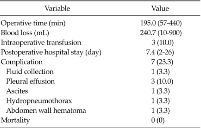
Table 2.Intraoperative and postoperative outcomes
Variable Value
Operative time (min) 195.0 (57-440)
Blood loss (mL) 240.7 (10-900)
Intraoperative transfusion 3 (10.0) Postoperative hospital stay (day) 7.4 (2-26)
Complication 7 (23.3)
Fluid collection 1 (3.3)
Pleural effusion 3 (10.0)
Ascites 1 (3.3)
Hydropneumothorax 1 (3.3)
Abdomen wall hematoma 1 (3.3)
Mortality 0 (0)
Values are presented as mean (range) or number (%).
performed to ablate primary or metastatic hepatic tumors in 30 patients. There were 21 patients with hepatocellular carcinoma (HCC), 8 patients with metastatic hepatic ma- lignancies and one patient with cholangiocarcinoma who was suspected of having HCC preoperatively. Of the 8 pa- tients with metastatic hepatic malignancies, the most com- mon primary malignancy was colorectal cancer (n = 5).
Other malignancies were breast cancer (n = 1), renal cell carcinoma (n = 1) and Klatskin tumor (n = 1). Of the 8 pa- tients with metastatic hepatic malignancies, metachro- nous metastasis was dominant (n = 7).
The Child-Pugh class of most patients with HCC was class A (85.7%). The most common cause of HCC was hep- atitis B (85.7%). All patients with HCC underwent trans- arterial chemoembolization or percutaneous RFA pre- operatively. Of the 30 patients, 14 patients (46.7%) under- went additional laparoscopic surgery simultaneously with LRFA procedures: hepatic resection in 5 (4 tumorectomies and 1 left lateral sectionectomy), lower anterior resection
in one and cholecystectomy in 8 patients (Table 1).
Nineteen patients underwent LRFA for only one lesion.
Another six and five patients had LRFA for 2 and 3 lesions, respectively. A median tumor size was 2.0 cm (range, 0.8 to 4.5 cm). New malignant lesions including 3 HCCs and 1 metastatic tumor were identified by LUS, which were not detected by preoperative ultrasound, CT or magnetic res- onance imaging (MRI). These newly detected nodules were ablated with LRFA.
Complications
The mean operation time was 195.0 minutes and the mean intraoperative blood loss was 240.7 mL. Transfusion was required in three patients (10.0%) during the opera- tion. The mean postoperative hospital stay was 7.4 days.
There was no operative mortality. Postoperative complica- tions were observed in 7 of 30 patients (23.3%) including right pleural effusion in 3 patients, and perihepatic fluid collection, mild ascites, hydropneumothorax and hema- toma at the puncture site in 1 patient respectively. All pa- tients with complications recovered well with con- servative management (Table 2).
Survival and recurrence
The median follow-up period was 18.2 months (range, 3.4 to 41.1 months). The overall success rate for LRFA was 95.7% and the three-year overall survival rates of patients with HCC and metastatic tumors were 83.7% and 64.0 %, respectively (Fig. 1). One patient with cholangiocarcinoma
Fig. 2. Cumulative recurrence rates after laparoscopic radiofrequency ablation for primary hepatocellular carcinoma (A) and liver metastases (B).
Fig. 1. Overall survival rates after laparoscopic radiofrequency ablation for primary hepatocellular carcinoma (A) and liver metastases (B).
3YRS, 3 year overall survival rates.
Table 3. Recurrence patterns for laparoscopic radiofrequency ablation in patients with hepatocellular carcinoma (HCC) and metastases
HCC patients (n = 21)
Metastasis patients (n = 8)
Intrahepatic recurrence 13 4
Incomplete ablation 2 1
Local 2 1
New lesion 12 3
Extrahepatic recurrence 4 2
In HCC patients, 4 patients had both intrahepatic and extrahepatic recurrences, 2 patients had both local and new lesion formation and 1 patient had both incomplete ablation and new lesion formation. In the metastasis patients, 1 patient had both incomplete ablation and local recurrence and 1 patient had a new lesion and extrahepatic recurrence.
died of multiple intraperitoneal metastases 10 months af- ter LRFA.
During follow-up in 21 patients with HCC, 13 patients (61.9%) experienced a recurrence. The overall recurrence rate at 3, 6, and 12 months after LRFA was 14.3%, 19.6%, and 47.4%, respectively (Fig. 2). The median disease-free survival time was 11.6 months (range, 1.0 to 36.3 months).
There were 2 cases of incomplete ablation, 2 cases of local tumor progression and 12 cases of new intrahepatic lesions. Two patients had both local tumor progression and development of a new lesion. One patient had both in- complete ablation and a new lesion (Table 3).
In the follow-up of 8 patients with metastatic hepatic malignancies, 5 patients (62.5%) had recurred lesions. The cumulative overall recurrence rate at 3, 6, and 12 months
after the LRFA was 12.5%, 37.5%, and 53.1%, respectively (Fig. 2). The median disease-free survival time was 9.4 months (range, 1.6 to 34.5 months). Of these 5 patients, 3 patients had tumor recurrences within the liver. One case had both incomplete ablation and local tumor pro- gression, and two cases had new tumor progression. One patient had extrahepatic tumor recurrence in the lung and the another patient had both intrahepatic tumor re- currence and extrahepatic lymph node metastasis.
DISCUSSION
RFA of primary and metastatic hepatic malignancies by laparoscopy can be a useful substitute for percutaneous RFA when the lesions are superficial or close to visceral or- gans [11]. In a study of LRFA for 66 patients with HCC by Berber et al. [12], the overall survival rates at 1, 2 and 3 years were 78%, 48% and 38%, respectively. Curley et al.
[13] reported a 1.8% of local tumor progression rate and a 21.9% of new tumor recurrence rate in 48 patients with HCC and 75 patients with metastatic hepatic malignancies during follow-up (mean, 15.0 months). In contrast, the mean overall survival rate for the patients with unresec- table metastatic malignancies was only 9 to 14 months [14,15]. In our study, 3-year survival rates for HCC and metastatic hepatic malignancies were 83.7% and 64.3%, re- spectively while one-year cumulative recurrence rates for HCC and metastatic hepatic malignancies were 47.4% and 53.1%, respectively. There was no operative mortality. Our results showed a better survival and a similar recurrence rate in comparison with the aforementioned other studies.
The LRFA procedure has been reported to be safe for treating hepatic malignancies [16-18]. In a study by Berber and Siperstein [19], a total of 521 RFA procedures were performed for 428 patients (286 men and 142 women);
these patients had a mean age of 61 years (range, 25 to 89 years). A total of 1,636 lesions (mean, 3.1 per patients with a range, 1 to 16) were ablated. The 30-day mortality rate was 0.4% (n = 2) and the morbidity rate was 3.8% (n = 20).
The post RFA complications were self-limited nausea, ileus, abdominal distention, abdominal pain, fevers and urinary retention in 6 patients and liver abscess in 4
patients. There were also trocar injuries, postoperative bleeding, pneumonia, wound infection and variceal bleed- ing. In our study, there was no postoperative mortality.
Seven patients had postoperative morbidity (pleural effu- sion, ascites, hydropneumothorax, fluid collection and hematoma), which were treated with conservative mana- gement. During the follow-up, other complications in- cluding liver abscess did not occur.
Jung et al. [9] introduced LRFA as a useful procedure for the treatment of HCC regardless of its location. According to their reports, serum transaminase levels were tran- siently elevated (P < 0.01) after RFA but returned to nor- mal within one week, and alpha-fetoprotein level de- creased significantly 1 month after RFA (P < 0.05). And the results showed that the LRFA procedure is a good treatment for severe cirrhosis patients with reducing dete- rioration of liver function. Our study showed that LRFA was good approach method for superficial lesions in the liver or lesions close to visceral organs with the result of 16.7% of local tumor progression rate. LUS is a useful and essential diagnostic tool in LRFA. Santambrogio et al. [20]
reported that during the LRFA for HCC, intraoperative LUS identified 26 new malignant lesions (25%) which had been missed by preoperative imaging. We detected new nodules in 4 patients (13.3%, 2 HCCs, and 2 metastatic tu- mors) using LUS and performed additional ablation for them. In particular, lesions which were small-sized and hard to detect with CT or MRI were able to be detected with LUS. By using LUS, both accurate staging and effec- tive treatment were able to be achieved.
One of the important roles of LRFA is simultaneous use of this procedure with laparoscopic liver resection. When a patient had multiple lesions, the main lesions were re- sected with laparoscopic liver resection and the remaining small lesions were treated with LRFA. Therefore, applica- tion of laparoscopic liver resection has been increasing and the role of LRFA is increased in the treatment of hep- atic malignancies [21-24]. In our study, 5 patients under- went laparoscopic liver resection and LRFA simultaneo- usly. With a mean follow-up time of 24.2 ± 14.3 months, they are all alive without specific complications.
Recently, RFA has been reported as a good bridge ther- apy prior to liver transplantation: the ablation success rate
with this procedure is 91.4%. In particular, the laparo- scopic approach is associated with a lower adhesion than open surgery and dissection around the liver hilum is not required in this approach [25]. Thus, LRFA would be a good bridge therapy for prevention of tumor progression and downstaging of multiple lesions.
In conclusion, in cases not amenable to surgical re- section, LRFA represents a safe and effective treatment for malignant liver tumors especially when a percutaneous approach to the lesions is deemed difficult.
CONFLICTS OF INTEREST
No potential conflict of interest relevant to this article was reported.
REFERENCES
1. Clark HP, Carson WF, Kavanagh PV, Ho CP, Shen P, Zagoria RJ. Staging and current treatment of hepatocel- lular carcinoma. Radiographics 2005;25 Suppl 1:S3-23.
2. Lau WY, Leung TW, Yu SC, Ho SK. Percutaneous local ablative therapy for hepatocellular carcinoma: a review and look into the future. Ann Surg 2003;237:171-9.
3. Wang XH, Cheng F, Zhang F, Li XC, Qian JM, Kong LB, et al. Living donor liver transplantation treatment of Wilson's disease complicated with neuropathy. Zhonghua Yi Xue Za Zhi 2003;83:1569-71.
4. Siperstein A, Garland A, Engle K, Rogers S, Berber E, String A, et al. Laparoscopic radiofrequency ablation of primary and metastatic liver tumors. Technical considera- tions. Surg Endosc 2000;14:400-5.
5. Lim HK, Choi D, Lee WJ, Kim SH, Lee SJ, Jang HJ, et al.
Hepatocellular carcinoma treated with percutaneous ra- dio-frequency ablation: evaluation with follow-up multi- phase helical CT. Radiology 2001;221:447-54.
6. Chung MH, Wood TF, Tsioulias GJ, Rose DM, Bilchik AJ.
Laparoscopic radiofrequency ablation of unresectable hep- atic malignancies: a phase 2 trial. Surg Endosc 2001;15:
1020-6.
7. Cuschieri A, Bracken J, Boni L. Initial experience with lapa- roscopic ultrasound-guided radiofrequency thermal abla- tion of hepatic tumours. Endoscopy 1999;31:318-21.
8. Goletti O, Lencioni R, Armillotta N, Puglisi A, Lippolis PV, Lorenzetti L, et al. Laparoscopic radiofrequency thermal ablation of hepatocarcinoma: preliminary experience. Surg Laparosc Endosc Percutan Tech 2000;10:284-90.
9. Jung MK, Lee JH, Kim TS, Kim HS, Cho CM, Tak WY, et al.
Laparoscopic and percutaneous ultrasound guided radio- frequency ablation for hepatocellular carcinoma: a pre- liminary study. Korean J Hepatol 2002;8:209-17.
10. Santambrogio R, Bianchi P, Pasta A, Palmisano A, Montorsi M. Ultrasound-guided interventional procedures of the liver during laparoscopy: technical considerations. Surg Endosc 2002;16:349-54.
11. Mulier S, Ni Y, Jamart J, Ruers T, Marchal G, Michel L.
Local recurrence after hepatic radiofrequency coagulation:
multivariate meta-analysis and review of contributing factors. Ann Surg 2005;242:158-71.
12. Berber E, Rogers S, Siperstein A. Predictors of survival af- ter laparoscopic radiofrequency thermal ablation of hep- atocellular cancer: a prospective study. Surg Endosc 2005;
19:710-4.
13. Curley SA, Izzo F, Delrio P, Ellis LM, Granchi J, Vallone P, et al. Radiofrequency ablation of unresectable primary and metastatic hepatic malignancies: results in 123 patients.
Ann Surg 1999;230:1-8.
14. Rosales J, Leong LA. Chemotherapy for metastatic color- ectal cancer. J Natl Compr Canc Netw 2005;3:525-9.
15. McMurrick PJ, Nelson H. Liver-directed therapies for gas- trointestinal malignancies. Curr Opin Oncol 1997;9:367-72.
16. Tateishi R, Shiina S, Teratani T, Obi S, Sato S, Koike Y, et al.
Percutaneous radiofrequency ablation for hepatocellular carcinoma: an analysis of 1000 cases. Cancer 2005;103:
1201-9.
17. Jiang HC, Liu LX, Piao DX, Xu J, Zheng M, Zhu AL, et al.
Clinical short-term results of radiofrequency ablation in liver cancers. World J Gastroenterol 2002;8:624-30.
18. Kim KH, Yoon YS, Yu CS, Kim TW, Kim HJ, Kim PN, et al.
Comparative analysis of radiofrequency ablation and sur- gical resection for colorectal liver metastases. J Korean Surg Soc 2011;81:25-34.
19. Berber E, Siperstein AE. Perioperative outcome after lapa- roscopic radiofrequency ablation of liver tumors: an analy- sis of 521 cases. Surg Endosc 2007;21:613-8.
20. Santambrogio R, Podda M, Zuin M, Bertolini E, Bruno S, Cornalba GP, et al. Safety and efficacy of laparoscopic ra- diofrequency ablation of hepatocellular carcinoma in pa- tients with liver cirrhosis. Surg Endosc 2003;17:1826-32.
21. Bleicher RJ, Allegra DP, Nora DT, Wood TF, Foshag LJ, Bilchik AJ. Radiofrequency ablation in 447 complex un- resectable liver tumors: lessons learned. Ann Surg Oncol 2003;10:52-8.
22. Vlastos G, Smith DL, Singletary SE, Mirza NQ, Tuttle TM, Popat RJ, et al. Long-term survival after an aggressive sur- gical approach in patients with breast cancer hepatic metastases. Ann Surg Oncol 2004;11:869-74.
23. Abdalla EK, Vauthey JN, Ellis LM, Ellis V, Pollock R, Broglio KR, et al. Recurrence and outcomes following hep- atic resection, radiofrequency ablation, and combined re- section/ablation for colorectal liver metastases. Ann Surg 2004;239:818-25.
24. Pawlik TM, Izzo F, Cohen DS, Morris JS, Curley SA.
Combined resection and radiofrequency ablation for ad- vanced hepatic malignancies: results in 172 patients. Ann
Surg Oncol 2003;10:1059-69.
25. Martin AP, Goldstein RM, Dempster J, Netto GJ, Katabi N, Derrick HC, et al. Radiofrequency thermal ablation of hep-
atocellular carcinoma before liver transplantation: a clin- ical and histological examination. Clin Transplant 2006;20:
695-705.