Biomedical Science Letters 2014, 20(3): 129~138 http://dx.doi.org/10.15616/BSL.2014.20.3.129 eISSN : 2288-7415
Inhibitory Effects of Water Extract from Rice Bran Due to cAMP-dependent Phosphorylation of VASP (Ser157) on
ADP-induced Platelet Aggregation
Hyun-Hong Kim1, Jeong Hwa Hong2, Pajaree Ingkasupart2, Dong-Ha Lee1,3 and Hwa-Jin Park1,†
1Department of Biomedical Laboratory Science, Inje University, Gyungnam 621-749, Korea
2Department of Smart Foods and Drugs, Inje University, Gyungnam 621-749, Korea
3Department of Biomedical Laboratory Science, Korea Nazarene University, Chungnam 331-718, Korea
In this study, we investigated the effect of water extract from rice bran (RB) on ADP (20 μM)-stimulated platelet aggregation. RB dose-dependently inhibited ADP-induced platelet aggregation, and its IC50 value was 224.0 μg/mL, which was increased by adenylate cyclase inhibitor SQ22536 and cAMP-dependent protein kinase (A-kinase) inhibitor Rp-8-Br-cAMPS. RB elevated the phosphorylation of VASP (Ser157) which was also inhibited by SQ22536 and Rp-8-Br-cAMPS. It is thought that RB-elevated cAMP contributed to the phosphorylation of VASP (Ser157) to inhibit ADP-induced platelet aggregation. Therefore, we demonstrate that RB has an antiplatelet effect via cAMP-dependent phosphorylation of VASP (Ser157), and RB may have preventive or therapeutic potential for platelet aggregation-mediated diseases, such as thrombosis, myocardial infarction, atherosclerosis, and ischemic cerebrovascular disease.
Key Words: Rice bran, Platelet aggregation, cAMP-dependent protein kinase, cAMP, Vasodilator-stimulated phosphoprotein- Ser157 phosphorylation
INTRODUCTION
Platelet aggregation is absolutely essential for the for- mation of a hemostatic plug when normal blood vessels are injured. However, it can also cause cardiovascular diseases such as thrombosis, atherosclerosis and myocardial infarction (Schwartz et al., 1990).
When platelets are stimulated by agonists such as adenosine diphosphate (ADP), thrombin and TXA2, phos- phatidylinositol 4, 5-bisphosphate is hydrolyzed by phos-
pholipase C-β via G-protein coupled receptor to inositol 1, 4, 5-trisphosphate and diacylglycerol (Guidetti et al., 2008;
Berridge et al., 1984; Jennings et al., 2009). In special, it is known that ADP binds to the Gq-coupled P2Y1 receptor, which mediates PLC-β, and the Gi-coupled P2Y12 receptor, which mediates inhibition of adenylate cyclase and amplifies platelet aggregation (Cattaneo, 2005).
Intracellular cyclic adenosine monophosphate (cAMP) as an antiplatelet regulator decrease the [Ca2+]i mobilization (Menshikov et al., 1993; Schwarz et al., 2001). The anti- platelet effects of cAMP are mediated via cAMP-dependent protein kinases (A-kinase), which phosphorylates substrate protein, vasodilator stimulated phosphoprotein (VASP) (Halbrügge et al., 1989; Halbrügge et al., 1990; Butt et al., 1994). VASP (Ser157 and Ser239) phosphorylation involves in inhibition of VASP affinity for contractile protein fila- mentous actin, and fibrinogen binding to glycoprotein IIb/
IIIa (αIIb/β3) to inhibit platelet aggregation (Laurent et al., Original Article
*Received: June 2, 2014 / Revised: September 16, 2014 Accepted: September 27, 2014
Hyun-Hong Kim and Jeong Hwa Hong contributed equally to this work.
†Corresponding author: Hwa-Jin Park. Department of Biomedical Laboratory Science, College of Biomedical Science and Engineering, Inje University, 197, Inje-ro, Gimhae, Gyungnam 621-749, Korea.
Tel: +82-55-320-3538, Fax: +82-55-334-3426 e-mail: [email protected]
○CThe Korean Society for Biomedical Laboratory Sciences. All rights reserved.
1999; Sudo et al., 2003). Therefore, elevating cAMP and phosphorylating VASP are very useful for evaluating the antiplatelet effect of substances or compounds. For instance, a major catechin analogue, (-)-epigallocatechin-3-gallate (EGCG) from green tea, is known to produce cAMP via adenylate cyclase activation and subsequently phosphorylates VASP-Ser157 through A-kinase activation to inhibit platelet aggregation (Ok et al., 2012). Furthermore, verapamil and theophylline have antiplatelet function that by elevating the cAMP level (Gasser et al., 1991), and are currently used to prevent and/or treat cardiovascular diseases as antiplatelet agents (Menshikov et al., 1993).
Rice bran is produced as a by-product in the rice milling process, a method in which the outer layer of the rice grain is removed. Rice bran has various biological effects like anti-inflammatory, cholesterol-lowering, antioxidant and anti-diabetic activities (Qureshi et al., 2002; Jun et al., 2012; Hou et al., 2010). In recent, it is reported that water extract (RB) from rice bran has a neuroprotective effect on ischemic brain injury (Baek et al., 2014).
In the present study, we investigated the effect of RB on VASP phosphorylation in ADP-induced platelet aggregation, and evaluated the anti-platelet effect.
MATERIALS AND METHODS Materials
ADP was purchased from Chrono-Log Co. (Havertown, PA., USA). Sodium ferulate was purchased from AK Scientific Inc. (Union City, CA., USA). cAMP enzyme immunoassay (EIA) kit was purchased from Cayman Chemical Co. (Ann Arbor, MI., USA). SQ22536, Rp-8-Br- cAMPS and other reagents were obtained from Sigma Chemical Co. (St. Louis, MO., USA). Anti-phosphor-VASP (Ser157), anti-rabbit IgG-horseradish peroxidase conjugate (HRP), and lysis buffer were obtained from Cell Signaling (Beverly, MA., USA). Polyvinylidene difluoride (PVDF) membrane was from GE Healthcare (Piseataway, NJ., USA).
Enhanced chemiluminesence solution (ECL) was from GE Healthcare (Chalfont St, Giles, Buckinghamshire, UK).
Preparation of rice bran water-extract (RB)
Rice bran was obtained from Gimhae Rice Processing Complex just after milling rice cultivar of Samkwang (Gimhae, Korea). To inactivate enzymes, rice bran was autoclaved at 121℃ for 30 min. After cooling to room temperature, rice bran was vacuum-packed and stored in the freezer until use.
RB was prepared according to the following methods;
100 g of rice bran was mixed with 900 mL distilled water followed by hot water extraction at 121.5℃ for 15 min.
After cooling to room temperature, the mixture was centri- fuged at 8,000 ×g for 10 min. The supernatant was filtrated and concentrated to 54 brix by vacuum evaporation.
The yield of water extract from RB was 2.6%. The resultant concentrate was designated as RB and kept in a refrigerator (4℃) until use. RB was dissolved in saline (0.9% NaCl) to investigate the effects on platelet aggregation.
Determination of total phenolic content of RB
Total phenolic content was determined by modified Singleton's method (Singleton et al., 1965). RB was dissolved 50% MeOH at the concentration of 0.1% (w/v). 0.2 mL of the dissolved sample was reacted with 1.0 mL of 10%
Folin-Ciocalteu reagent for 4 min at room temperature, and then 0.8 mL saturated sodium carbonate solution (about 75 g/L) was added into the reaction mixture. After incubation at room temperature for 30 min, the mixture was centrifuged 3,000 rpm, and the supernatant was taken. The absorbance readings of the supernatant were taken at 765 nm. Gallic acid was used as a reference standard, and the results were expressed as milligram gallic acid equivalent (mg gallic acid)/100 g RB.
Detection of phenolic compounds of RB with HPLC Because it is reported that rice bran contains phenolic compounds, we detected phenol compounds in RB with high performance liquid chromatography (HPLC) (Goufo and Trindade, 2014). RB was dissolved in distilled water (100 mg/mL), for the first time, and the pH of dissolved sample was set to 2~3 with 2N HCl, the sample was extracted three times with 0.5 mL ethylacetate, and was
concentrated by vacuum rotary and vacuum-dried, and then was dissolved with 0.5 mL methanol, and then it was analyzed by HPLC. An Agilent 1100 liquid chromatography system (Palo Alto, CA., USA), equipped with vacuum degasser, quaternary gradient pump, autosampler and diode array detector (DAD), connected to an Agilent ChemStation software. A TSKgel ODS-100V column (150 mm×4.6 mm id, 5 μm, Tosoh, Japan) was used at a column temperature of 40℃. The mobile phase consisted of methanol (A) and 50 mM NaH2PO4 (B), pH 2.5 with phosphoric acid using the following program: 0~20 min, 30% A and 70% B. The flow rate was at 1.0 mL/min and sample injection volume was 5 μL. The UV detection was operated at 310 nm.
Seven concentrations (12.5, 25, 50, 62.5, 125, 250, 500 μg/
mL) of authentic ferulic acid were injected in duplicate, then the calibration curve was constructed by plotting the peak area against the concentration of each analyte.
Preparation of washed human platelets
Human platelet-rich plasma (PRP) anti-coagulated with acid-citrate-dextrose solution (0.8% citric acid, 2.2% sodium citrate, 2.45% glucose) were obtained from Korean Red Cross Blood Center (Changwon, Korea). PRP was centri- fuged for 10 min at 125 ×g to remove a little red blood cells, and was centrifuged for 10 min at 1,300 ×g to obtain the platelet pellets. The platelets were washed twice with washing buffer (138 mM NaCl, 2.7 mM KCl, 12 mM NaHCO3, 0.36 mM NaH2PO4, 5.5 mM glucose, and 1 mM EDTA, pH 6.5). The washed platelets were then resuspended in suspension buffer (138 mM NaCl, 2.7 mM KCl, 12 mM NaHCO3, 0.36 mM NaH2PO4, 0.49 mM MgCl2, 5.5 mM glucose, 0.25% gelatin, pH 6.9) to a final concentration of 5×10⁸/mL. All of the above procedures were carried out at 25℃ to avoid platelet aggregation from any effect of low temperatures. The Korea National Institute for Bioethics Policy Public Institutional Review Board (Seoul, Korea) approved these experiments.
Measurement of platelet aggregation
Washed platelets (108/mL) were preincubated for 3 min at 37℃ in the presence of 2 mM CaCl2 with or without substances, then stimulated with ADP (20 μM) for 5 min.
Aggregation was monitored using an aggregometer (Chrono- Log Corporation, Havertown, PA., USA) at a constant stirring speed of 1,000 rpm. Each aggregation rate was calculated as an increase in light transmission. The suspen- sion buffer was used as the reference (transmission 0%).
RB was dissolved in saline (0.9% NaCl).
Measurement of cAMP
Washed platelets (108/mL) were preincubated for 3 min at 37℃ with or without substances in the presence of 2 mM CaCl2, and then stimulated with ADP (20 μM) for 5 min for platelet aggregation. The aggregation was terminated by the addition of 80% ice-cold ethanol. cAMP was measured with synergy HT multi-model microplate reader (BioTek Instruments, Winooski, VT., USA) using cAMP EIA kit.
Western blot for analysis of VASP-phosphorylation Washed platelets (108/mL) were preincubated with or without substances in the presence of 2 mM CaCl2 for 3 min and then stimulated with ADP (20 μM) for 5 min at 37℃. The reactions were terminated by adding an equal volume (250 μL) of lysis buffer (20 mM Tris-HCl, 150 mM NaCl, 1 mM Na2EDTA, 1 mM EGTA, 1% Triton X-100, 2.5 mM sodium pyrophosphate, 1 mM serine/threonine phosphatase inhibitor β-glycerophosphate, 1 mM ATPase, alkaline and acid phosphatase, and protein phosphotyrosine phosphatase inhibitor Na3VO4, 1 μg/mL serine and cysteine protease inhibitor leupeptin, and 1 mM serine protease and acetylcholinesterase inhibitor phenylmethanesulfonyl fluoride, pH 7.5). Platelet lysates containing the same protein (15 μg) were used for analysis. Protein concentrations were measured by using bicinchoninic acid (BCA) protein assay kit (Pierce Biotechnology, USA). The effects of substances on VASP-phosphorylation were analyzed by western blotting.
A 8~10% SDS-PAGE was used for electrophoresis and a PVDF membrane was used for protein transfer from the gel. The dilutions for anti-phosphor-VASP (Ser157), and anti-rabbit IgG-HRP were 1:1,000 and 1:10,000, respectively.
The membranes were visualized using ECL. Blots were analyzed by using the Quantity One, Ver. 4.5 (Bio-Rad, Hercules, CA., USA).
Statistical analyses
The experimental results are expressed as the mean ± S.E.M. accompanied by the number of observations. Data were assessed by analysis of variance (ANOVA). If this analysis indicated significant differences among the group means, then each group was compared by the Newman- Keuls method. P<0.05 was considered to be statistically significant.
RESULTS AND DISCUSSION The contents of total phenolics and ferulic acid in RB
When total phenolics was determined by using gallic acid as a standard, the contents of total phenolics in RB was contained, as shown in Table 1, 24.0 ± 1.0 mg in 100 g RB. Because it is reported that RB supplemented with ferulic acid has a synergistic neuroprotective effect in rat (Baek et al., 2014), we analyzed ferulic acid content in RB with HPLC. As shown in Fig. 1A, authentic ferulic acid was observed at 13.89 min, retention time, in HPLC chro- matogram. As shown in Fig. 1B, the retention time (13.69 min) of peak F was almost in accord with that of ferulic acid. Accordingly, it is thought that peak F is a compound Table 1. Total phenolic acid content of RB
Total phenolic (TP) content (mg / 100g RB)
Ferulic acid (FA) content (mg / 100g RB)
FA/TP (%)
RB 24.0 ± 1.0 21.1 87.9
Fig. 1. HPLC chromatograms of RB and ferulic acid. (A) The chromatogram of ferulic acid. (B) The chromatogram of RB. HPLC was performed as described in "Materials and Methods."
A
B
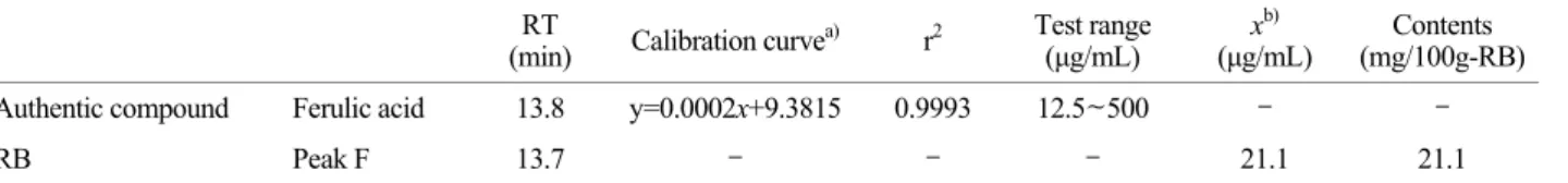
corresponding to ferulic acid. The content of ferulic acid calculated from calibration curve, the content of peak F corresponding to ferulic acid was 21.1 mg/100 g-RB (Table 2), which was corresponded to 87.9% of total phenolic contents (Table 1).
Effect of RB on ADP-induced platelet aggregation The concentration of ADP-induced maximal platelet aggregation was approximately 20 μM (Fig. 2A). Therefore, 20 μM of ADP was used as the platelet agonist in this study. When washed platelets (108/mL) were activated with
ADP (20 μM) in the presence of 2 mM CaCl2, the aggre- gation rate was increased up to 64.5 ± 3.0%. However, various concentrations of RB (50 to 1,000 μg/mL) signifi- cantly inhibited ADP-stimulated platelet aggregation in a dose-dependent manner (Fig. 2B), and its the half-maximal inhibitory concentration (IC50) was approximately 224.0 μg/mL (Fig. 2C).
Effect of RB on cAMP production
Intracellular cAMP is known as antiplatelet regulators.
cAMP is produced by adenylate cyclase from ATP. cAMP Table 2. Calibration curve and content of ferulic acid in RB
(min) RT Calibration curvea) r2 Test range
(μg/mL) xb)
(μg/mL) Contents (mg/100g-RB)
Authentic compound Ferulic acid 13.8 y=0.0002x+9.3815 0.9993 12.5~500 - -
RB Peak F 13.7 - - - 21.1 21.1
a, b) y, peak areas of analytes ; x, concentrations of analytes in 100 mg/mL RB (μg/mL).
C
B A
Fig. 2. Effect of RB on ADP-induced platelet aggregation. (A) The concentration threshold of ADP on platelet aggregation. (B) Effect of RB pretreatment on ADP-induced platelet aggregation. (B) IC50
value of RB on ADP-induced platelet aggregation. Measurement of platelet aggregation was carried out as described in "Materials &
Methods" section. Inhibition rate by RB was recorded as percentage of the ADP-induced aggregation rate. IC50 value of RB was cal- culated by 4-parameter log fit method. The data are expressed as the mean ± S.E.M. (n = 4). *P<0.05, **P<0.001 versus the ADP- stimulated platelets.
ADP (μM)
inhibits platelet aggregation via cAMP/A-kinase pathway.
If some substance enhances the production of cAMP, the substance could have anti-platelet effects via cAMP/A-kinase pathway. As shown in Fig. 3, ADP decreased intracellular cAMP level from 2.09 ± 0.17 pmoL/109 platelets (basal level) to 0.99 ± 0.28 pmoL/109 platelets. When platelets, however, were stimulated in the presence of both RB and ADP, the level of cAMP was increased to 2.38 ± 0.19 pmoL/109 platelets by RB (250 μg/mL) (Fig. 3). ADP induces platelet aggregation by decreasing cAMP level.
Therefore, the increase of cAMP by RB (Fig. 3) would contribute to the inhibition of ADP-induced platelet aggre- gation by RB (Fig. 2B).
Effect of RB on ADP-induced platelet aggregation in presence of adenylate cyclase inhibitor or cAMP- dependent protein kinase (A-kinase) inhibitor
If RB increased cAMP level by activating adenylate
cyclase to inhibit ADP-induced platelet aggregation (Fig.
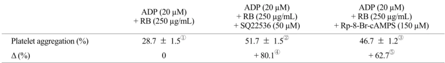
2B), then ADP-induced platelet aggregation would be increased in the presence of an adenylate cyclase inhibitor that inhibits the generation of cAMP. As shown in Fig. 4A, surprisingly, the platelet aggregation (28.7 ± 1.5%) by RB (250 μg/mL) plus ADP (20 μM) was increased by 51.7
± 1.5% in the presence of adenylate cyclase inhibitor SQ22536 (50 μM). This result means that RB may elevate the cAMP level via activation of adenylate cyclase to inhibit ADP-induced platelet aggregation. Otherwise, the platelet aggregation (28.7 ± 1.5%) by RB (250 μg/mL) plus ADP (20 μM) would not be increased to 80.1% in the presence of adenylate cyclase inhibitor SQ22536 (50 μM) (Table 3).
On the other hand, adenylate cyclase inhibitor SQ22536 did not significantly affect on ADP-induced platelet aggre- gation (Fig. 4A).
The inhibition of platelet aggregation by cAMP is caused via activation of cAMP-dependent protein kinase (A-kinase).
Therefore, platelet aggregation would be increased in the presence of A-kinase inhibitor, and would be accord with that by cAMP inhibitor SQ22536. Accordingly, we investi- gated whether RB-inhibited platelet aggregation is increased by A-kinase inhibitor, Rp-8-Br-cAMPS. As shown in Fig.
4B, the platelet aggregation by RB (250 μg/mL) plus ADP (20 μM) was increased by A-kinase inhibitor, Rp-8-Br- cAMPS (150 μM). These results suggest that the inhibitory mode of ADP-induced platelet aggregation by RB is dependent on cAMP/A-kinase pathway. Otherwise, the platelet aggregation (28.7 ± 1.5%) in the presence of RB (250 μg/mL) and ADP (20 μM) would not be increased to 62.7% in the presence of A-kinase inhibitor Rp-8-Br- cAMPS (150 μM) (Table 3). On the other hand, A-kinase inhibitor Rp-8-Br-cAMPS did not significantly affect on ADP-induced platelet aggregation (Fig. 4B).
Table 3. Changes of platelet aggregation in the presence of SQ22536 or Rp-8-Br-cAMPS ADP (20 µM)
+ RB (250 µg/mL)
ADP (20 µM) + RB (250 µg/mL) + SQ22536 (50 µM)
ADP (20 µM) + RB (250 µg/mL) + Rp-8-Br-cAMPS (150 µM)
Platelet aggregation (%) 28.7 ± 1.5① 51.7 ± 1.5② 46.7 ± 1.2③
Δ (%) 0 + 80.1④ + 62.7⑤
Platelet aggregations are from Fig. 4, B. Δ (%) ④, ②-①/①×100; Δ (%) ⑤, ③-①/①×100.
Fig. 3. Effect of RB on cAMP production. Measurement of cAMP was carried out as described in "Materials & Methods" section.
The data are expressed as the mean ± S.E.M. (n = 4). *P<0.05 versus the ADP-stimulated platelets.
Effect of RB on VASP phosphorylation
Downstream pathway of cAMP/A-kinase involves in phosphorylating VASP to inhibit platelet aggregation. Ser157 at 50 kDa of VASP is phosphorylated by the cAMP/
A-kinase pathway (Horstrup et al., 1994; Smolenski et al., 1998). Therefore, phosphorylation of Ser157 at 50 kDa of VASP is a useful indicator for monitoring cAMP/A-kinase pathway. ADP increased weakly the phosphorylation of VASP (Ser157) [p-VASP (Ser157)] at 50 kDa and the ratio of
p-VASP (Ser157) to β-actin (Fig. 5A lane 2). It reflects that ADP involves in a feedback inhibition by elevating p-VASP (Ser157 and Ser239) (Gambaryan et al., 2010). The ratio of p-VASP (Ser157) to β-actin was dose dependently increased in the presence of both ADP and RB (150 and 250 μg/mL) (Fig. 5A lane 3, 4). As shown in Fig. 5A lane 5, both ADP (20 μM) and RB (250 μg/mL)-phosphorylated VASP (Ser157) at 50 kDa [p-VASP (Ser157)] were inhibited by adenylate cyclase inhibitor SQ22536 (50 μM). In addition, the phosphorylation of VASP (Ser157) by both ADP and RB Fig. 4. Effects of RB with adenylate cyclase inhibitor or A-kinase inhibitor on ADP-induced human platelet aggregation (A) Effect of RB in the presence of adenylate cyclase inhibitor SQ22536. (B) Effect of RB in the presence of A-kinase inhibitor Rp-8-Br-cAMPS. Measure- ment of platelet aggregation was carried out as described in "Materials & Methods" section. The data are expressed as the mean ± S.E.M.
(n = 4). **P<0.001 versus the ADP-stimulated platelets, ‡P<0.001 versus the ADP-stimulated platelets in the presence of RB (250 μg/mL).
A
B
was also decreased in the presence of A-kinase inhibitor Rp-8-Br-cAMPS (Fig. 5A lane 6). These results indicate that RB phosphorylates VASP (Ser157) through adenylate cyclase activation, cAMP elevation, and A-kinase activation.
Otherwise, the ratio of VASP (Ser157) phosphorylation to
β-actin by both RB (250 μg/mL) and ADP (20 μM) would not be decreased in the presence of SQ22536 (50 μM), adenylate cyclase inhibitor, or Rp-8-Br-cAMPS (150 μM), A-kinase inhibitor, which were returned to the basal level, intact platelets (Fig. 5B). It is established that cAMP/
A-kinase/VASP (Ser157) phosphorylation is involved in inhibition of ADP-induced platelet aggregation (Halbrügge et al., 1989; Halbrügge et al., 1990; Butt et al., 1994). In addition, both adenylate cyclase inhibitor SQ22536 and A-kinase inhibitor Rp-8-Br-cAMPS inhibited ADP-induced VASP (Ser157) phosphorylation. These results suggest that SQ22536 and Rp-8-Br-cAMPS inhibited cAMP/A-kinase- dependent VASP (Ser157) phosphorylation by inhibiting cAMP production and A-kinase activity.
With regard to the regulatory effects of VASP (Ser157) phosphorylation by phenolic compounds on platelet aggre- gation, epigallocatechin-3-gallate (Ok et al., 2012) and caffeic acid (Lee et al., 2014) also elevated cAMP level and phosphorylated VASP via cAMP/A-kinase pathway to inhibit platelet aggregation. RB contains ferulic acid, a phenolics, and seems to involve in cAMP-dependent phos- phorylation of VASP (Ser157) to inhibit platelet aggregation.
In the present study, however, it is unknown whether ferulic acid in RB directly involved in cAMP-dependent phosphorylation of VASP (Ser157). These should be study in the future. There are reports that ferulic acid, and its derivatives has an antiplatelet effects (Yasuda et al., 2003;
Wang and Ou-Yang, 2005), but its antiplatelet mechanism is unknown. Therefore, because RB contains 87.9% of ferulic acid among total phenolics (Table 1), the inhibition of ADP-induced platelet aggregation by RB (Fig. 2B) may be resulted from the action of ferulic acid in RB.
Effect of ferulic acid on ADP-induced platelet aggre- gation
It is inferred that ferulic acid in RB (Fig. 1B) may have inhibitory effect on ADP-induced platelet aggregation.
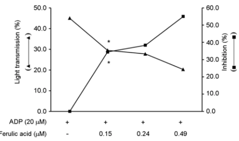
Therefore, we investigated whether authentic ferulic acid (FA) has an antiplatelet activity on ADP-induced platelet aggregation. To investigate the antiplatelet activity of FA, we used 0.15, 0.24, 0.49 μM of sodium ferulate (MW.
216.17) corresponding to ferulic acid concentration that A
B
Fig. 5. Effect of RB on VASP phosphorylation. (A) Lane 1, Intact platelets (base); Lane 2, ADP (20 μM); Lane 3, ADP (20 μM) + RB (150 μg/mL); Lane 4, ADP (20 μM) + RB (250 μg/mL). Lane 5, ADP (20 μM) + RB (250 μg/mL) + SQ22536 (50 μM); Lane 6, ADP (20 μM) + RB (250 μg/mL) + Rp-8-Br-cAMPS (150 μM). (B) Lane 1, Intact platelets (base); Lane 2, ADP (20 μM); Lane 3, ADP (20 μM) + SQ22536 (50 μM); Lane 4, ADP (20 μM) + Rp-8-Br- cAMPS (150 μM). Western blotting was performed as described in "Materials and Methods." The data are expressed as the mean
± S.E.M. (n = 4). **P<0.001 versus the ADP-stimulated platelets,
‡P<0.001 versus the ADP-stimulated platelets in the presence of RB (250 μg/mL).
contains in RB (150, 250, 500 μg/ml). As shown in Fig. 6, ferulic acid (FA) dose dependently inhibited ADP-induced platelet aggregation. These concentration of FA that inhibited ADP-induced platelet aggregation is very low as compared with that by caffeic acid, an analogue of FA (Lee et al., 2014). These results suggest that antiplatelet effect of RB may be resulted from ferulic acid (Fig. 1B) in RB.
Antiplatelet drugs such as thienopyridine derivatives (i.e.
ticlopidine, clopidogrel) have characteristics that phospho- rylate VASP, which is mediated by cAMP or cGMP (Barragan et al., 2003). Therefore, it is thought that RB as well as thienopyridine derivatives may represent a useful tool in the therapy and prevention of vascular diseases associated with platelet aggregation.
Acknowledgements
This study was supported by a grant from the High Value-added Food Technology Development Program [112074-3], iPET (Korea Institute of Planning and Evaluation for Technology in Food, Agriculture, Forestry and Fisheries).
REFERENCES
Barragan P, Bouvier JL, Roquebert PO, Macaluso G, Commeau P, Comet B, Lafont A, Camoin L, Walter U, Eigenthaler M.
Resistance to thienopyridines: clinical detection of coronary stent thrombosis by monitoring of vasodilator-stimulated phosphoprotein phosphorylation. Catheter Cardiovasc Interv.
2003. 59: 295-302.
Berridge MJ, Irvine RF. Inositol trisphosphate, a novel second
messenger in cellular signal transduction. Nature. 1984. 312:
315-321.
Butt E, Abel K, Krieger M, Palm D, Hoppe V, Hoppe J, Walter U.
cAMP- and cGMP-dependent protein kinase phosphorylation sites of the focal adhesion vasodilator stimulated phospho- protein (VASP) in vitro and in intact human platelets. J Biol Chem. 1994. 269: 14509-14517.
Cattaneo M. The P2 receptors and congenital platelet function defects. Semin Thromb Hemost. 2005. 31: 168-173.
Gasser JA, Betterridge DJ. Comparison of the effects of carvedilol, propranolol, and verapamil on in vitro platelet function in healthy volunteers. J Cardiovasc Pharmacol. 1991. 8: S29-S34.
Goufo P, Trindade H. Rice antioxidants: phenolic acids, flavonoids, anthocyanins, proanthocyanidins, tocopherols, tocotrienols, γ-oryzanol, and phytic acid. Food Sci Nutr. 2014. 2: 75-104.
Guidetti GF, Lova P, Bernardi B, Campus F, Baldanzi G, Graziani A, Balduini C, Torti M. The Gi-coupled P2Y12 receptor regulates diacylglycerol-mediated signaling in human platelets.
J Biol Chem. 2008. 283: 18352-18363.
Halbrügge M, Walter U. Purification of a vasodilator-regulated phosphoprotein from human platelets. Eur J Biochem. 1989.
185: 41-50.
Halbrügge M, Friedrich C, Eigenthaler M, Schanzenbächer P, Walter U. Stoichiometric and reversible phosphorylation of a 46-kDa protein in human platelets in response to cGMP- and cAMP-elevating vasodilators. J Biol Chem. 1990. 265: 3088 -3093.
Hou Z, Qin R, Ren G. Effect of anthocyanin-rich extract from black rice (Oryza sativa L. Japonica) on chronically alcohol- induced liver damage in rats. J Agric Food Chem. 2010. 58:
3191-3196.
Fig. 6. Effect of ferulic acid on ADP-induced platelet aggregation. Measurement of platelet aggre- gation was carried out as described in "Materials &
Methods" section. The data are expressed as the mean ± S.E.M. (n = 4). *P<0.05 versus the ADP- stimulated platelets.
Jennings LK. Role of platelets in atherothrombosis. Am J Cardiol.
2009. 103: 4A-10A.
Jun HI, Song GS, Yang EI, Youn Y, Kim YS. Antioxidant activities and phenolic compounds of pigmented rice bran extracts. J Food Sci. 2012. 77: C759-C764.
Laurent V, Loisel TP, Harbeck B, Wehman A, Gröbe L, Jockusch BM, Wehland J, Gertler FB, Carlier MF. Role of proteins of the Ena/VASP family in actin-based motility of Listeria monocytogenes. J Cell Biol. 1999. 144: 1245-1258.
Lee DH, Cho HJ, Kim HH, Rhee MH, Ryu JH, Park HJ. Inhibitory effects of total saponin from Korean red ginseng via vasodilator-stimulated phosphoprotein-Ser(157) phospho- rylation on thrombin-induced platelet aggregation. J Ginseng Res. 2013. 37: 176-186.
Lee DH, Kim HH, Cho HJ, Bae JS, Yu YB, Park HJ. Antiplatelet effects of caffeic acid due to Ca2+ mobilization-inhibition via cAMP-dependent inositol-1, 4, 5-trisphosphate receptor phos- phorylation. J Atheroscler Thromb. 2014. 21: 23-37.
Menshikov Myu, Ivanova K, Schaefer M, Drummer C, Gerzer R.
Influence of the cGMP analog 8-PCPT-cGMP on agonist- induced increases in cytosolic ionized Ca2+ and on aggre- gation of human platelets. Eur J Pharmacol. 1993. 245: 281 -284.
Ok WJ, Cho HJ, Kim HH, Lee DH, Kang HY, Kwon HW, Rhee MH, Kim M, Park HJ. Epigallocatechin-3-gallate has an anti-platelet effect in a cyclic AMP-dependent manner. J
Atheroscler Thromb. 2012. 19: 337-348.
Qureshi AA, Sami SA, Khan FA. Effects of stabilized rice bran, its soluble and fiber fractions on blood glucose levels and serum lipid parameters in humans with diabetes mellitus Types I and II. J Nutr Biochem. 2002. 13: 175-187.
Schwartz SM, Heinmark RL, Majesky MW. Developmental mechanisms underlying pathology of arteries. Physiol Rev.
1990. 70: 1177-1209.
Singleton VL, Rossi JA. Colorimetry of total phenolics with phosphomolybdicphosphotungstic acid reagents. Am J Enol Vitic. 1965. 16: 144-158.
Smolenski A, Bachmann C, Reinhard K, Honig-Liedl P, Jarchau T, Hoschuetzky H, Walter U. Analysis and regulation of vasodilator-stimulated phosphoprotein serine 239 phospho- rylation in vitro and in intact cells using a phosphospecific monoclonal antibody. J Biol Chem. 1998. 273: 20029-20035.
Sudo T, Ito H, Kimura Y. Phosphorylation of the vasodilator- stimulated phosphoprotein (VASP) by the anti-platelet drug, cilostazol, in platelets. Platelets. 2003. 14: 381-390.
Wang BH, Ou-Yang JP. Pharmacological actions of sodium ferulate in cardiovascular system. Cardiovasc Drug Rev. 2005. 23:
161-172.
Yasuda T, Takasawa A, Nakazawa T, Ueda J, Ohsawa K. Inhibitory effects of urinary metabolites on platelet aggregation after orally administering Shimotsu-To, a traditional Chinese medicine, to rats. J Pharm Pharmacol. 2003. 55: 239-244.