1
산양유의 체세포수에 영향을 미치는 비병원성 요인
김민경․최아리․한기성․정석근․오미화․장애라․설국환․함준상*
농촌진흥청 국립축산과학원
Non-Pathogenic Factors Affecting Somatic Cell Counts of Goat Milk
Min-Kyung Kim, Ari Choi, Gi-Sung Han, Seok-Geun Jeong, Mi-Hwa Oh, Aera Jang, Kuk-Hwan Seol and Jun-Sang Ham*
National Institute of Animal Science, RDA, Suwon 441-350, Korea
ABSTRACT
Somatic cell counts (SCCs) of goat milk can vary widely depending on the counting methods used and non-pathogenic factors; the goat milk industry can be threatened by establishment of a legal standard based on the findings in cow milk.
In Korea, SCCs have been excluded from the items that are analyzed under the “Livestock Products Processing and Composition Standards” in accordance with a recent NVRQS Notice amendment. From April to October, SCCs of 150 goat milk samples from 2 farms were analyzed using a Somascope calibrated with standard goat milk samples. Average SCCs of the samples was 598,000/mL, and significant differences were not found between farms and between breeds. SCCs increased from 3 to 8 months after delivery.
Keywords : goat milk, somatic cell counts (SCCs)
* Corresponding author: Jun-Sang Ham, National Institute of Animal Science, RDA, Suwon 441-350, Korea. Tel: +82-31-290-1692, Fax:
+82-31-290-1697, E-mail: [email protected]
서 론
축산물가공처리법 제2조 4항에서 “원유”라 함은 판매 또 는 판매를 위한 처리․가공을 목적으로 하는 착유상태의 우 유와 양유를 말하고, 제4조 2항의 규정에 의거 “원유의위생 등급기준”이 고시되어 있다(국립수의과학검역원, 2002). 산 양유의 체세포수는 이를 준용하기 어려우므로 산양유 체세 포 기준 설정이 주장된 바 있으나(Shin 등, 2008), 최근 “축 산물의가공기준및성분규격”이 개정(국립수의과학검역원, 2010) 되면서 원유의 검사 기준에서 양유는 체세포수가 제외되었 다. 따라서, 우리나라 산양유 생산자는 식품위생당국의 위 협에서 체세포 문제는 벗어난 것으로 생각되나, 외국에서도 산양유 산업의 생존이 위협 당할 수 있는 법적 기준의 적용 우려 때문에(Kapture, 1985; 1991) 산양유의 과학적 체세포
품질 기준 개발의 필요성을 많은 연구자들이 주장한 바 있 다(Hinckley, 1990; de Cremoux, 1998; Baudry & Mercier, 1998;
Toussaint, 1998). 소의 유즙 분비와 달리(merocrine) 산양 유 즙 분비(apocrine)에서는 많은 세포질 입자가 존재하기 때문
에 정상 우유의 지표로 인정되는SCC(체세포수)가 산양유에
서는 적절하지 못하며(Maisi, 1990; Atherton, 1992), 대부분의 실험실에서 일반적인 것처럼 우유로 보정한Fossomatic으로 산양유의 체세포 측정시24% 정도 높게 평가된다(Haenlein, 2002). 신 등(2008)은 국내 12개 산양 농가 시료의 체세포수 분석 결과(Somacount 500), 체세포수 평균은 1,559×103이라 고 보고한 바 있다. 또한, 이 등(2010)은 8개 목장의 48 시료 를 분석한 결과, pyronin Y-methyl green stain 측정시 전체 평 균이 7.3×10⁵cells/mL인 반면 ADAM-SCC와 Somacaount 500 으로 측정시 각각 4.9×10⁵과 11.6×10⁵cells/mL로 과소 또는 과대평가됨을 보고한 바 있다. 본 고에서는 산양유 체세포 관련 비병원성 요인과 체세포수와 생산성의 관계를 고찰하 고 산양유 표준시료로 보정한 체세포 분석장비(Somascope)로
분석한 국내 산양유의 체세포수를 보고하고자 한다.
본 론
1. 산양유 체세포 관련 비병원성 요인
1) 비유기
유방염에 걸리지 않은 Spanish Verata 100마리 유산양에 대한 연구에서, 비유 초기 평균 920,000(n=25)이었던 체세포 수가 580,000으로 감소하였고, 210일에는 1,810,000으로 증 가하였다(Rota et al., 1993b). 비록 비유 말기 값은 훨씬 높았 으나 유사한 결과가 많은 다른 연구자들에 의해 보고되었으 며(Dulin et al., 1983; Maisi, 1990; Kalogridou-Vassiliadou et
al., 1992; Droke et al., 1993; Rota et al., 1993a; Wilson et al.,
1995; Zeng & Escobar, 1995; 1996; Galina et al., 1996; Zenget al., 1997), 비유말기에는 체세포수가 2,000,000 이상이어
Fig. 1. SCCs (1000 mL−.1) in relationship to stage of lactation (days in milk) in milk from individual US dairy goats (n=2276~
3978) on DHIA from February 1992 to January 1994 (Haenlein &
Hinckley, 1995)
Fig. 2. SCCs above 1,130,000/mL in percentage of all goats sampled monthly on DHIA in relationship to stage of lactation (Haenlein & Hinckley, 1995).
도 유방 감염으로 생각되기 어렵다. 비유기에 따른 체세포 의 회귀식이 계산되었고(Rota et al., 1993b), 이는 위생공무 원에 의한 법적 허용치 및 산양 개체 관리 목적으로 목장 탱크내 산양유 평가에 매우 유용하다. 미국 유산양 공식 기 록 프로그램(DHIA)의 많은 개체(n=2276~3978) 자료에서도 체세포의 계절성이(Haenlein & Hinckley, 1995) 나타나며, 비 유 말기에 법적 한계를 넘는 비율이 증가하였다(Fig. 1, 2).
2) 발정
착유우에 에스트로겐 주입시 유량 변화에 관계없이 발정 기동안 체세포 증가가 관찰되었다(Haenlein & Krauss, 1974).
이는 산양유에서도 경험적으로 확인되고 있으나(Wilson et
al., 1995), 연구는 많이 되어 있지 않다(Rubino, 1996). Aleandri et al.(1994)은 무리에 숫 산양을 도입하여 발정이 시작된 자
아넨 무리에서 체세포가 233,000 높아졌다고 보고하였다.3) 착유단계
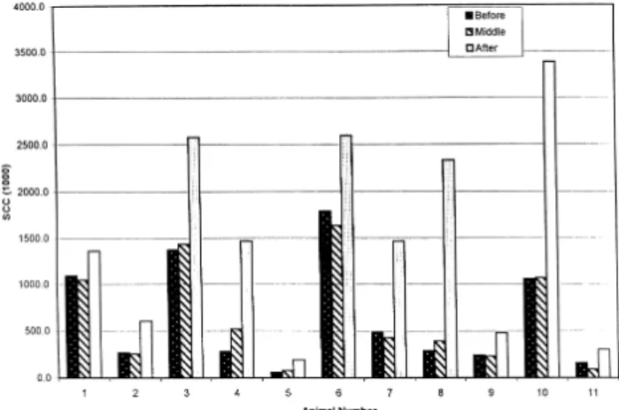
Delaware 대학(Haenlein, 1999) 연구에 따르면 손 착유나 기계 착유에 관계없이 착유 후반기에 체세포수가 유의적으 로 높았다. 유지방 조성도 유의적으로 차이나 나타난 반면
단백질 함량은 차이를 나타내지 않았다.
4) 산차
산양유의 평균 체세포수는 산차에 유의적으로 영향 받는 다(Maisi, 1990; Wilson et al., 1995; Contreras et al., 1999). 1산 차에52%인 다형핵호중구(polymorphonuclear neutrophilic leuk- ocytes, PMN)가 4산차에 69%로 증가한 반면 macrophage는 20%에서 14%로, lymphocytes는 12%에서 5%로 감소하였다 (Rota et al., 1993a). Spanish Verata 산양 100마리 평균 체세포 수는1산차에 1,270,000에서 4산차에 2,020,000으로 증가하였
Fig. 3. SCCs in milk of the same 11 Alpine goats in late lactation
before, middle and after portable bucket machine milking for 6
days(Haenlein, 1999, unpublished data).
고(Rota et al., 1993b), 미국의 138마리 유산양은 1산차에 942,000에서 2산차에 1,552,000, 3산차 이상에서 2,247,000으 로 증가하였다.
5) 스트레스
Acidosis와 높은 곡물 사양 같은 영양적 스트레스가 체세 포를 증가시키며(Lerondelle et al., 1992), 백신도 체세포를 증가시킨다.
2. 체세포수와 생산성의 관계
비유 단계의 진행 및 유방염 감염에 의한 결과의 혼동으 로 체세포수의 증가로 인한 산양유 생산량 감소를 보고한 논문은 거의 없다(Zeng & Escobar, 1995; Zeng et al., 1997).
비록 연질chevre type 치즈 제조시 체세포수 250,0000 이 하에서 수율 7.7 kg 우유/kg 치즈이고, 1,000,000 이상에서 7.2 kg 우유/kg 치즈의 수율을 얻었으나 체세포와 수율 사이 의 유의적 관계는 관찰되지 않았다(r=-0.25)(Galina et al., 1996). CMT 점수와 치즈 수율 사이에 유의적인 음의 상관 관계(r=-0.32)가 관찰되었다.
미국에서 유우의 유방염으로 인한 생산성 손실은20억 US$
(Harmon, 1994)에 달하고, 이러한 손실은 체세포수와 200,000 에서0%, 500,000에서 6%, 1,000,000에서 18%, 그리고 1,500,000 에서29%에 달한다고 보고되었다(DHIA). 유산양에서 유사 하게 분석한 결과, 비유단계 및 계절번식으로 인한 혼동때 문에 생산성 손실과의 관계는 결정되지 않았다(Haenlein &
Hinckley, 1995). 확실히, 준임상형 및 임상형 유방염의 각 단계에서 진정한 산양유 체세포수와 생산성 수준과의 상관 관계에 대한 연구가 필요하다.
3. 국내산 산양유의 체세포수
4월부터 10월까지 경기도 양주 및 충북 영동 목장에서 수 집한150개 산양 개체유 시료의 체세포수는 평균 598,000±
71,800이었으며, 목장간 유의적 차이는 관찰되지 않았다(Fig.
4). 이는 신 등(2008)이 보고한 1,559,000에 비해 대단히 낮 은 수치이나, 분석 장비의 보정, 착유 계절 및 비유기에 의해 영
Fig. 4. Average SCCs of 150 goat milk samples from two farms.
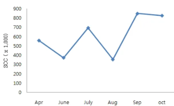
Fig. 5. SCCs of 150 goat milk samples according to month.
Fig. 6. SCCs of 110 goat milk samples from MGEN farm.
Fig. 7. SCCs of 150 goat milk samples according to breed(Alpine=
8, Togenberg=19, Saanen=123).
향을 받은 것으로 생각된다. 본 실험에서는 Combiscope FTIR (Delta Instrument)을 산양유 ELS sample로 보정하였으며, 겨
울철 시료가 없기 때문에 낮게 나타난 것으로 생각된다.
월별 체세포 분석 결과를 보면6월과 8월에 낮게 나타났
지만4월에 558,000±106,000에서 7월에 695,000±212,000, 9 월에848,000±308,000으로 증가하는 경향을 나타내었다(Fig.
5). 그런데, 개체관리를 한 엠젠목장 110개 시료에 대해 출 산 후4개월부터 8개월까지 분석한 결과는 307,000±64,000 에서926,000±199,000까지 지속적으로 증가하는 결과를 나 타내었다(Fig. 6). 이는 Fig. 1과 일치하는 결과이나, 수치는 다소 낮은 것으로 생각된다.
한편, 산양유 시료의 체세포 분석 결과를 알파인(N=8), 토겐버그(N=19), 자아넨(N=123)의 종에 따라 분류한 결과
는 Fig. 7과 같다. 토겐버그가 222,000±41,000으로 가장 낮 은 수치를 나타내었으며, 알파인이 463,000±155,000, 자아넨 이 665,000±86,000으로 가장 높은 수치를 나타내었다.
결 론
산양유의 체세포수는 측정방법 및 비병원성 요인에 따라 크게 차이가 날 수 있으며, 우유에서 밝혀진 사실을 근거로 법적 기준 제정시 유산양 산업의 존립을 위협받을 수 있다.
국내의 경우, 최근 축산물의가공기준및성분규격이 개정되 어 원유의 검사기준에서 산양의 체세포 검사제외를 명확히 하였다. 이는 영세한 국내 유산양 농가를 위해 바람직하다 고 생각된다. 금년 4월부터 10월까지 두 개 목장에서 수집
한 150개 개체유를 표준시료로 보정한 체세포 분석장비로
분석한 결과, 체세포수는 평균 598,000이었으며 농장간, 유 산양 품종간 차이는 관찰되지 않았다. 4월부터 10월까지 착 유 월에 따라 체세포수는 증가하는 경향이었으며, 분만일이 기록된 개체의110개 시료를 비유일수에 따라 분석한 결과, 분만120일부터 240일까지 30만에서 90만으로 직선적 증가 를 나타내었다.
참고문헌
1. Aleandri, M., Fagiolo, A., Calderini, P., Colafrancesco, R., Giangolini, G., Rosati, R. and De Michelis, F. 1994. Studies conducted on somatic cells count of goat milk. In: Pro- ceedings of the Symposium on Somatic Cells and Milk of Small Rumminants. EAAP Publication No. 77, Bella, Italy, 25-27 September 1994, Wageningen Pers, The Netherlands, pp. 65-70.
2. Atherton, H. V. 1992. Using somatic cells and antibiotic tests for determining the quality of goat milk. In: Gipson, T. A., et al. (Eds.), Proceedings of the National Symposium on Dairy Goat Production and Marketing. Langston University, Langston, OK, pp. 128-135.
3. Baudry, C. and Mercier, P. 1998. Interpreter les numerations cellulaires de tank. Reussir La Chevre. No. 226, May-June, pp. 21-22.
4. de Cremoux, R. 1998. Les numerations cellulaires. Reussir La Chevre. 226(May-June):18-20.
5. Droke, E. A., Paape, M. J. and di, A. L. 1993. Prevalence of high somatic cell counts in bulk tank goat milk. J. Dairy Sci. 76:1035-1039.
6. Dulin, A. M., Paape, M. J., Schultz, W. D. and Weinland, B.
T. 1983. Effect of parity, stage of lactation and intramam- mary infection on concentration of somatic cells and cyto- plasmic particles in goat milk. J. Dairy Sci. 66:2426-2433.
7. Galina, M. A., Morales, R., Lopez, B. and Carmona, M. A.
1996. Effect of somatic cell count on lactation and soft cheese yield by dairy goats. Small Rumin. Res. 21:251-257.
8. Haenlein, G. F. W. 2002. Relationship of somatic cell counts in goat milk to mastitis and productivity. Small Rumin.
Res. 45:163-178.
9. Haenlein, G. F. W. and Hinckley, L. 1995. Goat milk somatic cell count situation in USA. Int. J. Anim. Sci. 10:305-310.
10. Haenlein, G. F. W. and Krauss, W. C. 1974. Effects of single injections of diethylstilbestrol on milk composition and counts of leucocytes in milk of Holstein-Riesian cattle. Z.
Tierphysiol. Tierernaehr. Futtermittelkd. 34:50-60.
11. Hinckley, L. S. 1990. Revision of the somatic cell count standard for goat milk. Dairy Food Environ. Sanitat. 10:
548-549.
12. Kalogridou-Vassiliadou, D., Manolkidis, K. and Tsigoida, A.
1992. Somatic cell counts in relation to infection status of the goat udder. J. Dairy Res. 59:21-28.
13. Kapture, J. 1985. Profile of goat milk marketing in the US.
United Caprine News. 6:25-27.
14. Kapture, J. 1991. Milk conference focus: Somatic cell counts.
Dairy Goat J. 6/7(370):415.
15. Lee, S. G, Kim, M. K., Lee, Y. J., Jeong, S. G., Oh, M.
H., Kim, D. H., Park, K. W., Lee, W. K. and Ham, J. S. 2010.
Comparison of measuring methods for somatic cell count in goat milk. Korean J. Food Sci. Ani. Resour. 30(1):120-123.
16. Maisi, P. 1990. Analysis of physiological changes in caprine milk with CMT, NAGase and antitrypsin. Small Rumin.
Res. 3:485-492.
17. MIFAFF/NVRQS. 2002. Raw milk sanitation grading Stan- dards (NVRQS Notice 2002-4).
18. MIFAFF/NVRQS 2010 Livestock products processing and composition Standards (NVRQS Notice 2010-13).
19. Rota, A. M., Gonzalo, C., Rodriguez, P. L., Rojas, A. I., Martin, L. and Tovar, J. J. 1993a. Somatic cell types in goats milk in relation to total cell count, stage and number of lactation. Small Rumin. Res. 12:89-98.
20. Rota, A. M., Gonzalo, C., Rodriguez, P. L., Rojas, A. I., Martin, L. and Tovar, J. J. 1993b. Effects of stage of lactation and parity on somatic cell counts in milk of Verata goats and algebraic models of their lactation curves.
Small Rumin. Res. 12:211-219.
21. Rubino, R. (Ed.), 1996. Proceedings of the Symposium on Somatic Cells and Milk of Small Ruminants. EAAP Publication No. 77, Bella, Italy, 25-7 September 1994, Wageningen Pers, The Netherlands, pp. 384.
22. Shin, J. H., Jeong, S. G., Han, G. S., Jang, A., Chae, H. S., Yoo, Y. M., Ahn, J. N., Woo, K. T., Choi, S. H., Lee, W.
K. and Ham, J. S. 2008. Study on the somatic cell count grade of goat milk in Korea. Korean J. Food Sci. Ani.
Resour. 28:574-579.
23. Toussaint, G. 1998. UCAL: Politique de qualite et cellules.
Reussir La Chevre. 226(May-June):23-24.
24. Wilson, D. J., Stewart, K. N. and Sears, P. M. 1995. Effects of stage of lactation, production, parity and season on somatic
cell counts in infected and uninfected dairy goats. Small Rumin. Res. 16:165-169.
25. Zeng, S. S. and Escobar, E. N. 1995. Effect of parity and milk production on somatic cell count. Small Rumin. Res.
17:269-274.
26. Zeng, S. S. and Escobar, E. N. 1996. Effect of breed and milking method on somatic cell count. Small Rumin. Res.
19:169-175.
27. Zeng, S. S., Escobar, E. N. and Popham, T. 1997. Daily variations in somatic cell count, composition, and production of Alpine goat milk. Small Rumin. Res. 26:253-260.
(2010년 11월 1일 접수; 2010년 11월 15일 채택)