* 이 논문은 2007년도 정부재원(교육인적자원부 학술연구조성사업비)으로 한국학술진흥재 단의 지원을 받아 연구되었음(KRF-2007-354-G00027).
†교신저자: 박혜주, 영남대학교 생활과학대학 체육과, 연구세부분야: 스포츠 심리학 E-mail: [email protected]
경기 승패에 따른 야구팬들의 정서경험: fMRI연구 *
박 혜 주
†류 호 상
영남대학교 체육학부
본 연구는 경기장에서 스포츠 팬들이 자신이 응원하는 팀의 경기 승패에 따라 경험하는 긍 정, 부정 정서가 정서의 편측화를 보이는 지를 fMRI 실험을 통하여 살펴보는 것이다. 이를 위해 S 프로야구 구단 팬 12명을 대상으로 S구단이 승리한 장면과 패배한 장면을 보여주었 다. 측정결과, 승리장면을 지켜본 피험자들의 좌우 쐐기소엽, 우하후두이랑, 우하전두이랑, 좌편도체, 우해마옆이랑, 좌갈고리, 좌대상이랑, 좌하측두이랑, 우중측두이랑, 좌소뇌비탈, 좌 소뇌정상 영역에서 활성화 반응이 나타났다. 반면에 패배장면을 지켜본 피험자들의 우중간 전두이랑, 좌전대상회, 좌소이랑, 좌조가비핵, 좌창백핵, 좌배쪽전핵, 좌시상, 좌전장, 좌뇌섬 엽 영역에서 활성화 반응이 나타났다. 본 연구결과 정서가의 반구비대칭 증거가 뚜렷하게 나타나지 않았으며 승리자극에서는 나타난 편도체의 활성이 패배자극에서는 나타나지 않아 패배정서 유발에 보다 심층적인 연구가 필요할 것으로 보아졌다.
주제어 : 승패, 야구팬, 정서경험, 긍정정서, 부정정서, 기능성자기공명영상, 뇌활성
서 론
스포츠 팬들은 스포츠에 대한 열정과 관심이 높아 선수나 팀과의 일체감이 형 성되어 스포츠를 즐기는 사람들로[1], 이들은 단순한 시청이 아닌 직접 경기장을 찾아가는 사람들로써 일반관중과는 차이가 있다. 이들은 자신들이 응원하는 팀이 승리했을 때는 자신감이 넘치고 긍정적인 감정이 강해지지만 패배했을 때는 분노 나 우울과 같은 부정적인 감정을 경험하게 된다[8][10]. 이와 같은 현상은 스포츠 팬들이 가지는 특성으로 선수와 자신을 동일시하면서 선수가 느끼는 감정과 거의 일치되는 경험을 하기 때문이다[22]. 팬들이 경험하는 승리와 패배정서는 기존의 정서연구에서 다루어지는 긍정과 부정정서로 볼 수 있다. 정서의 긍정과 부정적인 감정에 대한 연구는 얼굴표정에서 나타난 쾌감(pleasant)과 불쾌감(unpleasant)[9][47]
혹은 행복(happy)과 슬픔(sad 혹은 unhappy)[16][60]과 같은 상반된 감정경험에 관한 연구가 대부분이었다.
스포츠에서 상반된 감정을 불러일으키는 경기 승패에 따른 관중의 정서경험에
관한 연구들은 그동안 주로 설문지를 통해 많이 이루어져 왔으나[38][58] 최근에는
뇌파나 fMRI를 이용하여 보다 객관적이고 과학적인 접근을 통해 관중의 정서경험
을 심리생리학적으로 살펴보려는 연구가 시도되고 있다. 이인희(2006)는 접근-철회
정서이론(approach-withdrawal emotional valence hypotheses)[11][19][21]을 근거로 경기
승패에 따른 관중의 정서경험을 EEG를 통해 조사한 결과 관중의 긍정적 정서 경
험은 좌반구 활동과 관련이 있으며, 부정적 정서 경험은 우반구 활동과 관련이 있
음을 증명하였다. 이 후 Park 등(2009)은 2002년 월드컵 축구경기장면을 보여주고
fMRI를 측정한 결과 자신이 응원하는 팀이 승리했을 때는 좌우측 후두엽, 좌측 측
두엽, 좌측 변연계 영역이 활성화되었으며, 패배했을 때는 우측 전두엽, 우측 변연
계 영역이 활성화됨을 보여 주어 긍정과 부정 정서 경험에 대한 정서의 편측화를
지지하였다. 하지만 이 연구는 사용된 영상이 2002년 월드컵 국가 간의 경기장면
으로 참여한 피험자들은 한국 국적의 일반인이어서 스포츠 팬을 대상으로 한 연구
라고 하기에는 한계가 있다. 게다가 한국인에게는 4강 신화의 달성이라는 기쁨의
기억으로 인해 승리 장면과 패배 장면의 강도가 동일하지 않아 패배 장면에서의
부정정서가 덜 유발된 것으로 보인다.
정서를 유발하는 fMRI 연구의 초기에는 사진을 주로 사용하였으나[21][32][42]
[47], 최근에는 비디오 영상을 사용하는 경우가 많아지고 있다. 사진과 같은 시 각유발은 피험자로 하여금 정서 경험을 회상시켜 ‘내적으로 생성된 정서경험 (internally generated emotional experiences)’을 하게 하는 것으로[9], 단순히 피험자들이 수동적으로 사진의 얼굴표현들을 바라보게 함으로써 정서의 인식만 조사하게 되어 [20] 피험자들의 주관적인 정서 경험을 유발하는 데 한계가 있다는 지적이 있다 [5][7][52]. 이에 비해 비디오 자극은 정서적 장면을 그대로 노출시켜 ‘외적으로 생 성된 정서경험(externally generated emotional experiences)’을 하게 함으로써 의도된 정 서를 수행하는데 효과적인 자극이 될 수 있다[4][6][49]. 또한 사진자극에 비해 비 디오 자극은 유발된 정서를 오래 지속시킬 수 있으며[27], 더 역동적으로 진행되는 맥락을 제공할 수 있을 뿐만 아니라, 목표 정서를 유발하는데 용이하며[29] 개인 정서 유발에 비교적 높은 생태학적 타당도를 가진다[27]. 본 연구에서는 스포츠 팬 들이 자신이 응원하는 팀의 경기장면을 지켜볼 때 느껴지는 정서경험에 관하여 살 펴보고자 함으로 동영상 자극이 더 타당한 것으로 보아진다.
본 연구는 Park 등(2009)의 추후연구로 일반 관중이 아닌 스포츠 팬들을 대상으 로 하였을 때, 경기 승패에 따른 상반된 정서 경험이 정서의 편측화를 보이는 지 를 살펴보는 것이다. 현장에서 역동적인 스포츠 장면을 지켜볼 때 경험되는 정서 를 유발하기 위해 정지된 장면이 아닌 비디오 영상 자극을 사용하여 경기승패에 따라 스포츠 팬들의 정서가 어떻게 나타나는지를 뇌신경생리학적인 측면에서 살펴 보고자 한다. 스포츠에서의 승리의 경험은 팬들의 좌반구를 활성화시켜 긍정정서 를 유발시킴으로써 공격성향을 감소시키고 정서 조절력을 향상시킨다고 한다.
fMRI를 통한 팬들의 정서 경험에 관한 생리심리학적인 정보는 스포츠가 지니고 있 는 긍정적인 측면을 증명하고 스포츠 문화를 활성화시키는데 기여할 수 있을 것으 로 보아진다.
연구방법
그림 1. 실험 디자인
연구대상
본 실험에 참가한 피험자는 S구단 야구 팬 12명으로 하였다(모두 남학생이며 평 균나이는 25.2세). 이들 피험자는 교정시력이 1.0이상이며, 안질환, 신경질환 그리고 뇌질환 병력이 없는 자들로 모두 오른손잡이다. 피험자 전원은 소정의 비용을 받 고 실험에 참가하였다.
과제 및 절차
본 실험에 사용된 자극은 한국야구위원회 홈페이지에서 팬들의 조회율이 가장
높았던 장면중에서 S구단이 승리한 장면과 패한 장면을 편집한 다음 S구단 팬 5명
을 대상으로 승리와 패배로 인한 감정유발이 되는지 파일럿 테스트를 통해 수정하
여 사용하였다. 승리장면은 S구단 우승 장면들로 투수가 마지막 타자를 잡자 포수
가 기뻐 뛰어나오고 모든 선수들이 얼싸안고 기쁨을 나누는 장면, 우승 축포가 경
기장 안 밖에서 터지며 선수들이 하이파이브와 포옹을 하는 장면과 한국시리즈 우
승 현수막이 운동장을 가득 메우고 경기장을 꽉 메운 관중들의 열광장면으로 구성
되었다. 패배장면은 상대팀 타자의 홈런과 환호에 S구단 팬은 침울한 표정을 짓고
안타까움에 얼굴을 모자에 묻어버리는 장면, 홈런친 타자를 환호하는 상대구단 선
수들과 양볼을 손으로 감싸고 절망스러워하는 팬의 표정과 상대팀의 안타에 고개
를 떨구는 투수장면으로 구성되었다. 통제자극은 고교야구 시합 장면으로 수비수
들이 송구하여 타자를 아웃시키고 쓰리아웃 체인지하는 장면으로 구성되었다. 실 험 디자인은 블락 디자인으로 구성되었다[43][52]. 자극은 fMRI 스캐너 내에 설치되 어 있는 LCD 프로젝터(IFIS-SA)를 통해 제시되었으며, 제시된 자극은 dummy 6초 이후에 통제 장면 18초, 승리 장면 18초, 패배 장면 18초를 3회 반복하여 모두 168 초 동안 제시되었다(그림 1). 참가자들에게는 실험 시작 전에 실제로 경기장에서 경기를 지켜볼 때처럼 관람토록 지시를 하였다.
영상자료의 획득
fMRI 자료는 3T MRI scanner (ISOL Technology, Korea)에 의해 수집되었다. fMRI는 혈액산소포화도(Blood Oxygen Level Dedendent: BOLD) 효과를 최대로 하기 위해 초 고속영상 기법인 gradient-echo planar imaging(EPI) 기법을 사용하였다. 해부학적 뇌 영상은 T1 강조영상(TE=16 ms, TR=2800 ms, flip angle=60°, FOV=20 cm, 256×256 matrix, 4 mm slice thickness)을 사용하여 자료를 수집하였으며, 기능적 뇌 영상인 T2(TE=35 ms, TR=3000 ms, flip angle = 60°, FOV=20 cm, 64×64 matrix, 4mm slice thickness)영상이 수집되었으며 한 피험자에게서 찍힌 스캔은 56scan이었다.
영상 자료의 분석
fMRI 자료 분석은 MATLAB(Mathworks Inc., Sherborn, MA) 적용이 가능한 SPM2 (Welcome Department of Cognitive Neurology, London, UK) 소프트웨어를 사용하였다.
Dummy영상에 해당하는 처음 2scan의 기능적 영상은 T1 균형효과를 위해 분석에서
제외시켰으며, 나머지 영상을 첫 번째 영상에 재배열시켜 기능적 영상과 해부학적
영상을 상관 정립시켰다. 피험자간의 뇌 형태차이를 교정하기 위해 모든 영상은
뇌의 내부 공간에 PRESTO template(Montreal Neurologic Institute=MNI)를 적용시켜
뇌의 차이를 표준화시켰다. 모든 기능적 영상은 full-width at half-maximum(FWHM
Gaussian Kernel)을 사용해 편평화 시켰다. 표준화된 개인의 뇌 영역을 집단에 따라
그룹 분석하여 일차체성감각피질(primary somatosensory cortex=S1), 일차피질(primary
cortex=M1), SMA, 전운동영역(premotor area=PM), 뇌교, 그리고 소뇌의 평균치를 구
하고 Z-score에 따라 각 부위의 색채 지도화 부호를 만들어 승패에 따른 피험자 들의 정서경험이 어떠한 뇌 활성화를 보이는지를 분석하였다. 모든 좌표 값은 Talairach 프로그램을 사용하여 브로드만 영역(Brodmann's areas: BA)으로 표시하였다.
분석은 whole-brain 분석을 하였다.
결 과
승리장면시와 패배장면시의 피험자들의 fMRI분석에 대한 결과는 다음과 같다.
승리 > 통제 시의 뇌 활성 영역
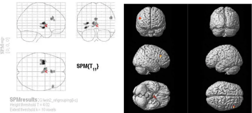
승리 장면에서는 좌우 쐐기소엽, 우하후두이랑, 우하전두이랑, 좌편도체, 우해마 옆이랑, 좌갈고리, 좌대상이랑, 좌하측두이랑, 우중측두이랑, 좌소뇌비탈, 좌소뇌정 상 영역에서 유의하게 활성화 되었다(uncorrected p < .001, 표 1, 그림 2).
그림 2. 승리 > 통제 시의 뇌 활성 영역
Region Winning > Control
L/R
x
y z BA z-value쐐기소엽(Cuneus) L -2 -74 20 18 4.41
R 4 -80 22 18 3.81
하후두이랑(Inferior occipital gyrus) R 18 -92 -5 17 3.22 하전두이랑(Inferior frontal gyrus) R 16 10 -14 47 3.96
편도체(Amygdala) L -22 0 18 4.37
해마옆이랑(Parahippocampal gyrus) R 28 4 -14 34 4.10
갈고리(Uncus) L -38 -12 -30 20 4.04
대상이랑(Cingulate gyrus) L -6 -54 26 31 3.49 하측두이랑(Inferior temporal gyrus) L -50 -5 -30 20 3.51 중측두이랑(Middle temporal gyrus) R 40 2 -30 21 3.92
소뇌비탈(Declive) L -32 -88 -20 3.82
소뇌정상(Culmen) L -24 -24 -26 3.89
Uncorrected p level of 0.001: x, y, z correspond to calculated Talairach space.
BA = Brodmann's area.
표 1. 승리장면에서의 뇌 활성 영역
패배 > 통제 시의 뇌 활성 영역
패배장면에서는 우중간전두이랑, 좌전대상회, 좌소이랑, 좌조가비핵, 좌창백핵,
좌배쪽전핵, 좌시상, 좌전장, 좌뇌섬엽 영역에서 유의하게 활성화 되었다(uncorrected
p < .001, 표 2, 그림 3).
Region Losing > Control
L/R x y z BA z-value
중간전두이랑(Middle frontal gyrus) R 54 28 28 46 3.69 전대상회(Anterior cingulate) L 0 32 20 32 3.47
소이랑(Sub-gyral) L -38 -6 -8 21 4.57
조가비핵(Putamen) L -28 -15 -4 3.50
창백핵(Lateral globus pallidus) L -12 4 -2 3.52 배쪽전핵(Ventral anterior nucleus) L -10 -2 10 4.14
시상(Thalamus) L -2 -4 5 3.49
전장(Claustrum) L -30 -4 10 4.61
뇌섬엽(Insula) L -38 0 5 13 3.38
Uncorrected p level of 0.001: x, y, z correspond to calculated Talairach space.
BA = Brodmann's area.
표 2. 패배 장면에서의 뇌 활성 영역
그림 3. 패배 > 통제 시의 뇌 활성 영역
논 의
본 연구는 비디오 영상 자극을 사용하여 경기 승패에 따라 스포츠 팬들의 긍정, 부정 정서 경험이 정서의 편측화를 보이는 지를 살펴보는 것이다. 일반 관중을 대 상으로 하였을 때 보여졌던 이인희(2006)와 Park 등(2009)의 결과와 달리 정서가 (emotional valence)의 반구비대칭(hemispheric asymmetry) 증거가 뚜렷하게 나타나지 않 았다. 물론 단 두 편의 선행연구 결과와 비교해서 단정 짓기는 어렵지만, 이에 대 해 Canli 등(1998)은 정서유발 fMRI연구에서는 정서가의 반구비대칭이 잘 나타나지 않는다고 하였다. 그 이유는 정서유발 자극이 피험자들로 하여금 단지 수동적으로 자극을 바라보는 것이어서 정서의 인식과정인지 경험과정인지 그 차이점이 구분되 지 않는 다는 것과[7] 성격특성으로 인한 개인차 변인[15]과 게다가 정서는 많은 외적, 내적 요인들에 의해 영향을 받기 쉽기 때문인 것으로[28] 지적된다. 이는 상 반된 정서 자극에 대한 fMRI 연구에서 정서가 이론이 적용되지 않은 Killgore와 Yurgelun-Todd(2007), Rodway 등(2003)의 연구 결과와도 일치한다.
경기 승패에 따른 피험자들의 뇌활성 영역을 살펴보면, 승리장면을 지켜볼 때에 변연계 영역의 활성이 두드러졌다. 이 영역의 편도체와 해마옆이랑의 활성은 Beck(2001)과 Breiter 등(1996)의 연구에서 즐거움을 경험했을 때 활성화된 영역과 일 치함을 보이고 있다. 해마 영역은 회상(recollection)에 필수한 영역으로[62], 기억이 통합되도록 영역간의 정보를 조직하거나 재조직화하도록 돕는 역할을 하며[45] 주 관적인 정서 경험동안에 발생하는 것으로 보고된다[25]. 또한 편도체와 해마 사이 의 상호작용을 통하여[23][54] 인간의 정서 정보에 대한 기억을 향상시킨다고 한다 [49]. 편도체는 즐거운 장면과 행복한 얼굴 표정과 같은 긍정정서를 처리할 때[63]
특히 정서적으로 중요한 사건에 대한 기억을 조절할 때 중요한 역할을 하며[30],
그 감정도 생생하게 기억하는 역할을 한다[23][36][56]. 대상이랑 역시 기억이나 이
미지를 떠올리게 하는 정서적 자극에 반응하는 영역으로[9], 행복한 얼굴표정에서
활성화 되는 영역이다[24][50]. 본 연구에서 제시한 승리장면은 시즌 우승장면으로
본 실험에 참가한 S구단 팬들에게는 가장 감격적이고 기뻤던 기억으로 회상되어
당시의 승리로 인한 강한 긍정 감정이 불러 일으켜 진 것으로 보아진다. 그 외 활
성화 된 하후두이랑은 얼굴 이미지 분석[33]과 얼굴 형상 인식[31]과 관련되는 영
역으로 알려지며, 쐐기소엽은 행복한 얼굴사진을 지켜보는 동안 행복한 사건들을 회상할 때 활성화가 훨씬 더 잘 일어났다는 Prohovnik, Skudlarski, Fulbright, Gore 그 리고 Wexler(2004)의 연구와 행복한 얼굴표정으로 인식할 때 활성화 된다는 Kilts 등(2003)의 연구에서도 동일하게 보여진 영역이다. 하후두이랑과 쐐기소엽 영역의 활성화는 선수들의 기쁨의 표정에서 행복했던 기억이 회상되었기 때문인 것으로 보아진다. 우측 중측두이랑은 움직이는 시각 자극을 지켜볼 때[13], 주의를 유지하 거나 초점을 맞출 때 중요하게 관여하는 영역으로 즐거운 정서를 생성하는 장면을 지켜보는 동안에 보여진다고 보고된 바 있다[42][46]. 하전두이랑은 정서 인식 혹은 평가[13][57], 정서적 공감과[34][55] 관련되는 영역으로 연속된 행동을 지켜보는 동 안에 활성화된다고 보고된다[35]. 우측 중측두이랑과 하전두이랑의 활성화는 본 연 구에서 제시된 동영상 승리 장면이 우승의 순간이 포착된 장면으로 S구단의 모든 선수들이 일제히 마운드로 뛰어나오는 행동과 경기장의 환호에 피험자들의 주의가 모아졌기 때문이라고 여겨진다.
패배장면을 지켜볼 때에 활성화 된 중간전두이랑은 인지와 정서의 통합과 감정 의 생생한 의식적인 경험과 관련된다[17][42]. 전대상회피질은 주의 과정화 (attentional processing) 측면에서 중요한 역할을 하며[20] 주관적인 정서적 반응에 주 의를 귀울일 때 활성화되는 영역으로[42] 분노조건과[2][41] 슬픔 조건에서[44] 활 성화 되는 것으로 보고되었다. 시상은 특히 슬픈 얼굴 표정 인식과[18] 분노 조건 에서[2] 활성화 된다고 알려져 있다. 본 연구에서 제시된 패배장면은 상대팀의 승 리타구로 인한 S구단 팬의 허탈하고 씁쓸한 안타까운 표정에 초점이 맞춰져 있어 피험자들로 하여금 자신이 응원하는 팀이 졌을 때 속상하고 안타까운 감정이 유발 되어 몰입시켰기 때문인 것으로 본다.
본 연구에서는 승리장면에서는 활성화된 편도체 영역이 패배장면에서는 활성화
되지 않았다. 편도체는 정서 기억과[6] 정서 유발의[28] 중요한 영역으로, 부정정서
특히 공포 처리에 깊이 관여한다고 알려져 있다[12]. 하지만 본 연구에서 유발된
패배 장면에 대한 팬들의 부정 정서는 슬픈 장면에서 반응하는 정대상회 피질의
BA32 영역이 활성화됨으로써[44] 오히려 슬픔에 가깝다고 할 수 있다. 본 연구와
유사한 연구 결과로 Killgore 등은(2004) 행복한 얼굴 표정과 슬픈 얼굴 표정을 바
라보게 하는 실험에서 행복한 표정에서는 편도체의 활성이 나타났지만 슬픈 표정
에서는 편도체의 활성이 나타나지 않았다. 이는 슬픔이 공포, 분노, 행복에 비해 표현되는 각성 정도가 낮고[59] 편도체가 정서 각성 차원에 즉각적으로 반영하기 때문인 것으로 설명할 수 있다[61]. 승리와 패배로 인해 유발되는 관중의 부정정서 (분노, 슬픔, 안타까움)는 기존 정서 연구에서 유발되는 부정정서(혐오, 공포, 분노, 메스꺼움)와는 차이가 있다고 보아진다. 사회적 관계에서 경험하는 분노의 감정은 지속적이며 격해지지만 매 순간 변화되는 스포츠 장면에서 관중이 경험하는 분노 의 감정은 일시적이며 오히려 안타까움이 동반된 슬픔에 가깝다고 할 수 있다.
fMRI 연구에서 슬픔의 표현은 다른 정서에 비해 뇌활성의 한계가 있다고 지적됨으 로써[37][50] 추후연구에서는 패배로 인한 팬들의 정서가 보다 명확하고 구체적으 로 제시될 필요성이 있을 것으로 사료된다. 본 연구는 자극 유발에서 실제 경기장 에서 스포츠 팬들이 경험할 수 있는 정서를 유발시키는데 한계점을 지니고 있지만 스포츠 팬들을 대상으로 한 뇌신경생리학적인 연구가 기존에 없었다는 점에서 이 러한 시도는 의미가 있다고 보아진다.
참고문헌
[1] 박상윤(2005). 스포츠 팬 일체성에 따른 스폰서십 효과 및 구매의도에 관한 연 구. 성균관대학교 대학원 석사학위논문.
[2] 이옥현, 석지아, 박미숙, 엄진섭, 권애란, 손진훈 (2006). 부적 정서와 관련된 뇌 활성화 영역. Proceedings of KFIS Spring Conference, 16(1), 351-364.
[3] 이인희 (2006). 승패에 따른 정서가 관중의 대뇌반구 비대칭에 미치는 영향. 경 북대학교 체육학과 박사학위논문.
[4] 장은혜, 석지아, 엄진섭, 손진훈 (2005). 시청각 동영상(Audio-visual Film Clips)을 이용한 정서유발프로토콜 개발. 한국심리학회지: 실험, 17(1), 69-84.
[5] Aalto, S., Näätänen, P., Wallius, E., Metsähonkala, L., Stenman, H., Niemi, P. M., Karlsson, H. (2002). Neuroanatomical substrata of amusement and sadness: a PET activation study using film stimuli. NeuroReport , 13(1), 67-73.
[6] Adolphs, R., Tranel, D., & Buchanan, T. W. (2005). Amygdala damage impairs
emotional memory for gist but not details of complex stimuli. Nature Neuroscience , 8, 512–518.
[7] Beck, E. D. (2001). The perception, experience and regulation of emotion: Emotional dynamics of soccer fans at winning and losing games. Doctoral dissertation. University of California, San Diego.
[8] Bernhardt, P. C., Dabbs, J. M., Fielden, J. A., & Lutter, C. D. (1998). Testosterone changes during vicarious experiences of winning and losing among fan at sporting events. Physiology & Behavior , 65(1), 59-62.
[9] Berthoz, S., Blair, R. J. R., Le Clec'h, G. & Martinot, J.-L. (2002). Emotions: From neuropsychology to functional imaging. International Journal of Psychology , 37(4), 193-203.
[10] Bizman, A., & Yinon, Y. (2002). Engaging in distancing tactics among sport fans:
effects on self-esteem and emotional responses. The Journal of Social Psychology , 142(3), 381-392.
[11] Blackhart, G. C., Minnix, J. A., Kline, J. P. (2006). Can EEG asymmetry patterns predict future development of anxiety and depression? A preliminary study. Biological Psychology , 72(1), 46-50.
[12] Breiter, H. C., Etcoff, N. L., Walen, P. J., Kennedy, W. A., Rauch, S. L., Buckner, R. L., Stauss, M. M., Hyman, S. E., Rosen, B. R. (1996). Response and habituation of the human amygdala during visual processing of facial expression, Neuron , 17(5), 875-887.
[13] Carr, L., Iacoboni, M., Dubeau, M. C., Mazziotta, J. C., Lenzi, G. L.(2003). Neural mechanisms of empathy in humans: a relay from neural systems for imitation to limbic areas. Proceedings of the National Academy of Sciences of the United States of Americ a, 100, 5497–5502.
[14] Cornette, L., Dupont, P., Spileers, W., Sunaert, S., Michiels, J., Van Hecke, P., Mortelmans, L., Orban, G. A., (1998). Human cerebral activity evoked by motion reversal and motion onset: a PET study. Brain, 121(Pt 1), 143-157.
[15] Canli, T., Zhao, Z., Desmond, J. E., & Kang, E. (2001). An fMRI study of
personality influences on brain reactivity to emotional stimuli. Behavioral Neuroscience , 115(6), 33-42.
[16] Canli, T., Desmond, J. E., Zhao, Z., Glover, G., & Gabrieli, J. D. E. (1998).
Hemispheric asymmetry for emotional stimuli detected with fMRI. NeuroReport , 9(14), 3233-3239.
[17] Chávez-Eakle, R. A., Graff-Guerrero, A., García-Reyna, J.-C., Vaugier, V., &
Cruz-Fuentes, C. (2007). Cerebral blood flow associated with creative performance: A comparative study. NeuroImage , 38(3), 519-528.
[18] Cheung, C. C. Y., Lee, T. M. C., Yip, J. T. H., King, K. E., & Li, L. S. W.
(2006) The differential effects of thalamus and basal ganglia on facial emotion recognition. Brain and Cognition , 61(3), 262–268.
[19] Davidson, R. J. (1995). Cerebral asymmetry, emotion, and affective style. In:
Davidson, R. J., Hugdahl, K. (Eds.), Brain Asymmetry. MIT, Cambridge, pp. 361-387.
[20] Davidson, R. J., & Irwin, W. (1999). Functional MRI in the study of emotion. In C.
Moonen & P. A. Bandettini (Eds), Medical Radiology - Diagnostic Imaging and Radiation Oncology: Functional MRI. pp. 487-499. Heidelberg: Springer.
[21] Davidson, R. J., Pizzagalli, D., Nitschke, J. B., Putnam, K. M. (2002). Depression:
perspectives from affective neuroscience. Annual Reviews of Psychology , 53(5), 45-574.
[22] Dimmock, J. A., & Gorve, J. R. (2006). Identification with sport teams as a function of the search for certainty. Journal of Sports Sciences , 24(11), 1203-1211.
[23] Dolcos, F., LaBar, K. S., Cabeza, R. (2004) Interaction between the amygdala and the medial temporal lobe memory system predicts better memory for emotional events.
Neuron , 42, 855-863.
[24] Dolan, R. J., Fletcher, P., Morris, J., Kapur, N., Deakin, J. F. W., & Frith, C. D.
(1996). Neural activation during covert processing of positive emotional facial expressions. NeuroImage, 4, 194–200.
[25] Garrett, A. S. & Maddock, R. J. (2006). Separating subjective emotion from the perception of emotion-inducing stimuli; An fMRI study. NeuroImage , 33(1), 263-274.
[26] Gray, J. R., Braver, T. S., Raichle, M. E. (2002). Intergration of Emotion and
Cognition in the Lateral Prefrontal Cortex. Proceedings of the National Academy of Sciences of the United States of Americ a, 99(6), 4115-4120.
[27] Gross, J. J., & Levenson, R. W. (1995). Emotion elicitation using films. Cognition and Emotion , 9(1), 87-108.
[28] Habel, U., Klein, M., Kellermann, T., Shah, N. J., & Schneider, F. (2005). Same or different? Neural correlates of happy and sad mood in healthy males. NeuroImage , 26(1), 206-214.
[29] Hagemann, D., Naumann, E., Maier, S., Becker, G., Lürken, A., Bartussek, D.
(1999). The assessment of affective reactivity using films: validity, reliability and sex differences. Personality and Individual Differences . 26(4), 627-639.
[30] Hamann, S. B., Ely, T. D., Grafton, S. T, & Kilts, C. D. (1999). Amygdalaa ctivity related to enhanced memory for pleasant and aversive stimuli. Nature Neuroscience, 2, 289-293.
[31] Haxby, J. V., Hoffman, E. A., & Gobbini, M. I. (2000). The distributed human neural system for face perception. Trends in Cognitive Sciences ., 4, 223–233.
[32] Hofer, A., Siedentopf, C. M., Ischebeck, A., Rettenbacher, M. A., Verius, M., Felber, S., & Fleischhacker, W. W. (2006). Gender differences in regional cerebral activity during the perception of emotion: A functional MRI study. NeuroImage , 32(2), 854-862.
[33] Ishai, A., Ungerleider, L. G., Martin, A., Haxby, J. V.(2000). The representation of objects in the human occipital and temporal cortex. Journal of Cognitive Neuroscience , 12, 35–51.
[34] Jabbi, M., Swart, M., Keysers, C. (2007). Empathy for positive and negative emotions in the gustatory cortex. NeuroImage , 34, 1744–1753.
[35] Kaplan, J. T., & Iacoboni, M. (2006). Getting a grip on other minds: mirror neurons, intention understanding, and cognitive empathy. Social Neuroscience , 1, 175–183.
[36] Kensinger, E. A., Corkin, S. (2004). Two routes to emotional memory: Distinct neural processes for valence and arousal. Proceedings of the National Academy of Sciences of the United States of Americ a, 101, 3310–3315.
[37] Kesler-West, M.L., Andersen, A.H., Smith, C.D., Avison, M.J., Davis, C.E., Kryscio,
R.J. (2001). Neural substrates of facial emotion processing using fMRI. Brain Res.
Cogn. Brain. Res. 11(2), 213-226.
[38] Kerr, J. H., Wilson, G. V., Nakamur, I., & Sudo, Y. (2005). Emotional dynamics of soccer fans at winning and losing games. Personality and Individual Differences , 38(8), 1855-1866.
[39] Killgore, W. D. S. & Yurgelun-Todd, D. A. (2007). The right-hemisphere and valence hypotheses: could they boty be right (and sometimes left)?. SCAN , 2, 240-250.
[40] Kilts, C. D., Egan, G., Gideon, D. A., Ely, T. D., Hoffman, J. M., (2003).
Dissociable neuronal pathways are involved in the recognition of emotion in static and dynamic facial expressions. NeuroImage , 18(1), 156-168.
[39] Kringelbach, M. L., Rolls, E. T. (2003). Neural correlates of rapid reversal learning in a simple model of human social interaction. NeuroImage , 20(3), 1371-1383.
[41] Lane, R. D., Fink, G. R., Chau, P. M.-L., & Dolan, R. J. (1997a). Neural activation during selective attention to subjective emotional responses. NeuroReport , 8(18), 3969-3972.
[42] Lane, R. D., Reiman, E. M., Bradley, M. M., Lang, P. J., Ahern, G. L., Davidson, R. J., & Schwartz, G. E. (1997b). Neuroanatomical correlates of pleasant and unpleasant emotion. Neuropsychologia , 35, 1437-1444.
[43] Levesque, J., Eugene, F., Joanette, Y., Paquette, V., Mensour, B., Beaudoin, G., (2003). Neural circuitry underlying voluntary suppression of sadness. Biological Psychiatry , 53(6), 502- 510.
[44] McGaugh, J. L. (2000). Memory-a century of consolidation. Science , 287(5451), 248-251.
[45] Morris, J. S., Frith, C. D., Perrett, D. I., Rowland, D., Young, A. W., Calder, A. J., Dolan, R. J. (1996). A differential neural response in the human amygdala to fearful and happy facial expressions. Nature, 383, 812-815.
[46] Paradiso, S., Johnson, D. L., Andreasen, N. C., O’'Leary, D. S., Watkins, G. L.,
Boles Ponto, L. L., & Hichwa, R. D. (1999). Cerebral blood flow changes associated
with attribution of emotional valence to pleasant, unpleasant, and neutral visual stimuli
in a PET study of normal subjects. American Journal of Psychiatry , 156(10), 1618–
1629.
[47] Park, H. J., Li, R. X., Kim, J. G., Kim, S. W., Moon, D. H., Kwon, M. H. &
Kim, W. J. (2009). Neural correlates of winning and losing while watching soccer matches. International Journal of Neuroscience , 119(1), 76-87.
[48] Phelps, E. A. (2004) Human emotion and memory: interactions of the amygdala and hippocampal complex. Current Opinion Neurobiology , 14, 198–202.
[49] Phillips, M. L., Bullmore, E. T., Howard, R., Woodruff, P. W. R., Phillips, E.T.
Bullmore, R. Howard, P. W. R. Woodruff, I. C. David, A. S. (1998). Investigations of facial recognition memory and happy and sad facial expression perception: an fMRI study, Psychiatry Res. NeuroImaging, 83, 127–138.
[50] Prohovnik, I., Skudlarski, P., Fulbright, R. K., Gore, J. C., & Wexler, B. E. (2004).
Functional MRI changes before and after onset of reported emotions. Psychiatry Reserach: NeuroImaging , 132(3), 239-250.
[51] Robins, D., Hunyadi, E., & Schultz, R. T. (2009). Superior temporal activation in response to dynamic audio-visual emotional cues. Brain and Cognition , 69(2), 269-278.
[52] Rodway, P., Wright, L., Hardie, S. (2003). The valence-specific laterality effect in free viewing conditions: the influence of sex, handedness, and response bias. Brain Cognition , 53, 452–463.
[53] Richardson, M. P., Strange, B. A., & Dolan, R. J. (2004). Encoding of emotional memories depends on amygdala and hippocampus and their interactions. Nature Neuroscience, 7(3), 278– 285.
[54] Schulte-Rüther, M., Markowitsch, H. J., Fink, G. R., Piefke, M. (2007). Mirror neuron and theory of mind mechanisms involved in face-to-face interactions: a functional magnetic resonance imaging approach to empathy. Journal of Cognitive Neuroscience , 19(8), 1354-1372.
[55] Sharot, T., Delgado, M. R,, Phelps, E. A. (2004). How emotion enhances the feeling of remembering. Nature Neuroscience , 7, 1376–1380.
[56] Seitz, R. J., Schafer, R., Scherfeld, D., Friederichs, S., Popp, K., Wittsack, H. J.
(2008). Valuating other people’s emotional face expression: a combined functional magnetic resonance imaging and electroencephalography study. Neuroscience , 152, 713–
722.
[57] Wann, D. L., Dolan, T. J., McGeorge, K. K., & Allison, J. A. (1994). Relationships between spectator identification and spectators' perception of influence, spectators' emotions and competition outcome. Journal of Sport and Exercise Psychology , 16(4), 347-364.
[58] Watson, D., Tellegen, A. (1985). Toward a consensual structure of mood. Psychological Bulletin, 98(2), 219-235.
[59] William, D. S., Killgore, Deborah, A. Yurgelun-Todd. (2003). Activation of the amygdala and anterior cingulate during nonconscious precessing of sad versus happy faces. NeuroImage , 21, 1215-1223.
[60] Yang, T. T., Menon, V., Eliez, S., Blasey, C., White, C. D., Reid, A. J. (2002).
Amygdalar activation associated with positive and negative facial expressions.
NeuroReport, 13(14), 1737-1741.
[61] Yonelinas, A. P., Hopfinger, J. B., Buonocore, M. H., Kroll, N. E. A. & Baynes, K.
(2001). Hippocampal, parahippocampal and occipital-temporal contributions to associative and item recognition memory: an fMRI study. Brain Imaging , 12(2), 359-363.
[62] Zalla, T., Koechlin, E., Pietrini, P., Basso, G., Aquino, P., Sirigu, A., & Grafman, J.
(2000). Differential amygdala responses to winning and losing: a functional magnetic resonance imaging study in humans. European Journal of Neuroscience, 12(5), 1764-1770.
1차원고접수 : 2010. 5. 24 2차원고접수 : 2010. 8. 5 최종게재승인 : 2010. 8. 13
(