J. Exp. Biomed. Sci. 10 (2004) 269–273
Effect of Ferulic Acid on Cell Viability and Cell Adhesion Activity in Normal Human Gingival Fibroblasts
Joo-Hyun Lee, Byung-Jo Jin, Il-Hong Son
1and Du-Seok Han
†College of Dental Medicine, 1School of Medicine, Wonkwang University, Jeonbuk 570-749, Korea
This study was designed to investigate the effect of ferulic acid on cell viability and cell adhesion activity in normal human gingival fibroblasts. The cell viability and cell adhesion activity of ferulic acid was measured by MTT assay or XTT assay, respectively, after normal human gingival fibroblasts were treated with or without ferulic acid for 48 hours.
The cell viability of ferulic acid on normal human gingival fibroblasts did not show any decreasement by MTT assay and also, cell adhesion activity did not decreased by XTT assay, respectively, compared with control after cells were treated with various concentrations of ferulic acid for 48 hours. MTT
50and XTT
50were 2,130.0 µM and 1,773.7 µM ferulic acid, respectively. These results suggest that ferulic acid is non-toxic to normal human gingival fibroblasts by showing no significant differences in the cell viability and the adhesion activity compared with control by colorimetric assay.
Key Words: Normal human gingival fibroblast, Ferulic acid, Cell viability, Cell adhesion
서 론
페놀산은 페놀화합물의 일종으로 주로 과일을 비롯하여 채소나 녹차와 같은 식물성분에 다량 포함되어 있다 (De Heredia et al., 2001). 페놀산들은 효용도 다양하여 여러 용도 로 사용되어 지고 있는데 예를 들면 식품첨가제를 비롯하여 국소적 수렴제, 해독제, 항염증제 등으로 사용되고 있으며 또한 양조용수와 포도주의 생산과정 중 정화제 및 제빵과정 에서의 향미제, 및 냉동디저트 등 매우 폭 넓게 사용되고 있 다 (Ferguson, 2001). 이들 대부분의 합성경로를 보면 주로 Shikimic acid pathway를 통해서 합성되나 일부는 malonic acid pathway를 통해서 합성되는 방향족 물질로서 모두 phen- ylalanine ammonia lyase (PAL)에 의해 촉매되는 반응을 통해 합성이 촉진된다 (Cao et al., 2002). 페놀산에 속하는 물질은 지금까지 수많은 종이 알려져 있으나 그 중 비교적 구조식이 간단한 대표적인 것을 살펴보면 gallic acid를 비롯하여 syr- ingic acid 및 ferulic acid 등 다수의 페놀산이 알려져 있다 (Sakagami et al., 2000: Isuzugawa et al., 2001). 위에서 본 페놀 산들의 구조식을 보면 한 개 내지는 수개의 수산기를 비롯 하여 질산기, 아실기, 카르복실기 및 메틸기 등 다양한 구조
적 분자들을 형성하고 있다 (Goldberg et al., 1999). 따라서 이와 같은 화학 구조적 조성이 화학약제나 약물과 반응하면 각각 서로 다른 독특한 약리활성을 보인다는 것은 잘 알려져 있다 (Sakagami et al., 2000: Ferguson, 2001). 이러한 현상은 페놀산의 구조를 이루고 각 체인들이 반응물질에 따라 구조 적 변화가 각각 다르게 형성되기 때문에 나타나는 현상의 하 나로 알려져 있다 (Xu et al., 1988: Sakagami et al., 2000). 예를 들면 gallic acid는 화학적 구조식에 세 개의 수산기와 한 개 의 카르복실기를 가지고 있어 항산화나 항균과 같은 약리활 성을 나타낸다고 한다 (Shahrzad et al, 2002). 이에 비하여 syringic acid는 gallic acid에 비하여 한 개의 수산기와, 메틸 화된 두 개의 수산기 및 한 개의 카르복실기를 가지고 있어 gallic acid에 비하여 적은 수산기로 인하여 세포에 항산화작 용을 비롯하여 세포의 DNA 분절능이 매우 감소된다고 한다 (Heilmann et al., 2000). 이러한 약리활성은 약물의 화학적 구 조와 밀접한 관련이 있다는 것을 말해주고 있다 (Shim et al., 1995). 한편, ferulic acid는 이의 화학 구조에 한 개의 수산기 를 비롯하여 한 개의 메틸화된 수산기 및 한 개의 카르복실 기를 가지고 있다 (양, 2003). 아직까지 이의 약리작용에 대 해서는 많이 알려져 있지 않으나 지금까지 밝혀진 약리활성 을 살펴보면 ferulic acid는 syringic acid 처럼 항산화작용과 DPPH radical-scavenging activity가 있다고 보고된 바 있다 (Kolodziej et al., 2001: 양, 2003). 이는 syringic acid와 ferulic acid는 모두 한 개의 수산기와 한 개의 카르복실기를 가지고 있으며 (Cha et al., 1996: Heilmann et al., 2000), syringic acid가 두 개의 메틸화를 가지고 있는 반면 ferulic acid는 한 개의
*논 문 접 수: 2004년 8월 17일 수정재접수 : 2004년 9월 15일
†교신저자: 한두석, (우) 570-749 전북 익산시 신룡동 344-2, 원광대학교 치과대학 구강해부학교실
Tel: 063-850-6853, Fax: 063-255-3462 e-mail: [email protected]
메틸기를 가지고 있다는 것만 다르다 (Cha et al., 1996: 양, 2003). 따라서 산소라디칼과 치환될 수 있는 수산기의 수는 같아 항산화작용은 거의 비슷한 반면, 해독작용에 관여하는 수산기의 메틸화와 카르복실의 에스테르화는 서로 달라 이 들 두 종류간의 페놀산의 독성경감효과는 서로 동일하지 않 은 것을 알 수 있다 (De Heredia et al., 2001). 더욱이 두 페 놀산간의 화학적 구조가 완전하게 일치되지 않기 때문에 이 의 구조적 차이에 의한 약리활성이 같지 않음을 제시하고 있 다 (Sharma et al., 1994). Gallic acid, syringic acid, ferulic acid를 대상으로 한 정상세포와 항암세포의 실험에서 이들이 기존 항암제보다 독성이 적고 세포생존율이 더 높다고 보고된 바 있다 (양, 2003). 한편, ferulic acid는 항산화작용을 비롯하여 항혈관작용 및 항암작용 등의 약리활성이 보고된 바 있다 (한 등, 1999: 양, 2003). 특히, 항암작용과 관련하여 ferulic acid는 선택적 독성효과를 나타낸다고 제시된 바 있다 (Hu- sniye et al., 1988: 양, 2003). 즉, ferulic acid는 암세포와 정상 세포에 대한 독성효과가 다르게 나타난다고 한다 (Wattenberg, 1985: 양, 2003). 그러나 정상세포에 대한 세포독성효과는 물 론 정상세포에 대한 세포생존과 세포부착에 대한 연구는 매 우 빈약하다. 따라서 본 연구는 ferulic acid가 정상세포에 미 치는 독성효과를 세포생존율과 세포부착능의 측면에서 조사 하기 위하여 정상인체치은섬유모세포를 배양한 후 일정 시 간 동안 ferulic acid를 처리한 다음 정량분석을 하였다.
재료 및 방법
1. 시 약
본 실험의 세포배양에 사용한 약제로는 Dulbecco's modi- fied eagle medium (DMEM), fungizone 및 fetal bovine serum (FBS)으로서 이들은 모두 Gibco 사에서 구입하였으며, 또한, 3-[4,5-dimethylthiazol-2-yl]-2,5-diphenyltetrazolium bromide (MTT)를 비롯한 2,3-bis-[2-methoxy-4-nitro-5-sulfophenyl]-2H- tetrazolium-5-caboxanilide (XTT), phosphate buffered salin (PBS) 및 phenolic compound인 syringic acid는 Sigma Chemical 사 에서 각각 구입하였다.
2. 분석기기
정상인체치은섬유모세포의 배양은 CO2 항온기 (Shellab Co., Cornelius, U.S.A.)를 사용하였으며 세포수의 산정은 Turk 형 혈구계산기 (Marienfeld Co., Mergentheim, Germany)를 사용 하였다. 또한, MTT와 XTT 정량분석은 ELISA reder (Spectra max 250, Molecular Devices, Sunnyvale, U.S.A.)를 사용하였다.
3. 세포배양
정상인체치은섬유모세포의 배양은 MEM 배지에 10% FBS
를 비롯하여 penicillin (25 unit/ml), fungizone을 첨가하여 사 용하였다. 세포연속배양은 일차배양한 flask의 세포를 0.25%
trypsin-EDTA로 처리한 후 Turk 형 혈구계산기로 세포를 5×
104 cells/ml가 되도록 세포부유액을 만들었다.
4. 세포생존율의 분석
MTT 분석은 Mosmann (1983)의 방법에 의하여 정상인체 치은섬유모세포를 5×10⁴ cells/ml 세포수로 산정하여 배양액 에 넣고 24시간 배양한 후 1~100 µM의 ferulic acid를 각각 의 농도로 처리한 다음 48시간 동안 배양하였다. 배양 완료 후 MTT (Sigma Co.) 50 µg/ml가 포함된 배양액을 배양용기당 1 ml씩을 넣어 3시간 동안 배양하였다. 배양 완료 후 배양액 을 버리고 dimethylsulfoxide (DMSO)를 배양용기당 2 ml/well 씩을 넣어 5분간 실온에서 방치하여 MTT formazan을 용해 한 다음 분광광도계 ELISA reader로 MTT 흡광도를 측정하 여 대조군과 비교 조사하였다.
5. 세포부착능의 분석
1 mg laminin을 PBS에 용해시킨 저장액을 냉장고에 보관 한 후 실험당일 필요한 양을 희석한 다음 24 well plate에 200 µl씩 분주하여 하룻밤 동안 건조시켰다. 건조 완료 후 PBS로 두세 번 세척한 다음 3% BSA (bovine serum albumin, Sigma Co.)를 각 well당 200 µl씩 첨가하여 잘 진탕한 후 다 시 이를 PBS로 두세 번 세척하였다. 배양된 정상인체치은섬 유모세포를 5×10⁴ cells/ml 밀도로 배양용기에 넣고 24시간 동안 배양하였다. 배양 완료 후 1~100 µM의 ferulic acid를 각각의 농도를 처리하여 48시간 동안 배양한 다음 PBS로 두세 번 세척하였다. 세척 완료 후 XTT와 혼합 후 각 배양 용기에 200 µl씩을 주입하여 4시간 동안 배양한 다음 450 nm에서 ELISA reader로 흡광도를 측정하였다.
6. 세포생존율과 세포부착능의 IC50 측정
Ferulic acid의 독성에 대한 IC50의 측정은 배양 중인 정상 인체치은섬유모세포를 각 용기당 5×10⁴ cells/ml씩 넣어 24 시간 동안 배양한 다음 100~3,000 µM의 각각의 농도로 gallic acid를 첨가하여 12~72시간 동안 배양하였다. 배양 완 료 후 MTT 정량 및 XTT 정량을 한 후 이들 각각에 대한 IC50 값을 측정하였으며 이 때 처리한 농도들이 배양 48시간 에서 IC50 값을 나타냄으로서 모든 실험은 48시간 동안 배양 한 후 측정하였다.
7. 통계처리
실험결과에 대한 통계처리는 students' t-test에 준하였고 p-value가 0.05 미만일 경우 유의한 것으로 판정하였다.
결 과
1. 세포생존율 분석
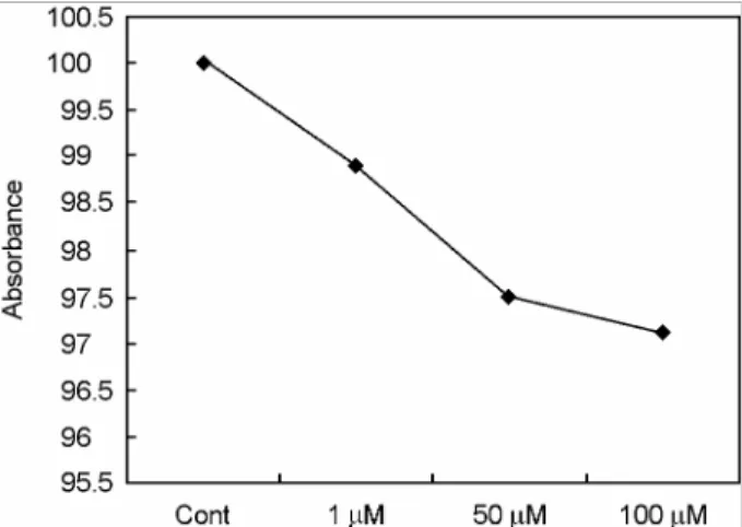
세포생존율 정량을 위해 ferulic acid가 1 µM, 50 µM, 100 µM 농도로 각각 포함된 배양액에서 정상인체치은섬유모세 포를 48시간 동안 배양한 다음 MTT를 정량분석하였다. 그 결과 MTT 흡광도는 대조군 [100% (3.92±0.16)]에 비하여 ferulic acid 1 µM의 농도에서는 98.9% (3.88±0.07)로 나타났 으며 50 µM의 농도에서는 97.5% (3.82±0.09)로 나타났다.
또한 100 µM의 ferulic acid의 처리에서는 대조군에 비하여 97.1% (3.80±0.10)로 나타났으나 모두 유의성은 나타나지 않 았다 (Table 1, Fig. 1).
2. 세포부착율 측정
세포부착율 정량을 위해 ferulic acid가 1 µM, 50 µM, 100 µM 농도로 각각 포함된 배양액에서 정상인체치은섬유모세 포를 48시간 동안 배양한 다음 XTT를 정량분석하였다. 그 결과 1 µM ferulic acid의 농도에서는 XTT 흡광도는 대조군 인 100% (3.98±0.13)에 비하여 99.9% (3.98±0.13)로 나타났 으며 50 µM의 농도에서는 97.7% (3.90±0.09)로 나타났다.
또한 100 µM의 ferulic acid의 처리에서는 대조군에 비하여 97.3% (3.88±0.06)으로 나타났으나 모두 유의성은 나타나지 않았다 (Table 2, Fig. 2).
3. 생존율의 MTT IC50 측정
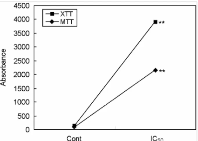
MTT50을 측정하기 위하여 정상인체치은섬유세포에 100 µM 이상의 ferulic acid가 각각의 농도로 포함된 배양액에서 48시간 동안 배양한 다음 MTT 정량을 측정하였다. 그 결과 MTT50은 2,130.0 µM에서 나타났다 (Table 3, Fig. 3).
4. 부착율의 XTT IC50 측정
XTT50을 측정하기 위하여 정상인체치은섬유세포에 100~
3,000 µM 이상의 ferulic acid가 각각의 농도로 포함된 배양 액에서 48시간 동안 배양한 다음 XTT 정량을 측정하였다.
Table 1. The cell viability of ferulic acid by MTT assay on normal human gingival fibroblast
Group MTT Concentration of
ferulic acid (µM) Mean ± S.D. (% of control) control 3.92±0.16 100
1 3.88±0.07 98.9
50 3.82±0.09 97.5
100 3.80±0.10 97.1 The normal human gingival fibroblasts were incubated with or without ferulic acid for 48 hours. The value represent the mean ± SD for triplicate experiments
Table 2. The adhesion activity of ferulic acid by XTT assay on normal human gingival fibroblasts
Group XTT Concentration (µM) Mean ± S.D. (% of control)
control 4.02±0.20 100
1 3.86±0.04 96.0
50 3.31±0.15 82.4**
100 2.93±0.06 72.9**
The normal human gingival fibroblasts were incubated with or without ferulic acid for 48 hours. The value represent the mean ± SD for triplicate experiments. Asterisks indicate significant diffe- rence from control. **P<0.01
Fig. 1. Cell viability was measured by MTT assay in normal human gingival fibroblasts after cells were treated with 1, 50, 100 µM picric acid for 48 hours, respectively.
Fig. 2. Cell viability was measured by XTT assay in normal human gingival fibroblasts after cells were treated with 1, 50, 100 µM picric acid for 48 hours, respectively.
그 결과 XTT50은 1,773.7 µM에서 나타났다 (Table 3, Fig. 3).
고 찰
Ferulic acid와 같은 천연성분은 식물로부터 추출되며 이러 한 식물성분은 세포에 대한 독성이 적기 때문에 각종 치료의 약품의 원료가 되고 있다 (Goldberg et al., 1999: De Heredia et al., 2001). 최근, 암의 예방이나 치료를 위한 신약개발을 각 종 천연물을 대상으로 활발히 진행되고 있는 이유도 여기에 있다 (Husniye et al., 1988: De Heredia et al., 2001). 근래에는 암의 치료보다는 암의 예방이나 암 전이에 대한 억제에 더 많은 연구가 이루어지고 있는데 그 이유는 일단 발암이 되면 완치가 어렵고 또한 치료 후에도 각종 부작용과 심각한 후유 증에 시달리게 됨으로서 무엇보다도 암의 예방이 최우선이 라는 차원에서 많은 투자와 노력이 가중되고 있다 (Ireland et al., 1987). 따라서 암 발생이나 전이에 대한 효과적인 억제 물질의 연구가 최대의 관심사로 부각되면서 신약개발도 이 에 호응하여 이루어지고 있다 (Ireland et al., 1987: 한 등, 1999: Heilmann et al., 2000). 천연물을 이용한 암 치료에 대한 약물개발에는 암의 진행과정에서 단계별로 특이적으로 생성 되는 물질, 즉 생화학적 표식자가 이용되고 있다 Mosmann, 1983: Ferguson, 2001). 이는 암의 촉진단계와 진행단계를 억 제하는 것으로서 여기에는 발암과정의 개시단계를 억제하 는 cytochrome P450효소의 억제물질을 비롯하여 carcinogen- DNA adduct 형성저해제, GSH 생성유도물질 등이 있으며 (양, 2003), 전이억제물질로는 laminin, fubronectin 및 CD44와 같은 부착단백과 세포신호전달억제제 등이 있다 (Xu et al., 1988: Sharma et al., 1994). 따라서 ferulic acid와 같은 페놀산 들의 항암효과는 이들 기전 중 일치되는 약리활성물질을 가 지고 있다는 것을 알 수 있다 (Husniye et al., 1988: Ferguson, 2001). 한편, 페놀화합물의 일종인 ferulic acid가 암세포에 대 한 항암작용에 유효한 약리작용을 가지고 있다고 알려져 있 어 암세포에 대한 독성효과가 있는 것으로 제시된 바 있으나 정상세포에 대한 세포독성효과에 대한 연구는 희소하다 (양,
2003). 따라서 본 연구에서는 ferulic acid가 정상세포인 인체 치은섬유모세포에 미치는 독성을 세포생존율과 세포부착능 의 측면에서 조사하였다. 먼저 MTT 정량에 의한 세포생존 율의 조사에서는 1~100 µM ferulic acid의 각각에 정상인체 치은섬유모세포를 48시간 동안 노출시킨 결과 농도에 비례 하여 대조군에 비하여 약간의 흡광도감소에 따른 세포생존 율이 저하된 것으로 나타났으나 통계적으로 거의 차이가 나 타나지 않아 유의성이 없는 것으로 나타났다. 이러한 실험결 과는 ferulic acid가 정상인체치은섬유모세포의 세포생존율에 아무런 영향을 미치지 않았다는 것을 말해주고 있으며 이는 세포독성을 가지고 있지 않다는 것을 증명해 주었다 (한 등, 2001: 양, 2003). 이와 동시에 MTT50 값을 측정한 결과 2,130.0 µM로 나타남으로서 이는 Borenfreud 등 (1988)의 독 성판정기준에 의하여 무독성인 것으로 나타났다. 이러한 이 유는 Borenfreud 등 (1988)은 검체물질의 MTT50 값이 100 µM 이하이면 고독성이라고 하였으며 1,000~2,000 µM이면 중간독성으로, 2,000 µM 이상이면 무독성으로 각각 판정하 였다. 한편, ferulic acid가 정상인체치은섬유모세포의 세포부 착능에 미치는 영향을 조사하기 위하여 MTT 정량분석에서 와 같이 1~100 µM ferulic acid가 각각 포함된 배양액에서 정 상인체치은섬유모세포를 48시간 동안 배양한 후 XTT 정량 분석을 한 결과 처리한 ferulic acid의 농도가 증가하였더라 도 흡광도는 대조군에 비하여 거의 차이가 없는 것으로 나타 남으로서 세포부착능에 유의성이 없는 것으로 나타났다.
본 실험의 결과는 ferulic acid가 정상인체치은섬유모세포 의 부착능에 별 영향을 미치지 않는 다는 것을 알 수 있으며 이는 또한 ferulic acid가 세포독성을 가지고 있지 않는 것으 로 나타났다 (De heredia et al., 2001: 양, 2003). 이의 결과와 연관하여 세포부착능에 대한 XTT50 값을 측정한 결과 본 실 Table 3. The of IC50 of cell viability and adhesion activity in
ferulic acid by MTT or XTT assays in normal human gingival fibroblasts
IC50 (µM) Cell line Cell viability
(MTT)
Adhesion activity (XTT) Normal human
gingival fibroblast 2,130.0 1,773.7 Cells were incubated with ferulic acid for 48 hours. IC50 of cell viability and cell adhesion activity were measured by MTT assay or XTT assay. The values represent the mean ± SD for triplicate experiments
Fig. 3. Cell viability and cell adhesion activity of IC50were measured by MTT and XTT assays in normal human gingival fibroblasts after cells were treated with ferulic acid for 48 hours, respectively.
험에서 1,773.7 µM로 나타났는데 이는 Borenfreud 등 (1988) 의 독성판정기준에 의하여 중간독성인 것으로 나타났다. 이 상의 실험결과들은 양 (2003)이 ferulic acid를 정상섬유모세 포에 처리한 결과 세포독성이 없었다는 연구결과와 일치하 였으며 동시에 본 연구에서도 페놀산의 일종인 ferulic acid 가 정상인체치은모세포에 세포생존율이나 세포부착능에 통 계적으로 유의한 영향을 미치지 않은 것으로 나타남으로서 정상세포에 독성을 보이지 않는 것으로 나타났다.
감사의 글
이 논문은 2003년도 원광대학교의 교비 지원에 의해서 수 행되었음.
REFERENCES
Borenfreund E, Babichi H, Matin-Alcuacil N. Comparisons of two in vitro cytotoxicity assay. The neutral red (NR) and tetrazolium MTT tests. Toxicol In Vitro. 1988. 2: 1-8.
Cao Y, Lou C, Fang Y, Ye J. Determination of active ingredients of Rhododendron dauricum L. by capillary electrophoresis with electrochemical detection. J Chromatogr A. 2002. 943:
153-157.
Cha HJ, Bae SK, Lee HY, Sato H, Seiki M, Park BC, Kim KW.
Anti-invasive activity of ursolic acid correlates with the reduced expression of matrix metalloproteinase-9 (MMP-9) in HT1080 human fibroblastoma cells. Cancer Res. 1996.
56: 2281-2284.
De Heredia JB, Torregrosa J, Dominguez JR, Peres JA. Kinetic model for phenolic compound oxidation by Fenton's reagent.
Chemosphere. 2001. 45: 85-90.
Ferguson LR. Role of plant polyphenols in genomic stability.
Mutat Res. 2001. 18: 89-111.
Goldberg DM, Hoffman B, Yang J, Soleas GJ. Phenolic consti- tuents, furans, and total antioxidant status of distilled spirits.
J Agric Food Chem. 1999. 47: 3978-3985.
Heilmann J, Calis I, Kirmizibekmez H, Schuhly W, Harput S, Sticher O. Radical scavenger activity of phenylethanoid gly- cosides in FMLP stimulated human polymorphonuclear leu- kocytes: structure-activity relationships. Planta Med. 2000.
66: 746-748.
Husniye Dilek F, Kucukali T. Mucin production in carcinomas of the uterine cervix. Eur J Obstet Gynecol Reprod Biol. 1998.
79: 149-151.
Ireland D, Cole S, Kelly P, Monaghan JM. Mucin production in cervical intraepithelial neoplasma and in stage 1b carcinoma of cervix with pelvic lymph node metastases. Br J Obstet Gynaecol. 1987. 94: 467-472.
Isuzugawa K, Ogihara Y, Inoue M. Different generation of inhi- bitors against gallic acid-induced apoptosis produces different sensitivity to gallic acid. Biol Pharm Bull. 2001. 24: 249-253.
Kolodziej H, Kayser O, Kiderlen AF, Ito H, Hatano T, Yoshida T, Foo LY. Antileishmanial activity of hydrolyzable tannins and their modulatory effects on nitric oxide and tumour necrosis factor-alpha release in macrophages in vitro. Planta Med.
2001. 67: 825-832.
Mosmann T. Rapid colorimetric assays for cellular growth and survival: Application to proliferation and cytotoxicity assays.
J Immunol Methods. 1983. 65: 55-63.
Sakagami H, Jiang Y, Kusama K, Atsumi T, Ueha T, Toguchi M, Iwakura I, Satoh K, Ito H, Hatano T, Yoshida T. Cytotoxic activity of hydrolyzable tannins against human oral tumor cell lines--a possible mechanism. Phytomedicine. 2000. 7: 39 -47.
Shahrzad S, Aoyagi K, Winter A, Koyama A, Bitsch I. Pharmaco- kinetics of gallic acid and its relative bioavailability from tea in healthy humans. J Nutr. 2001. 131: 1207-1210.
Sharma S, Jill DS, Kelloff GJ. Vernon ES. Screening of potential chemopreventive agents using biochemical markers of car- cinogensis. Cancer Res. 1994. 54: 5848-5855.
Shim JS, Kang MH, Kim YH, Roh JK, Roberts C. Chemopreven- tive effects of green tea (camellia sinensis) among cigarette smokers. Cancer Epidermiol Biomarkers Prev. 1995. 4: 387 -391.
Wattenberg LW. Chemoprevention of cancer. Cancer Res. 1985.
45: 1-8.
Xu L, Liu J, Min D, Wang S, Zhang Z, Guo D, Zheng K. Che- mical constituents of Conyza blinii Levl. Zhongguo Zhong Yao Za Zhi. 1988. 23: 293-295.
Yang HS. In Vitro Evaluation of Ferulic Acid and Vitamin A. The Korean J Anat. 2003. 27: 1-10.
Han DS, Lee SR, Jung WY. Studies on the cytotoxicity and Anti- neoplastic Activity of Methyl Gallate. The Korean J Anat.
2001. 34: 131-139.
Han SS, Lo SC, Choi YH, Kim MJ, Kwak SS. Antioxidative Compounds in Extracts of Accr ginnala Max. KJMCS. 1999.
7: 51-57.