TWO COLORIMETRIC ASSAYS VERIFY THAT CALCIUM SULFATE PROMOTES PROLIFERATING ACTIVITY OF HUMAN GINGIVAL FIBROBLASTS
Min Chae, D.D.S.1, Su-Yeon Kim, M.S., Ph.D.2, Soo-Yeon Kim, D.D.S., M.S.D.1, Suk-Won Lee, D.D.S., M.S.D.1
1Department of Dentistry, College of Medicine, The Catholic University of Korea
2Research Institute of Medical Science, St. Vincent’s Hospital
Statement of problem. The role of calcium sulfate in stimulating the growth of gingival soft tissue has been reported in few studies. Such a unique property of calcium sulfate could serve as a trouble-solving broker in compensating for the lack of soft tissues in various oral surgeries.
Purpose. The purpose of this study was to compare the proliferating activities of human gin- gival fibroblasts seeded on various bone graft barrier materials of calcium sulfate, collagen, and polytetrafluorethylene (PTFE).
Material and methods.Two calcium sulfates (CAPSET�and CalForma�, Lifecore Biomedical Inc., St. Paul, Minnesota, USA), a resorbable natural collagen (Bio-Gide�, Geistlich Pharma Ag., Wolhusen, Switzerland), and a non-resorbable PTFE (TefGen-FD�, Lifecore Biomedical Inc., St. Paul, Minnesota, USA) served as the human gingival fibroblasts’substrates and comprised the four experimental groups, whereas the untreated floors of culture plastics were used in the control group, in this study. Cells were trypsinized, seeded, and incubated for 48 h. The pro- liferating activities of fibroblasts were determined by XTT and SRB assay and absorbance (opti- cal density, OD) was measured. One-way ANOVA was used to analyze the differences in the mean OD values between the groups of CAPSET, CalForma, Bio-Gide, TefGen, and the con- trol (p<0.05).
Results. From the XTT assay, the mean OD value of the control group, the highest, was sig- nificantly greater than that of any of the four experimental groups followed by CalForma, CAPSET, TefGen, and Bio-Gide. Further, the mean OD value of CalForma, was significantly greater com- pared to that of Bio-Gide. From the SRB assay, Calforma showed the highest mean OD value, which was significantly greater than that of any other groups, followed by the control, CAPSET, Bio-Gide, and TefGen. The mean OD values of both the control and CAPSET were significantly greater compared to that of TefGen (p<0.05).
Conclusion.Assessment of the viability and proliferation of cultured fibroblasts seeded and incu- bated for 48 h on various barrier-material substrates using XTT and SRB assay showed that calci- um sulfate CalForma�promotes the proliferating activity of human gingival fibroblasts.
Key Words
Calcium sulfate, Human gingival fibroblast, XTT assay, SRB assay
J Korean Acad Prosthodont : Volume 45, Number 3, 2007
※This study was supported by SUHCHUN MDS Corporation, Seoul, Korea.
M
edically graded calcium sulfate hemi- hydrate (MGCSH), as a bone-graft material, has been widely used in oral surgery. However, few studies have reported the use of calcium sulfate as a barrier between the gingival soft tissue and the underlying bone graft material. Among those, one study reported the outstanding role of calcium sulfate in stimulating the growth of gingival soft tissue1. In the field of soft tissue esthetics in Dental Implantology, such a unique property of calcium sulfate could serve as a trouble-solv- ing broker for augmenting soft tissue implant coverage or in preventing peri-implant soft tissues from degenerating under various unfavorable clinical conditions. The purpose of this study was to compare the proliferating activities of human gingival fibroblasts plated on various bone graft barrier materials of calcium sulfate, resorbable collagen, and non-resorbable polyte- trafluorethylene (PTFE).MATERIAL AND METHODS CELL CULTURE
Healthy gingival tissues were obtained from patients who underwent oral surgery for remov- ing impacted wisdom teeth at St. Vincent’s Hospital Department of Dentistry. In all cases, tis- sues were obtained from subjects following informed consent as prescribed in an approved St.
Vincent’s Hospital Institutional Review Board (IRB) protocol. Tissues were incubated for 16-22 h in Hank’s balanced salt solution (HBSS, Gibco BRL, Life Technologies, Grand Island, NY, USA) at 4℃ for the purpose of separating connective tis- sue from epithelium. Obtained connective tis- sues were cut into small pieces and placed in Petri dishes (direct explant method) in Dulbecco’
s modified Eagle’s medium (DMEM, Gibco BRL,
Life Technologies, Grand Island, NY, USA) sup- plemented with penicillin G sodium (50 IU/ml), streptomycin sulfate (50mg/ml), and ampho- tericin B and were kept overnight at 4℃.
Cells or explants were washed 3 times in phos- phate-buffered salines (PBS, Gibco BRL, Life Technologies, USA) and suspended in DMEM sup- plemented with 10 % fetal bovine serum (FBS, Sigma-Aldrich Co., St. Louis, MO, USA), penicillin G sodium (50 IU/ml), streptomycin sulfate (50mg/ml), and amphotericin B. The composition and concentration of the solution were main- tained to be used as the culture medium through the entire procedure of this study (DMEM sup- plemented with 10% FBS and antibiotics).
Suspended fibroblasts were seeded into a T-75 flask (enzymatic dissociation) and incubated in a humidified incubator at 37℃ with 5% CO2in 95% air. When cells reached 80% confluence (about once per week), they were removed and sus- pended using a trypsin-EDTA solution (0.25%
trypsin and 0.1% glucose dissolved in 1 mM of EDTA-saline, Sigma-Aldrich Co., St. Louis, MO, USA), washed, centrifuged and resuspended.
Finally, human gingival fibroblasts were seeded for subculture at a cell population density of 2×
104cells/ml in 6-well plastic culture dishes in DMEM supplemented with 10% FBS and antibi- otics. In all experiments in this study, the culture medium was changed every second day after seeding.
PREPARATION OF SUBSTRATES
Powders of medically graded calcium sulfate hemi-hydrate bone graft barriers (CAPSET�and CalForma�, Lifecore Biomedical Inc., St. Paul, Minnesota, USA) were mixed with hydrated liq- uids according to the manufacturers’recom- mendations and solidified for 1 h on the floors of 24-well plates. The bone graft barrier membranes
of resorbable natural collagen (Bio-Gide�, Geistlich Pharma Ag, Wolhusen, Switzerland) and non- resorbable PTFE (TefGen-FD�, Lifecore Biomedical Inc., St. Paul, Minnesota, USA) were trimmed to an appropriate size and shape to be attached onto the floors of 24-well plates using a cyano- acrylate adhesive and a silicone bonding agent. The commercially available barrier materials served as the fibroblasts’substrates and comprised the four experimental groups, whereas the untreat- ed floors of 24-well plates were used in the con- trol group, in this study.
XTT ASSAY
The viability and proliferation of fibroblasts were determined by an XTT assay (Cell Proliferation Kit II, Roche Applied Science, Mannheim, Germany) as described by Roehm et al.2 Cultured fibroblasts (3rd-4th passage) were trypsinized and seeded on the prepared sub- strates of CAPSET, CalForma, Bio-Gide, and TefGen as well as on the control substrates at a cell population density of 4×10⁴cells/ml in DMEM supplemented with 10% FBS and antibiotics.
Fibroblasts were incubated in a humidified incu- bator at 37℃ with 5% CO2in 95% air for 48 h.
XTT (soium 3’-[1-[(phenylamino)-carbonyl]- 3,4-tetrazolium]-bis(4-methoxy-6- nitro)benzene- sulfonic acid hydrate) labeling reagent and elec- tron coupling reagent (N-methyl dibenzopy- razine methyl sulfate, PMS in phosphate-buffered salines, PBS) were thawed. Each vial was thor- oughly mixed and a clear solution was obtained.
An XTT labeling mixture was prepared by mix- ing 50 μl of XTT labeling reagent and 1 μl of electron coupling reagent. 50 μl of XTT labeling mixture was added per well and incubated for 2 h in a humidified incubator at 37℃ with 5% CO2 in 95% air. Absorbance (optical density, OD) of pro- duced formazan transferred to 96-well plates
was measured using ELISA analyzer (Spectra MAX 250, Molecular Devices Co., Sunnyvale, CA, USA) at 470 nm with a reference wave- length at 650 nm. All experiments were inde- pendently repeated in triplicate using newly prepared substrates.
SRB ASSAY
To assess the proliferation of fibroblasts by measuring their total protein content, a sul- forhodamine B (SRB) assay was performed (sul- forhodamine B sodium salt, Sigma-Aldrich, St.
Louis, MO, USA). Materials and methods used in fibroblast plating and incubation on five different substrates were identical to those used in the XTT assay. 50 ml of cold (4℃) 50% TCA (trichloroacetic acid) were gently added in each well. Plates were left for 30 min at 4℃ and sub- sequently washed 5 times with distilled water.
Plates were then left to dry at room tempera- ture for 24 h. 100 μl 0.4% SRB dye solution (0.4%
SRB dissolved in 1% acetic acid solution) was added to each well and left at room temperature for 30 min. SRB was removed and the plates were washed 5 times with 1% acetic acid before air drying. Bound SRB was solubilized with 150 μl 10 mM unbuffered Tris-base solution and transferred to 96-well plates. Plates were left on a plate shaker for 10 min. Absorbance was mea- sured using ELISA analyzer (Spectra Max 250, Molecular Devices Co., Sunnyvale, CA, USA) at 470 nm with a reference wavelength at 650 nm. All experiments were repeated independently in triplicate using newly prepared substrates.
STATISTICAL ANALYSIS
The mean OD values and the standard deviations of the data from both the XTT and SRB assays were calculated. One-way analysis of variance (ANO-
VA) was used to analyze the differences in the mean OD values between the groups of CAPSET, CalForma, Bio-Gide, TefGen, and the control (p<0.05).
SCANNING ELECTRON MICROSCOPY
Adhesion and morphology of fibroblasts on the surfaces of various bone graft barriers was ana- lyzed under scanning electron microscopic (SEM) observations. At 24 h incubation, samples were fixed in 4% paraformaldehyde for 2 h and rinsed twice in 0.1M PBS for 10 min. Samples were again fixed for another 2 h in 1% osmium tetrox- ide. Samples were then dehydrated by immers- ing for 10 min in each of 20, 50, 60, 70, 80, 90, and 100% ethanol dilutions followed by drying with a critical point dryer (Jumb, Bio-Rad Hercules. CA, USA). Samples were sputter coated with a 10-nm gold film (SEM coating system, E 5150, Bio-Rad Hercules. CA, USA) and imaged with JSM-5410 LV�SEM (Jeol, Tokyo, Japan).
RESULTS
ANALYSIS OF VARIANCE
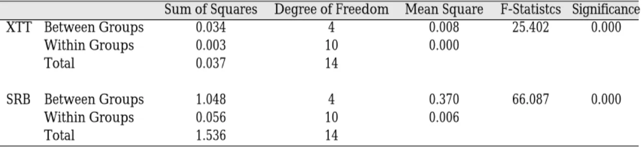
In ANOVA, the mean OD values from both the XTT and SRB assays at 48 h incubation were significantly different between and within all
groups (p<0.05). It was noted that, according to the data using different substrates, the results of the two colorimetric analyses were signifi- cantly related (Table I).
MULTIPLE COMPARISON TESTS
Multiple comparisons of the fibroblasts’via- bility/proliferation data from the XTT assay showed the mean OD value of the control group (0.22), the highest among all groups, to be sig- nificantly greater than that of any of the four experimental groups using barrier-material sub- strates such as CalForma (0.16), CAPSET (0.12), TefGen (0.11), and Bio-Gide (0.08) (p<0.05) (Fig.
1). Further, the mean OD value of CalForma was significantly greater compared to that of Bio-Gide (p<0.05). All other comparisons between groups were not statistically significant.
Comparative analysis on the fibroblasts’total protein content from the SRB assay revealed that CalForma showed the highest mean OD value (1.07), which was significantly greater than that of any other groups (p<0.05) such as the con- trol (0.43), CAPSET (0.38), Bio-Gide (0.35), and TefGen (0.15) (Fig. 2). Also, the mean OD values of both groups of control and CAPSET were sig- nificantly greater compared to that of TefGen (p<0.05). All other comparisons between groups were not statistically significant.
Table I. Analysis of variance (p<0.05)
Sum of Squares Degree of Freedom Mean Square F-Statistcs Significance
XTT Between Groups 0.034 4 0.008 25.402 0.000
Within Groups 0.003 10 0.000
Total 0.037 14
SRB Between Groups 1.048 4 0.370 66.087 0.000
Within Groups 0.056 10 0.006
Total 1.536 14
SCANNING ELECTRON MICROSCOPY
Human gingival fibroblasts plated on CAPSET (Fig. 3), CalForma (Fig. 4), and Bio-Gide (Fig. 5) appeared nearly ovoid. Roughness was observed on the surfaces of CAPSET and CalForma with the latter appearing rougher. Collagen fibers were observed on the surface of Bio-Guide.
DISCUSSION
The two colorimetric assays, XTT and SRB, used in this study are methods well established in investigating the influence of specific sub- stance and/or material on cell survival. The Fig. 1. Mean optical density values from XTT assay. Fig. 2. Mean optical density values from SRB assay.
Fig. 3. SEM view of human gingival fibroblasts adhered to CAPSET.
Fig. 5. SEM view of human gingival fibroblasts adhered to Bio-Gide.
Fig. 4. SEM view of human gingival fibroblasts adhered to CalForma.
results of the XTT assay for cell viability and proliferation are influenced not only by the num- ber but also by the metabolic activity of cells.
Cleavage of XTT by dehydrogenase enzymes of metabolically active cells yields a highly colored formazan product which is water soluble. This fea- ture enables the product to be diluted in medium, eliminates the need for formazan crystal solubi- lization prior to absorbance measurements as is required in MTT assay, protects cellular membranes from being damaged, and leads the cells to sur- vive throughout the test process. The development of sulforhodamine B (SRB) protein staining assay for the in vitro measurement of cellular protein con- tent of adherent and suspension cultures was established by Skehan et al. 3. The dye binds to basic amino acids of cellular proteins and colorimetric evaluation provides an estimate of total protein mass which is closely related to cell number.
The advantages of SRB assay over tetrazoli- um-based assays include higher sensitivity as well as better linearity to cell number and allow a correct estimation of cell proliferation.
Tetrazolium-based assays such as the XTT assay used in this study tend to exhibit a slightly hyper- bolic cell number against the optical density curve in adherent cells, leading to an error in the results. Since adherent fibroblasts are fre- quently used in in vitro assays, they, too, can display such an error. The results from SRB assay could be considered more reliable than those from XTT assay in relation to changes in pro- liferation (cell number) rather than changes in meta- bolic activity (viability). Indeed, our result from SRB assay strongly suggests that calcium sul- fate CalForma�stimulates human gingival fibrob- lasts’proliferation related to cell number. On the other hand, in SRB assay, an addition of 50 μl 50%
cold trichloroacetic acid (TCA) to the medium is necessary for cell fixation and this TCA fixes both cells and proteins such as fetal bovine serum contained in the medium, a phenomenon called
background staining. Together with any shearing force generated to dislodge the adherent cells at any step in SRB assay, background staining caus- es an error in the results4.
PTFE, Teflon, was originally developed in the industry to replace polyamides (nylons) in that it displays superior resistance to chemicals and friction. These characteristics are considered to yield another characteristic related to a decrease in wettability. Barely adhered cells to the poorly wettable PTFE surface were assumed to be easi- ly dislodged by forces generated from any manip- ulation such as adding or stirring up liquid solu- tions to/in the wells. Indeed, fibroblasts were rarely discovered on the surface of PTFE barrier mem- brane in SEM which is again, verified from XTT and SRB assays. On the contrary, cells were assumed to be strongly adhered to the highly wet- table calcium sulfate substrate, basically a gypsum product, which easily absorbs water even after solidification. These assumptions are based on the hypothesis corresponding with Altankov et al. that cell proliferation increases with increasing substrate surface wettability5.
The results of this study provide further evidence to support the findings by Hanein et al. that even subtle differences in inorganic substrata can have major effects on cell adhesion6. In this study, these differences can be considered in several ways. A thorough review by Discher et al.
made it clear that the contractile state of a cell can be strongly influenced by the stiffness of the anchoring substrate and that the contractile trac- tion forces exerted by a cell tend to increase with the stiffness of its substrate7. These intracellular forces, also called cytoskeletal tension per se or cytoskeletal prestress, together with extracellular matrix (ECM) and cytoskeletal structure are con- sidered to play decisive roles in the control of var- ious biological activities, including cell prolif- eration and growth8. Among the materials used as the substrates for cell culture in this study, the
floors of untreated culture plastics were consid- ered to be the stiffest followed by the solidified cal- cium sulfate materials. Results from both assays support the role of the substrate stiffness in alter- ing fibroblasts’viability and proliferation.
Considering cell migration as one of the essentials in proliferation, our result from the XTT assay cor- responds with that of a previous study9. Using var- ious barrier materials as substrates, the study concluded that human gingival fibroblasts migrat- ed most extensively over the normal culture plastic followed by calcium sulfate compared with PTFE or polylactic acid.
A hypothesis of cellular mechanotransduction, where alterations in substrate surface topography may lead to changes in the probability of gene tran- scription, is another point to be considered in this study10. The fact that a significant difference in the mean OD values from the SRB assay was present between CAPSET and CalForma, which are both calcium sulfates, suggests that substrate surface topography actually altered biological activities of cells which might have resulted from changes in gene expression. In SEM observa- tions, surface particles of CalForma appeared greater in size compared with those of CAPSET suggesting that the surface of CalForma is rougher.
Substrate chemistry, one of the important factors in cell-substrate interactions, is inappropriate for discussing the differences in the fibroblasts’pro- liferating activities between the groups because some of the materials used in this study were organ- ic while others were not. However, it would be very interesting to analyze the surface chemistry of various organic as well as inorganic barriers to determine the differences in the biological activities of cells that selectively adhere to such materials.
CONCLUSION
Cultured fibroblasts were plated and incubat- ed for 48 h on 1) two medically graded calcium sul-
fate hemi-hydrate bone graft barriers CAPSET� and CalForma�, 2) a resorbable natural collagen barrier membrane Bio-Gide�, and 3) a non- resorbable polytetrafluorethylene barrier mem- brane TefGen-FD�. Assessment of the viability and proliferation using XTT and sulforhodamine B assay showed that calcium sulfate CalForma� promotes the proliferating activity of human gingival fibroblasts.
REFERENCES
1. Anson D. Calcium sulfate-augmented soft tissue root coverage adjacent to connective tissue graft- ing: a new technique. Int J Periodontics Restorative Dent 2003;23:337-343.
2. Roehm NW, Rodgers GH, Hatfield SM, Glasebrook AL. An improved colorimetric assay for cell pro- liferation and viability utilizing the tetrazolium salt XTT. J Immunol Methods 1991;142:257-265.
3. Skehan P, Storeng R, Scudiero D, Monks A, McMahon J, Vistica D, Warren JT, Bokesch H, Kenney S, Boyd MR. New colorimetric cytotoxic- ity assay for anticancer-drug screening. J Natl Cancer Inst 1990;82:1107-1112.
4. Papazisis KT, Geromichalos GD, Dimitriadis KA, Kortsaris AH. Optimization of the sulforhodamine B colorimetric assay. J Immunol Meth 1997;208:151-158.
5. Altankov G, Grinnell F, Groth T. Studies on the bio- compatibility of materials: fibroblast reorganiza- tion of substratum-bound fibronectin on surfaces varying in wettability. J Biomed Mater Res 1996;30:385-391.
6. Hanein D, Geiger B, Addadi L. Differential adhesion of cells to enantiomorphous crystal surfaces.
Science 1994;263:1413-1416.
7. Discher DE, Janmey P, Wang YL. Tissue cells feel and respond to the stiffness of their substrate.
Science 2005;310:1139-1143.
8. Ingber DE. Tensegrity II. How structural net- works influence cellular information processing net- works. J Cell Sci 2003;116:1397-1408.
9. Payne JM, Cobb CM, Rapley JW, Killoy WJ, Spencer P. Migration of human gingival fibroblasts over guided tissue regeneration barrier materials.
J Periodontol 1996;67:236-244.
10. Dalby MJ. Topographically induced direct cell mechanotransduction. Med Eng Phys 2005;27:730-741.
Reprint request to:
SUK-WONLEE, D.D.S., M.S.D.
DEPARTMENT OFDENTISTRY,COLLEGE OFMEDICINE,
THECATHOLICUNIVERSITY OFKOREA, ST.VINCENT’S HOSPITAL
96-3, JI-DONG, PALDAL-GU,SUWON,442-723, KOREA