Received February 15, 2016, Revised March 25, 2016, Accepted for publication April 11, 2016
*These authors contributed equally to this study.
Corresponding author: Kyu Joong Ahn, Department of Dermatology, Konkuk University School of Medicine, 120 Neungdong-ro, Gwangjin-gu, Seoul 05029, Korea. Tel: 82-2-2030-5181, Fax: 82-2-2030-5179, E-mail: kjahn@
kuh.ac.kr
Su Young Kim, Department of Bioengineering, Graduate School of Engi- neering, Konkuk University, 120 Neungdong-ro, Gwangjin-gu, Seoul 05029, Korea. Tel: 82-2-450-4054, Fax: 82-502-770-2278, E-mail:
This is an Open Access article distributed under the terms of the Creative Commons Attribution Non-Commercial License (http://creativecommons.
org/licenses/by-nc/4.0) which permits unrestricted non-commercial use, distribution, and reproduction in any medium, provided the original work is properly cited.
Copyright © The Korean Dermatological Association and The Korean Society for Investigative Dermatology
Ann Dermatol Vol. 28, No. 6, 2016 https://doi.org/10.5021/ad.2016.28.6.740
ORIGINAL ARTICLE
Pretreatment of Ferulic Acid Protects Human Dermal Fibroblasts against Ultraviolet A Irradiation
Hyung Jin Hahn, Ki Bbeum Kim1, Seunghee Bae1, Byung Gon Choi2, Sungkwan An1, Kyu Joong Ahn2,*, Su Young Kim1,*
Department of Dermatology, Konyang University College of Medicine, Daejeon, 1Department of Biological Engineering, Graduate School of Engineering, Konkuk University, 2Department of Dermatology, Konkuk University School of Medicine, Seoul, Korea
Background: Approximately 90%∼99% of ultraviolet A (UVA) ray reaches the Earth’s surface. The deeply penetrating UVA rays induce the formation of reactive oxygen species (ROS), which results in oxidative stress such as photo- products, senescence, and cell death. Thus, UVA is consid- ered a primary factor that promotes skin aging. Objective:
Researchers investigated whether pretreatment with ferulic acid protects human dermal fibroblasts (HDFs) against UVA-induced cell damages. Methods: HDF proliferation was analyzed using the water-soluble tetrazolium salt assay.
Cell cycle distribution and intracellular ROS levels were as- sessed by flow cytometric analysis. Senescence was eval- uated using a senescence-associated β-galactosidase assay, while Gadd45α promoter activity was analyzed through a luciferase assay. The expression levels of superoxide dis- mutase 1 (SOD1), catalase (CAT), xeroderma pigmentosum complementation group A and C, matrix metalloproteinase 1 and 3, as well as p21 and p16 were measured using quanti-
tative real-time polymerase chain reaction. Results: Inhibi- tion of proliferation and cell cycle arrest were detected in cells that were irradiated with UVA only. Pretreatment with ferulic acid significantly increased the proliferation and cell cycle progression in HDFs. Moreover, ferulic acid pretreat- ment produced antioxidant effects such as reduced DCF in- tensity, and affected SOD1 and CAT mRNA expression.
These effects were also demonstrated in the analysis of cell senescence, promoter activity, expression of senescent markers, and DNA repair. Conclusion: These results demon- strate that ferulic acid exerts protective effects on UVA-in- duced cell damages via anti-oxidant and stress-inducible cel- lular mechanisms in HDFs. (Ann Dermatol 28(6) 740∼748, 2016)
-Keywords-
Cell aging, DNA repair, Ferulic acid, Reactive oxygen spe- cies, Ultraviolet rays
INTRODUCTION
Solar radiation in the form of ultraviolet (UV) light is div- ided into three types; ultraviolet A (UVA, 320∼400 nm), ultraviolet B (UVB, 280∼320 nm), and ultraviolet C (UVC, 200∼280 nm)1. However, terrestrial solar UV radi- ation is comprised mostly of UVA and partially of UVB because wavelengths shorter than 295 nm are blocked by the ozone layer in the stratosphere2. Although UVA pho- tons are about 1,000 times less energetic than UVB, they can still penetrate the skin to cause skin aging in the der- mis by regulating the extracellular matrix (ECM)3-5. Furthermore, UVA has been reported to produce reactive oxygen species (ROS) which cause oxidative stress, lead-
ing to cell death6,7. Therefore, UVA is one of the major factors of photoaging in skin.
As the largest organ of the human body, skin surrounds and protects our bodies from the external environment1,3,4,8. Skin is composed of two layers, namely the epidermis and dermis layer, which is composed of connective tissue in- cluding fibroblasts, matrix proteins, and other substances.
Studies have shown that human skin fibroblasts are the main components of the dermis and are more receptive to UVA exposure than keratinocytes, resulting in skin re- construction9. This is one symptom of photoaging, which is caused by UVA photons and UVA-induced ROS in the fibroblasts10,11. More interestingly, UVA has also been re- ported to stimulate fibroblasts to synthesize metallopro- teinase 1 (MMP1), which degrades dermal collagen10,12. Due to the diverse function of skin biology, fibroblasts have been studied mainly to understand the pathology fol- lowing exposure to toxins, chemicals, and cosmetics.
Therefore, it is necessary to find safe and effective natural products for human skin protection.
Ferulic acid (4-hydroxy-3-methoxycinnamic acid) is wide- ly present in fruits, vegetables, and grains. According to previous studies, ferulic acid has antioxidant and anti- cancer properties13-15. Ferulic acid has been shown to im- part beneficial effects in diabetes, Alzheimer’s disease, and cardiovascular disease by regulating antioxidant en- zyme and caspase activities, COX-2, and hypertension16-18. In the skin, ferulic acid has been shown to have a pro- tective effect on UVB-induced erythema19. The current study is aimed at investigating how ferulic acid protects human dermal fibroblasts (HDFs) against UVA radiation.
MATERIALS AND METHODS
Cell culture
Normal human dermal fibroblasts (nHDF; Lonza, Basel, Switzerland) were cultured in Dulbecco’s modified Eagle medium (DMEM; Gibco/Life Technologies, Carlsbad, CA, USA) supplemented with 10% fetal bovine serum (FBS;
Sigma-Aldrich, St. Louis, MO, USA) and 1% penicillin/
streptomycin (Gibco/Life Technologies) at 37oC in an at- mosphere of 5% CO2. Ferulic acid was purchased from Sigma-Aldrich and dissolved in dimethyl sulfoxide.
UVA irradiation
The HDFs (1×10⁶/well) were seeded into 6-well plates and cultured until 80%∼90% confluent. Prior to irradi- ation, cells were washed twice with phosphate buffered saline (PBS). The cells were irradiated with UVA light (UVA lamp; UVP, Upland, CA, USA) in fresh PBS. The ra- diation intensity was monitored by a fiberoptic spec-
trometer system USB2000 (Ocean optics, Dunedin, FL, USA). Control cells were treated identically but were not exposed to UVA irradiation. After UVA radiation, fresh medium was added to the cells and the cells were in- cubated at 37oC for 24 h.
Cell viability assay
HDF cell toxicity due to ferulic acid was evaluated using a water-soluble tetrazolium salt (WST-1) assay (EZ-Cytox Cell Viability Assay kit; Itsbio, Seoul, Korea). HDF cells were seeded at a density of 3×10³ cells into 96-well plates and incubated for 24 h. Then, the cells were in- cubated with ferulic acid (0∼50 μM) for 24 h. A WST-1 solution was added to cultured cells, which were then in- cubated at 37oC for 1 h. Cell viability was evaluated by measuring the absorbance at 450 nm using an iMark mi- croplate reader (Bio-Rad, Hercules, CA, USA). In order to examine the protective effects of ferulic acid against UVA, nHDFs were pretreated with FA for 6 h, and then the cells were washed with PBS followed by UVA irradiation. After UVA irradiation, PBS was replaced with normal growth medium and incubated 24 h. After incubation, WST-1 assay was performed to analyze the UVA protection effect of FA on cell viability of nHDFs.
Analysis of cell cycle by flow cytometry
Cells were collected and fixed in cold 70% ethanol at 4oC for 1 h. Then the fixed cells were stained by incubation with a propidium iodide (PI) staining solution (50 μg/ml PI, 0.5% Triton X-100 [both from Sigma-Aldrich] plus 100 μg/ml RNase) at 37oC for 1 h. Changes in cell cycle were determined based on the intensity of fluorescent PI stain- ing of 10,000 cells using the FL2-H channel of a FACSCalibur flow cytometer (BD Biosciences, San Jose, CA, USA)20.
Reporter transfection and transient luciferase activity assay
The Gadd45α promoters were cloned into pGL3 to acti- vate the firefly luciferase reporter vector (Promega, Madison, WI, USA). HDFs seeded into 96-well plates were cotransfected with pGL3-Gadd45α together with the pSV-β-galactosidase plasmid (Promega) using Lipo- fectamine 2000 reagent (Invitrogen/Life Technologies, Carlsbad, CA, USA). At 24 h post-transfection, cells were exposed to UVA radiation with or without ferulic acid treatment. At 24 h post-UVA radiation, cells were lysed using Passive lysis buffer (Promega). Luciferin was then added and the luciferase activity of each cell lysate was analyzed using a Veritas Luminometer (Turner Designs, Sunnyvale, CA, USA). The luciferase signal was normal-
ized to β-galactosidase activity and is presented as a per- centage of the control with standard deviation (SD).
Results shown represent the average of three independent experiments.
ROS scavenging assay
Intracellular ROS scavenging assays were performed by measuring the fluorescence intensity of the 2',7'-dichloro- fluroescein diacetate (DCF-DA) probe, which was propor- tional to the amount of ROS produced. Cells pre-treated with and without ferulic acid were irradiated with UVA prior to harvest. The cells were then mixed with DCF-DA solution and incubated at 37oC for 1 h. Fluorescence in- tensity was measured using a BD FACSCalibur flow cy- tometer (BD Biosciences).
Senescence-associated β-galactosidase activity
β-galactosidase expression was used as a marker for sen- escence in HDFs. Expression levels were determined us- ing the senescence-associated-β-galactosidase staining kit (Biovision, Milpitas, CA, USA) according to the manu- facturer’s protocol. HDFs were seeded at a density of 1×10⁶ cells/well in 60-mm cell culture plates and in- cubated until cells reached 90% confluence. Then cells were pretreated with 10 or 20 μM ferulic acid for 6 h be- fore irradiation with 10 J/cm2 UVA. After UVA radiation, the cells were washed with fresh media and incubated for another 24 h. To evaluate cellular senescence, the cells were washed in PBS prior to incubation in 0.5 ml fixing solution (4% formaldehyde, 0.5% glutaraldehyde in PBS buffer, pH 7.2) for 1 h. Fixed cells were then incubated in a staining solution mixture (staining solution [470 μl], staining supplement [5 μl], 20 mg/ml X-Gal in dime- thylformamide [25 μl]) for 24 h at 37oC. Finally, 70%
glycerol (1 ml) was added to the cells and images were captured using an Olympus IX51 microscope (Olympus, Tokyo, Japan).
RNA isolation and quantitative real-time polymerase chain reaction analysis
Total RNA was isolated using the TRIzol reagent (Life Technologies) according to the manufacturer’s protocol.
RNA purity and concentration were evaluated using the MaestroNanoⓇ microspectrophotometer (Maestrogen, Las Vegas, NV, USA). All cDNAs for sensitive and specific miRNA detection were synthesized using the miScript II RT kit (Qiagen, Hilden, Germany) according to the manu- facturer’s protocol. To evaluate the expression of p21 (forward primer: 5'-GTCCAGCGACCTTCCTCATCCA-3', reverse primer: 5'-CCATAGCCTACTGCCACCATC-3'), p16 (forward primer: 5'-CCCAACGCCCCGAACT-3', reverse
primer: 5'-GCAGAAGAGCTGCTACGTGAA-3'), SOD1 (for- ward primer: 5'-GGGAGATGGCCCAACTACTG-3', reverse primer: 5'-CCAGTTGACATGCAACCGTT-3'), MMP1 (for- ward primer: 5'-TCTGACGTTGATCCCAGAGAGCAG-3', reverse primer: 5'-CAGGGTGACACCAGTGACTGCAC-3'), MMP3 (forward primer: 5'-ATTCCATGGAGCCAGGCTTTC- 3', reverse primer: 5'-CATTTGGGTCAAACTCCAACTGTG- 3'), xeroderma pigmentosum complementation group A (XPA, forward primer: 5'-CCAGGACCTGTTATGGAAT TTGA-3', reverse primer: 5'-GCTTCTTGACTACCCCAAA CTTC-3'), xeroderma pigmentosum complementation group C (XPC, forward primer: 5'-AGCAGCTTCCCACC TGTTC-3', reverse primer: 5'-GTGGGTGCCCCTCTAGTG- 3'), and CAT (forward primer: 5'-TGGAGCTGGTAACCCA GTAGG-3', reverse primer: 5'-CCTTTGCCTTGGAGTATT TGGTA-3'), quantitative real-time polymerase chain reaction (RT-PCR) was performed using EvaGreen dye (Solis BioDyne, Tartu, Estonia) and Line-Gene K software (BioER, Hangzhou, China). The CT value for each gene was normalized to that of β-actin (forward primer: 5'-GGATTCCTATGTGGGCG ACGA-3', reverse primer: 5'-CGCTCGGTGAGGATCTTCA TG-3'). The 2−ΔΔCt method was used to calculate the rela- tive expression level of each gene21.
Statistical analysis
Analysis was performed using SPSS for Windows ver. 21.0 (SPSS Inc., Chicago, IL, USA). All results are presented as the mean percentage±SD of three independent experi- ments. Differences with a p-value less than 0.05, as de- termined by the Student’s t-test, were considered statisti- cally significant.
RESULTS
Ferulic acid reduces UVA-induced cytotoxicity of HDFs Before evaluating the protective effects of ferulic acid in UVA-induced cell death and inhibition of proliferation, we examined the cytotoxicity of ferulic acid in HDFs. The cells were treated with ferulic acid (0∼50 μM) for 24 h, and viability was determined using the WST-1 assay (Fig.
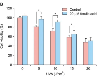
1A). Cell viability was greater than 90% at ferulic acid concentrations below 50 μM concentration. To determine whether ferulic acid affects the viability of HDFs under- going UVA-induced cellular stress, cultured cells were ir- radiated with various intensities of UVA (0∼20 J/cm2) and incubated for 24 h. As shown in Fig. 1B, HDF viability de- creased drastically after UVA radiation. However, pretreat- ment with 20 μM ferulic acid exhibited a protective effect up to 10 J/cm2 (Fig. 1B). This protective effect of ferulic acid on UVA-induced cell death was dose-dependent at 10 J/cm2 UVA intensity (Fig. 1C).
Fig. 1. Effects of ferulic acid on human dermal fibroblast (HDF) cell proliferation. HDF cells (3×10³ cells/well) were seeded into 96-well plates and treated with the indicated concentrations of ferulic acid for 24 h. Proliferation was measured using the water-soluble tetrazolium salt assay. (A) Cell viability following ferulic acid treatment in HDFs. (B) Protective effects of treatment with 20 μM ferulic acid against cytotoxicity induced by varying intensities of ultraviolet A (UVA) radiation in HDFs. Cells were pretreated with ferulic acid for 6 h prior to UVA radiation. (C) Protective effects of ferulic acid treatment against 10 J/cm2 UVA radiation. Cells were treated with the indicated concentration of ferulic acid for 6 h before UVA radiation. *p<0.05 by Student’s t-test.
Ferulic acid reduces UVA-induced G1 cell cycle arrest and DNA repair on HDFs
We investigated the effects of ferulic acid on cell cycle progression via PI staining followed by flow cytometric analysis. Our data demonstrate that treatment with 10 J/cm2 of UVA led to G1-phase arrest, and that pretreat- ment with ferulic acid protected cell cycle progression in a dose-dependent manner. Cells treated with UVA only exhibited significantly increased G1-phase cells (up to 67.9%) relative to untreated cells. This increase in G1- phase cells was reduced by pretreatment with ferulic acid at the indicated concentrations (Fig. 2A). Next, we exam- ined Gadd45α expression using a promoter luciferase ac- tivity assay. Gadd45α, a stress-inducible gene, has been implicated in cell cycle arrest, apoptosis, and DNA re- pair22,23. HDFs treated with UVA only showed increased luciferase activity (up to 4.7-fold) compared with un- treated cells. However, cells pretreated with ferulic acid for 6 h before UVA radiation exhibited significantly de- creased Gadd45α luciferase activity in a dose-dependent
manner (Fig. 2B). p21 has been reported to be a key regu- lator of G1- and G2-phase arrest, as well as aging and photoaging24,25. In addition, p21 is involved in Gadd45α as cell cycle regulation22,26. Therefore, we examined the relative expression of p21 mRNA in ferulic acid-pretreated HDFs irradiated with UVA using quantitative RT-PCR. As shown in Fig. 2C, cells only irradiated with UVA (10 J/cm2) displayed a greater than 5-fold increase in p21 mRNA expression relative to non-irradiated cells. However, cells pretreated 10 and 20 μM ferulic acid before UVA radiation showed a 4.8 (±0.5)- and 2.7 (±0.2)-fold in- crease, respectively. We also assessed the expression of DNA repair genes in HDFs following UVA radiation and ferulic acid pretreatment. XPA and XPC are core nucleo- tide excision repair (NER) factors expressed during UV-in- duced DNA lesion repair27. Studies have also reported that photolyase enzymes such as XPA and XPC can repair UV-induced DNA lesions efficiently28-31. Our data demon- strate that UVA irradiation reduced the expression of both XPA and XPC, whereas cells pretreated with ferulic acid showed significantly increased expression of both genes
Fig. 2. Effects of ferulic acid on ultraviolet A (UVA)-induced cell cycle arrest and DNA repair in human dermal fibroblasts (HDFs).
(A) UVA-induced cell cycle arrest was reduced by ferulic acid. Cell cycle progression was evaluated by propidium iodide staining followed by flow cytometric analysis. (B) UVA-induced upregulation of Gadd45α promoter activity was decreased by ferulic acid.
The Gadd45α promoter regions were cloned into the pGL3 luciferase reporter vector and then transfected into HDFs. Luciferase intensity was normalized against β-galactosidase activity. (C) Relative gene expression of p21 in human dermal fibroblasts (HDFs).
HDFs were irradiated with ultraviolet A (UVA) followed by ferulic acid. (D) Ferulic acid enhanced mRNA expression of nucleotide excision repair (NER) factors, xeroderma pigmentosum complementation group A (XPA) and xeroderma pigmentosum complementation group C (XPC). Pretreatment with ferulic acid promoted NER-associated genes expression. Gene expression was normalized against β-actin and the 2−ΔΔCt method was used to calculate the relative expression level. Results are expressed as the mean±standard deviation in triplicate. *p<0.05 by Student’s t-test.
(Fig. 2D). These results demonstrate that 10 J/cm2 UVA ra- diation resulted in G1-phase cell cycle arrest and DNA damage in HDFs, but that pretreatment with ferulic acid inhibited UVA-induced cell cycle arrest and DNA damage by regulating gene expression associated with cell cycle progression and DNA repair in a dose-dependent manner.
Ferulic acid reduces UVA-induced oxidative stress on HDFs
UVA induces intracellular ROS production that results in oxidative stress and DNA damage in cells32-34. Thus, we
evaluated the ROS scavenging effects of ferulic acid by an- alyzing DCF-DA fluorescent intensity. As shown in Fig.
3A, formation of intracellular ROS increased following ra- diation with 10 J/cm2 UVA. The increased intracellular ROS production was decreased by pretreatment with 10 and 20 μM of ferulic acid. Interestingly, treatment with N-acetylcysteine (NAC), a ROS scavenger used as a pos- itive control, showed no significant scavenging effects at 20 μM, which was the same concentration of ferulic acid used. Furthermore, we investigated the antioxidant effects of ferulic acid by relative gene expression analysis using
Fig. 3. Effects of ferulic acid on ultraviolet A (UVA)-induced oxidative stress in human dermal fibroblasts (HDFs). (A) Ferulic acid scavenged UVA-induced upregulation of intracellular reactive oxygen species (ROS) production. The 2',7'-dichloro- fluroescein diacetate (DCF-DA) probe was used to investigate intracellular ROS levels and 20 μM N-acetylcysteine (NAC) was used as a positive control for ROS scavenger. (B, C) Ferulic acid enhanced antioxidant expression of superoxide dismutase 1 (SOD1) and catalase (CAT). Relative gene expression of antioxidant, SOD1 and CAT, in ferulic acid pretreated cells.
Gene expression was normalized against β-actin and the 2−ΔΔCt method was used to calculate the relative expression level.
Results are expressed as the mean±standard deviation in triplicate. *p<0.05 by Student’s t-test.
qRT-PCR. Superoxide dismutases (SODs) and catalase (CAT) have been reported as the main enzymes involved in antioxidant defense against oxidative stress35. In this ex- periment, cells, irradiated UVA alone, showed a sig- nificant alteration in both SOD1 and CAT mRNA expression. Alterative SOD1 mRNA expression was in- creased and indicated 0.4 (±0.08)- and 0.8 (±0.04)- fold changes, by pretreatment with 10 and 20 μM of ferulic acid (Fig. 3B). The CAT mRNA expression was also in- creased by pretreatment with 10 and 20 μM of ferulic acid, respectively (Fig. 3C). These results verify that Ferulic acid pretreatment resulted in partial restoration of SOD1 and CAT mRNA level in a dose-dependent manner.
Ferulic acid inhibits cellular senescence by regulating senescence marker gene expression
UVA is widely accepted as a trigger of photoaging by reg- ulating ECM components such as MMPs and procolla- gens36,37. Therefore, in this study, we examined the rela- tive expression of p16 mRNA in ferulic acid-pretreated HDFs irradiated with UVA using quantitative real-time
PCR. p16 gene expression levels also changed under these conditions. As shown in Fig. 4A, relative p16 mRNA expression was increased in UVA-irradiated cells (4.3±
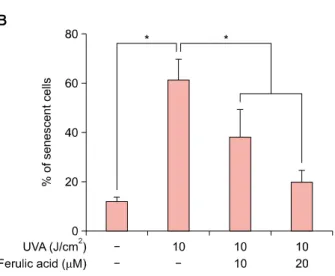
0.7 fold) and was reduced by ferulic acid in a dose-de- pendent manner. According to previous reports, UVA ra- diation can stimulate gene expression related with cell cy- cle arrest38,39. p16 are also known as cellular senescence markers40, leading us to examine cellular senescence us- ing a senescence-associated-β-galactosidase staining kit (BioVision). Pretreatment of cells with ferulic acid (10 and 20 μM) before UVA radiation resulted in significantly re- duced cellular senescence (38.2% and 19.6%, respecti- vely) compared with cells treated with UVA radiation only (61.2%, Fig. 4B). Further investigation showed that pre- treatment with ferulic acid regulated MMP1 and MMP3 gene expression in HDFs. As shown in Fig. 4C, UVA irra- diation alone increased MMP1 and MMP3 mRNA ex- pression, whereas pretreatment of ferulic acid prior to UVA radiation reduced expression. These results demon- strate the effects of ferulic acid in UVA-induced cellular senescence and reprogramming of ECM components.
Fig. 4. Effects of ferulic acid on cellular senescence and ex- tracellular matrix reconstruction. (A) Relative gene expression of p16, decreased with ferulic acid treatement prior to ultra- violet A (UVA) radiation. (B) Ferulic acid prohibited senescence in human dermal fibroblasts (HDFs) irradiated with UVA. The senescent cell population was analyzed using the senescence- associated β-galactosidase staining kit. (C) Relative gene ex- pression of metalloproteinase 1 (MMP1) and MMP3 in HDFs.
HDFs were irradiated with UVA followed by ferulic acid. Gene expression of MMP1 and MMP3 was reduced by feruilc acid pretreatment. Results are expressed as the mean±standard deviation in triplicate. *p<0.05 by Student’s t-test.
DISCUSSION
In this study, we investigated the protective effects of ferulic acid on UVA-induced proliferation, oxidation, sen- escence, and DNA damage in HDFs. Phenolic compounds in the plant are some of the most common water-soluble antioxidant compounds. Ferulic acid, one phenolic com- pound derived from cereal grains and vegetables, has been widely studied in biological aspects. Previous studies have demonstrated in skin biology that ferulic acid pro- tects UVB damages41.
Dermal fibroblasts, which mainly comprise the dermis lay- er of the skin, are established to contribute to the gen- eration of connective tissues42,43. During skin aging, MMPs, which are generated from dermal fibroblasts, can degrade all components in the ECM, including collagen, elastin, laminin, and proteoglycans44.
Results from our study indicate that pretreatment with ferulic acid decreases cell cycle arrest and DNA damage in HDFs. We verified that ferulic acid reduces Gadd45α promoter activity, cell cycle arrest, and expression of DNA
damage markers in a dose-dependent manner. Following analysis of Gadd45α promoter activity, we investigate gene expression of key regulator of cell cycle progression, p21. After then, we verified the effects of ferulic acid on NER by assessing XPA and XPC expression. Interestingly, expression of both XPA and XPC was significantly in- creased by pretreatment with ferulic acid. These findings suggest the protective effects of ferulic acid on UVA-in- duced cell cycle arrest and DNA damage in HDFs. Also, further studies are helpful to confirm the Gadd45α-de- pendent UVA protective effect of Ferulic acid in Gadd45α siRNA-mediated knockdown nHDFs.
Consistent with previous studies14,15, we demonstrated the antioxidant effects of ferulic acid against UVA radiation by analyzing intracellular ROS levels and relative expression of SOD1 and CAT. In this study, ferulic acid shows prop- erties about regulation of antioxidant gene expression and ROS scavenging effects. A previous study suggested that formation of intracellular ROS is the main cause of sen- escence and skin aging. Thus, we examined senescence marker expression and the frequency of senescent cells.
Our data show that UVA radiation increased the ex- pression of senescence marker, namely p16, and that marker is significantly decreased in ferulic acid-treated cells along with the frequency of senescent cells. These re- sults suggest the inhibitory effects of ferulic acid against UVA-induced cellular senescence in dermal fibroblasts.
Furthermore, UVA has been established as a factor that in- duces a decrease in type I collagen and inhibition of pro-collagen synthesis in dermal fibroblasts45-47. Our re- sults for MMP1 and MMP3 mRNA expression strongly suggest that ferulic acid not only regulates cellular sen- escence, but also reconstructs the ECM.
In conclusion, ferulic acid was found to regulate anti- oxidation, senescence, and DNA damage mechanisms through Gadd45α signaling pathways. Ferulic acid down- regulates MMP1 and MMP3 gene expression, thereby demonstrating the protective effects of ferulic acid on the dermis layer through ECM reconstruction. These findings indicate that ferulic acid can reverse the effects of aging in the skin by modulating its physiological structure, suggest- ing that ferulic acid is a potentially useful component for various cosmetic uses.
ACKNOWLEDGMENT
This study was supported by grants from the Ministry of Science, ICT and Future Planning (grant no. 2013R1A1A 1012205 to S. Bae and 20110028646 to S. An) of Republic of Korea.
REFERENCES
1. Lee JJ, An S, Kim KB, Heo J, Cho DH, Oh HM, et al. Extract of ettlia sp. YC001 exerts photoprotective effects against UVB irradiation in normal human dermal fibroblasts. J Microbiol Biotechnol 2016;26:775-783.
2. Faurschou A. Role of tumor necrosis factor-α in the regula- tion of keratinocyte cell cycle and DNA repair after ultraviolet-B radiation. Dan Med Bull 2010;57:B4179.
3. Bae S, Kim K, Cha HJ, Choi Y, Shin SH, An IS, et al.
Low-dose γ-irradiation induces dual radio-adaptive re- sponses depending on the post-irradiation time by altering microRNA expression profiles in normal human dermal fibroblasts. Int J Mol Med 2015;35:227-237.
4. Bae S, An IS, An S. Development of a high-throughput screening system for identification of novel reagents re- gulating DNA damage in human dermal fibroblasts. Acta Pharm 2015;65:331-341.
5. Mac-Mary S, Sainthillier JM, Jeudy A, Sladen C, Williams C, Bell M, et al. Assessment of cumulative exposure to UVA through the study of asymmetrical facial skin aging. Clin Interv Aging 2010;5:277-284.
6. Saito Y, Tsuruma K, Ichihara K, Shimazawa M, Hara H.
Brazilian green propolis water extract up-regulates the early expression level of HO-1 and accelerates Nrf2 after UVA irradiation. BMC Complement Altern Med 2015;15:421.
7. Bossi O, Gartsbein M, Leitges M, Kuroki T, Grossman S, Tennenbaum T. UV irradiation increases ROS production via PKCdelta signaling in primary murine fibroblasts. J Cell Biochem 2008;105:194-207.
8. Sjerobabski-Masnec I, Situm M. Skin aging. Acta Clin Croat 2010;49:515-518.
9. Zhou BR, Yin HB, Xu Y, Wu D, Zhang ZH, Yin ZQ, et al.
Baicalin protects human skin fibroblasts from ultraviolet A radiation-induced oxidative damage and apoptosis. Free Radic Res 2012;46:1458-1471.
10. Lan CC, Ho PY, Wu CS, Yang RC, Yu HS. LED 590 nm photomodulation reduces UVA-induced metalloproteinase- 1 expression via upregulation of antioxidant enzyme catalase.
J Dermatol Sci 2015;78:125-132.
11. Vile GF, Tyrrell RM. Oxidative stress resulting from ultraviolet A irradiation of human skin fibroblasts leads to a heme oxygenase-dependent increase in ferritin. J Biol Chem 1993;268:14678-14681.
12. Varani J, Spearman D, Perone P, Fligiel SE, Datta SC, Wang ZQ, et al. Inhibition of type I procollagen synthesis by damaged collagen in photoaged skin and by collagenase- degraded collagen in vitro. Am J Pathol 2001;158:931-942.
13. Janicke B, Hegardt C, Krogh M, Onning G, Akesson B, Cirenajwis HM, et al. The antiproliferative effect of dietary fiber phenolic compounds ferulic acid and p-coumaric acid on the cell cycle of Caco-2 cells. Nutr Cancer 2011;63:
611-622.
14. Xu X, Xiao H, Zhao J, Zhao T. Cardioprotective effect of sodium ferulate in diabetic rats. Int J Med Sci 2012;9:291- 300.
15. Roy S, Metya SK, Sannigrahi S, Rahaman N, Ahmed F.
Treatment with ferulic acid to rats with streptozotocin- induced diabetes: effects on oxidative stress, pro-inflamma- tory cytokines, and apoptosis in the pancreatic β cell.
Endocrine 2013;44:369-379.
16. Sgarbossa A, Giacomazza D, di Carlo M. Ferulic acid: a hope for Alzheimer's disease therapy from plants. Nutrients 2015;7:5764-5782.
17. Ardiansyah, Ohsaki Y, Shirakawa H, Koseki T, Komai M.
Novel effects of a single administration of ferulic acid on the regulation of blood pressure and the hepatic lipid metabolic profile in stroke-prone spontaneously hypertensive rats. J Agric Food Chem 2008;56:2825-2830.
18. Jung EH, Kim SR, Hwang IK, Ha TY. Hypoglycemic effects of a phenolic acid fraction of rice bran and ferulic acid in C57BL/KsJ-db/db mice. J Agric Food Chem 2007;55:9800- 9804.
19. Saija A, Tomaino A, Trombetta D, De Pasquale A, Uccella N, Barbuzzi T, et al. In vitro and in vivo evaluation of caffeic and ferulic acids as topical photoprotective agents.
Int J Pharm 2000;199:39-47.
20. Lee MJ, Cha HJ, Lim KM, Lee OK, Bae S, Kim CH, et al.
Analysis of the microRNA expression profile of normal human dermal papilla cells treated with 5α-dihydrotestos-
terone. Mol Med Rep 2015;12:1205-1212.
21. Livak KJ, Schmittgen TD. Analysis of relative gene expression data using real-time quantitative PCR and the 2(-Delta Delta C(T)) Method. Methods 2001;25:402-408.
22. Hildesheim J, Bulavin DV, Anver MR, Alvord WG, Hollander MC, Vardanian L, et al. Gadd45a protects against UV irradiation-induced skin tumors, and promotes apoptosis and stress signaling via MAPK and p53. Cancer Res 2002;62:7305-7315.
23. Fornace AJ Jr, Nebert DW, Hollander MC, Luethy JD, Papathanasiou M, Fargnoli J, et al. Mammalian genes coordinately regulated by growth arrest signals and DNA- damaging agents. Mol Cell Biol 1989;9:4196-4203.
24. Zhou BR, Xu Y, Wu D, Permatasari F, Gao YY, Luo D.
Ginsenoside Rg1 protects human fibroblasts against psoralen- and UVA-induced premature senescence through a telomeric mechanism. Arch Dermatol Res 2012;304:223- 228.
25. Chen A, Huang X, Xue Z, Cao D, Huang K, Chen J, et al.
The role of p21 in apoptosis, proliferation, cell cycle arrest, and antioxidant activity in UVB-irradiated human HaCaT keratinocytes. Med Sci Monit Basic Res 2015;21:86-95.
26. Kearsey JM, Coates PJ, Prescott AR, Warbrick E, Hall PA.
Gadd45 is a nuclear cell cycle regulated protein which interacts with p21Cip1. Oncogene 1995;11:1675-1683.
27. Park JM, Choi JY, Yi JM, Chung JW, Leem SH, Koh SS, et al.
NDR1 modulates the UV-induced DNA-damage checkpoint and nucleotide excision repair. Biochem Biophys Res Commun 2015;461:543-548.
28. Lo HL, Nakajima S, Ma L, Walter B, Yasui A, Ethell DW, et al. Differential biologic effects of CPD and 6-4PP UV- induced DNA damage on the induction of apoptosis and cell-cycle arrest. BMC Cancer 2005;5:135.
29. Chiganças V, Batista LF, Brumatti G, Amarante-Mendes GP, Yasui A, Menck CF. Photorepair of RNA polymerase arrest and apoptosis after ultraviolet irradiation in normal and XPB deficient rodent cells. Cell Death Differ 2002;9:1099-1107.
30. Chiganças V, Miyaji EN, Muotri AR, de Fátima Jacysyn J, Amarante-Mendes GP, Yasui A, et al. Photorepair prevents ultraviolet-induced apoptosis in human cells expressing the marsupial photolyase gene. Cancer Res 2000;60:2458-2463.
31. You YH, Lee DH, Yoon JH, Nakajima S, Yasui A, Pfeifer GP. Cyclobutane pyrimidine dimers are responsible for the vast majority of mutations induced by UVB irradiation in mammalian cells. J Biol Chem 2001;276:44688-44694.
32. Jaszewska E, Soin M, Filipek A, Naruszewicz M. UVA- induced ROS generation inhibition by Oenothera paradoxa defatted seeds extract and subsequent cell death in human dermal fibroblasts. J Photochem Photobiol B 2013;126:42-46.
33. Aroun A, Zhong JL, Tyrrell RM, Pourzand C. Iron, oxidative stress and the example of solar ultraviolet A radiation.
Photochem Photobiol Sci 2012;11:118-134.
34. Maverakis E, Miyamura Y, Bowen MP, Correa G, Ono Y, Goodarzi H. Light, including ultraviolet. J Autoimmun 2010;34:J247-J257.
35. Saify K, Saadat I, Saadat M. Down-regulation of antioxidant genes in human SH-SY5Y cells after treatment with morphine.
Life Sci 2016;144:26-29.
36. Kohl E, Steinbauer J, Landthaler M, Szeimies RM. Skin ageing. J Eur Acad Dermatol Venereol 2011;25:873-884.
37. Krutmann J. The role of UVA rays in skin aging. Eur J Dermatol 2001;11:170-171.
38. Lee KS, Cha HJ, Lee GT, Lee KK, Hong JT, Ahn KJ, et al.
Troxerutin induces protective effects against ultraviolet B radiation through the alteration of microRNA expression in human HaCaT keratinocyte cells. Int J Mol Med 2014;33:
934-942.
39. Assefa Z, Van Laethem A, Garmyn M, Agostinis P. Ultra- violet radiation-induced apoptosis in keratinocytes: on the role of cytosolic factors. Biochim Biophys Acta 2005;1755:
90-106.
40. Rayess H, Wang MB, Srivatsan ES. Cellular senescence and tumor suppressor gene p16. Int J Cancer 2012;130:1715- 1725.
41. Mancuso C, Santangelo R. Ferulic acid: pharmacological and toxicological aspects. Food Chem Toxicol 2014;65:
185-195.
42. Quan T, Qin Z, Xu Y, He T, Kang S, Voorhees JJ, et al.
Ultraviolet irradiation induces CYR61/CCN1, a mediator of collagen homeostasis, through activation of transcription factor AP-1 in human skin fibroblasts. J Invest Dermatol 2010;130:1697-1706.
43. Yang Y, Li S. Dandelion extracts protect human skin fibroblasts from UVB damage and cellular senescence.
Oxid Med Cell Longev 2015;2015:619560.
44. Cho S, Won CH, Lee DH, Lee MJ, Lee S, So SH, et al. Red ginseng root extract mixed with Torilus fructus and Corni fructus improves facial wrinkles and increases type I procollagen synthesis in human skin: a randomized, double-blind, placebo-controlled study. J Med Food 2009;
12:1252-1259.
45. Kim JA, Ahn BN, Kong CS, Kim SK. The chromene sargachromanol E inhibits ultraviolet A-induced ageing of skin in human dermal fibroblasts. Br J Dermatol 2013;168:
968-976.
46. Naylor EC, Watson RE, Sherratt MJ. Molecular aspects of skin ageing. Maturitas 2011;69:249-256.
47. Park SY, Park JY, Kim CH, Kang SU, Kim JH, Bark KM, et al.
Effects of xanthium stramarium and psoralea corylifolia extracts combined with UVA1 irradiation on the cell proliferation and TGF-β1 expression of keloid fibroblasts.
Ann Dermatol 2013;25:304-309.