藥 學 含 확
F e ru lic A cid오卜관련 페놀화합물의 암세포주에 대한 독성억제효과
한 두 석• 전 주 원• 전 성 우• 백승화#
원광대학교 치과대학 구강해부학교실 . 한의학전문대학원 한약자원개발학과 (Received May 6,2005; Revised June 24, 2005)
T h e I n h ib it o r y E f f e c t o f F e r u lic A c id a n d R e la t e d P h e n o lic C o m p o u n d s a g a in s t C a n c e r C e ll L in e s
Du Seok Han, Joo Won Chun, Sung Woo Jeon and Seung Hwa Baek#
Department of Oral Anatomy, School of Dentistry and Department of Herbal Resources,
Professional Graduate School of Oriental Medicine and Institute of Basic N atural Science, Wonkwang University, Iksan 570-749, Korea
Abstract — The inhibitory effect of ferulic acid and related phenolic compounds on the growth of normal cell lines and can
cer cell line was evaluated by the MTT and XTT methods. Ferulic acid decreased the cell viability of human skin melanoma cells by the MTT method and the cell adhesion activity of human oral epithelioid carcinoma cells by the XTT method. These results suggest that ferulic acid has a potential anticancer activity.
Keywords □ Inhibitory effect, phenolic compounds, MTT and XTT methods, ferulic acid, cell viability, cell adhesion activity
페놀화합물은치환될수 있는수산기를가진방향족고리 구 조를가지고있는이차대시산물의 총칭으로 화학적으로이질적 인 것이 많으며지용성, 수용성 및중합체등여러 가지 형태로 나타난다. Syringic acid는 shikimic acid pathway에의해서생합 성되는 페놀성화합물로서 최근에는세계각국에서 각종 약용식 물로부터분리하고 있다.1_6) Syringic acid의약리작용에관한연 구는거의없는실정이나항산화작용과 DPPH radical-scavenging activity에관한연구가있다.7’8) Syringic acid의에스테르유도체 인 phenylethanoid glycoside는강한 항산화작용을나타내는데 이 작용은 syringic acid의 방향족 메틸기(aromatic methoxy groups)의수, 수산기 (hydroxy groups) 및아실기의구조와주로 관련되며 acyl moiety 또는 phenylethanoid moiety에서 메틸기
에의한수산기의 대체나 sugar chain의변형시에는항산화작
용은감소한다고보고하였고,7) 페놀산인 gallic acid가건강세포 보다 암종세포에선택적으로세포독성이 강하게작용하는 기전
#본 논문에 관한 문의는 저자에게로 (전화) 063-850-6225 (팩스) (E-mail) [email protected]
을밝히기 위한연구에서 gallic acid는세포주기의 결과에 의하
여 선택성을나타내지 세포주기의 어느기에 영향을주지 않으며 gallic acid의구조의변화즉 phenolic hydroxyl group의메틸화
와 carboxyl group의에스텔화는 세포독성을분명히 감소시키며
각종세포에 대한 gallic acid의신호전달로는세포 내칼슘 이온 과활성산소(R0S)에의하여 변화하나 gallic acid에의하여 유도 되는세포사전달로는세포에 따라다르다고보고하였으며 또한 암종세포를간세포와함께 배양하면 세포사가유도되지 않으며 gallic acid에 저항하는 세포는 gallic acid의세포독성에 대하여 자신을보호하거나저항물질을생산할것으로추측하였다.wo) 방 향족 메틸기, 수산기 및아실기(C6-C3)의구조를 갖는 syringic acid가항산화작용이 있고 gallic acid와유시•한구조를갖고있으 므로 정상세포보다 암종세포에 세포독성이 강하게 나타날 것으 로판단되어 syringic acid를 NIH3T3 섬유모세포와인체피부섬 유모세포에 농도별로적용하고, 암종세포인 인체피부흑색종세 포와 인체구강유상피암종세포에도농도별로적용하고 48시간이
지난후세포생존률을알아보기 위하며 MTT 정량분석법을, 세
포부착능을알아보기 위하여 XTT 정량분석법을실시하고실험 과정의 모든세포를광학현미경적으로관칠하여보고하는바이다.
II
C — 0 H C — 0H C — 0H
CH qO
B enzoic acid G allic acid Syringic acid
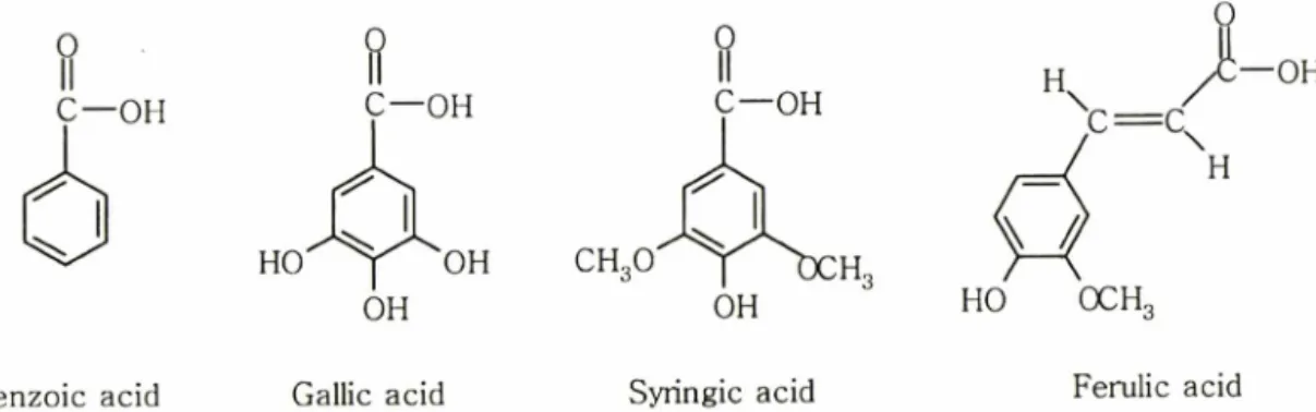
Fig. 1 - Molecular structures of benzoic acid, gallic acid, syringic acid and ferulic acid.
J -
H 人 一 O H
C = H
HO OCH3
Ferulic acid
실험재료 및 방법
시약
세포배양에 사용한 M inimum Essential M edium(MEM)과 Dulbecco's modified eagle medium(DMEM), RPM I 1640 배지, fetal bovine serum, penicillin, streptomycin, fungizone 入1 약 은 Gibco/BRL(Grand Island, NY, USA)제이었으며, 3-[4,5- Dimethylthiazol-2-yl] -2,5-diphenyltetrazolium bromide(MTT, Sigma Chemical Co.) 정량과 2,3-bis-[2-Methoxy-4-nitro-5- sulfophenyl]-2H-tetrazolium-5-caboxanilide(XTX Sigma Chemical Co.) 정량에 사용한 시약과 syringic acid, benzoic acid, gallic acid와 ferulic acid는(구조식 Fig. 1)는 Sigma Chemical Co.(ST.
Louis, MO, USA)사에서 구입하였다.
실험기기
세포의 배양은 C 02 incubator(Shellab. Co., Cornelius,
U.S.A.)를사용하였으며,세포수의 계산은 Turk형혈구계산기
(Marienfeld Co., Mergentheim, Germany)를이용하였다. MTT 정량, XTT 정량에는 ELISA reader(Spectra max 250, Molecular Devices, Sunnyvale, U.S.A.)를사용하였다.
세포배양
Syringic acid 자체의 세포독성과암세포에 대한세포독성을즉
정하기 위하여 원광대학교 의과대학에서 분양받은 NIH3T3 섬 유모세포주는 M EM 배지에 , 서울대학교세포은행에서분양받은 인체 피부섬유모세포(Detroit 551)는 DM EM 배지에, 서울대학 교암연구소에서 분양받은 인체피부흑색종세포(SK-MEL-3)와 인체 구강유상피암종세포(KB)는 RPM I 1640 배지에 10% fetal bovine serum, penicillin(25 unit/m/) 및 fungizone을 점 7]•石!•여 사용하였다. 세포의 배양은온도 37°(;습도 95%, 탄산가스농 도 5%의 배양기 (C 02 incubator, Shellab, Cornelius, U.S.A)를 사용하였다. 실험을위하여 일차배양한 flask의세포를 0.25%
trypsin-EDTA로처리하여 세포를분리한 후,Turk형혈구계산
기를이용하여 세포수가 5X10⁴ ceW m/가되도록세포부유액을
만들었다.
MTT 정량분석법
Mosmann의방법11>에의하며 , NIH3T3 섬유모세포와인체피 부섬유모세포, 인체 피부흑색종세포및인체 구강유상피암종세 포를 각배양용기에 5 x i0 4 cells/m/로 넣고 24시간 배양 후
ferulic acid와관련페놀화합물 자체의 세포독성과암종세포에
대한 syringic acid의세포독성을측정하기 우1하여 ferulic acid와 관련페놀화합물을 농도별(1, 25, 50 및 lO O ^M)로첨가한후 48시간배양한후분석당일조제한 MTT(Sigma Chemical Co.)
가포함된 배양액을배양용기당 lm /씩 넣어 3시간배 양하였다. 배양후배양액을버리고, dimethylsulfoxide(DMSO) 를 2 m//well씩넣어 5분간실온방치하여 MTT formazan을용 해한후,흡광도는분광광도계 ELISA reader로 MTT의흡광도
540 nm를측정하여 대조군과비교조사하였다.
XTT 정링분석법
Laminin-coated plate는 laminin lm g을 PBS 2 m/에용해하 여냉장고에 보관하면서 필요시에 laminin의농도(20 mg/m/)를 결정하여 찬 PBS 용액으로희석하고이용액을 24 well plate의 각 well에 200 |i/씩분주하여 하룻밤동안 건조시킨뒤 PBS로 두번세척하여 3% BSA를각 well에 200 씩첨가해잘흔들
어준다음 제거하고 PBS로두번정도세척하였다.
배양된 NIH3T3 섬유모세포,인체 피부섬유모세포,인체 구
강유상피암종세포 및인체 피부흑색종세포 5X 10⁴ cells/m/로 lam inin으로 coating한 배양용기에 넣고 24시간 배양한 후 MTT 정량분석법과 같은방법으로 ferulic acid와관련페놀화 합물을 넣고다시 48시간배양한 후 배지는조심스럽게 제거 하고 PBS로두번세척하였다. 여기에 XTT(Sigma Chemical
Co.)와 혼합하여 각배양용기에 200 씩 주입하고 4~6시간
동안 배양한 뒤 ELISA reader로 홉광도 450nm에서 측정하 였다.
/. Pharm. Soc. Korea
Ferulic Acid 와 관련 페놀화합물의 암세포주에 대한 독성억제효과 367
세포의광학현미경적 관찰
광학현미경적 관찰읍 위하여 NIH3T3 성유모세포와인체 피 부섬유모세포, 인체구강유상피암종세포및인체 피부흑색종세 포는각각세포분주후 ferulic acid와관련페놀화합물을처리한 후 MTT와 XTT를처리하기전에도립현미경 (Inverted microscope Olympus, Tokyo, Japan).0■로관찰하였다.
IC50 결정
니켈의 ICso 결정은배잉중인인체 치은섬유모세포를각배양
용기당 5X10⁴ cells/m/씩 넣고 24시간 배양후 1,25’ 50’ 100 HM의 ferulic acid 와관련페놀화합물을첨가하여 48시간배양 한후 MTT 정량과 XTT 정량을하여 ferulic acid이이들각각 에대한 50% 억제농도인 ICso을회귀방정식12)에의해구하였다.
통계처리
실험결과의 통계처리는 Students' t-test에준하였고, p-value
가 0.05 미만일경우유의한것으로판정하였다.
결과 및 고찰
페놀화합물의 세포독성은화합물의 분자구조에 따라,활성관 계가유의성있게 관찰되며, 페놀화합물의 치환기중에 수산기의 메틸화또는소심은 세포독성이 제거되기 때문에페놀성수산기
세포독성에필수적이라 생각된다.13) Table I에서 세포생존율
fr MTT 정량분석법으로측정한결과에 의하면,NIH 3T3 섬유
모세포와인체 피부섬유모세포(Detroit 551)에대한세포독성은 901 )iM~3,341 ]M 범위로관찰되었다. NIH 3T3 섬유모세포에 서세포생존율은 benzoic acid에서 1,226 ]iM농도로 가장강한
세포독성이었다. 그러나 benzoic acid의 3,4, 5번위치에수산기 가치환된 gallic acid에서는 세포독성이 약 2배정도약한활성 이측정되었지만,gallic acid의 3’ 5번위치에 methoxy기로치환 된 syringic acid(3,341 (iM)에서는 gallic acid의세포독성보다약
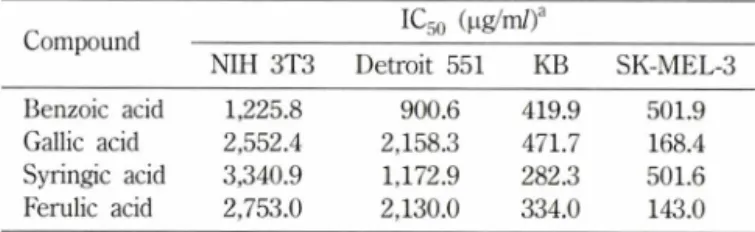
Table I - The cytotoxic activity of phenolic acid by the MTT method
Compound IC50 (나g/m/)a
NIH 3T3 Detroit 551 KB SK-MEL-3
Benzoic acid 1,225.8 900.6 419.9 501.9
Gallic acid 2,552.4 2,158.3 471.7 168.4
Syringic acid 3,340.9 1,172.9 282.3 501.6 Ferulic acid 2,753.0 2,130.0 334.0 143.0 aIC50 value of compound against each cancer cell line which was defined as a concentration (^g/m/) that caused 50% inhibition of cell growth in vitro. Compounds were examined in four concentrations in triplicate experiments. NIH 3T3: NIH 3T3 fibroblasts. Detroit 551: Human skin fibroblasts. KB: Human oral epithelioid carcinoma cells. SK-MEL-3: Human skin melanoma cells.
한세포독성이 관찰되었으며,이는 benzoic acid의세포독성보다 2.7배약한세포독성이 즉정되었다. 그러나 benzoic acid에서 3- methoxy, 4-hydroxy로치환된벤젠고리와 propenoic acid기로치 환된 ferulic acid는 syringic acid의세포독성 보다 강한 2,753
|iM 농도로 관찰되었다. 이와 같이 benzoic acid에서 electron- withdrawing group(hydroxy 그룹)이 치환된 phenolic acid는 electron- donating group(methoxy 그룹)이 치환된 phenolic acid보다세포독성이 강하게측정되었다. Benzoic acid의세포생 존율은 NIH 3T3 섬유모세포보다인체피부섬유모세포가 901 jiM 농도로강하게 관찰되었다. 인체 피부섬유모세포에서는 gallic acid의세포독성이 benzoic acid보다약 2.4배약하게관찰되었으
며,이는 NIH 3T3 섬유모세포보다강한세포독성으로관찰되었
다. 그러나 syringic acid에서는 gallic acid의세포독성보다오히 려강한세포독성(1,173 나M)이관찰되었으며,이는 benzoic acid 의세포독성보다 1.3배약한세포독성으로 NIH 3T3 섬유모세포 보다강한세포독성이측정되었다. 그러나 ferulic acid는 syringic acid의세폭독성 보다 약한 2,130 (iM 농도로 관찰되었다. 일반
적으로 MTT 정량분석법에서 인체피부섬유모세포는 901
2,158 농도범위로 NIH 3T3 섬유모세포보다 세포독성이 강
하게측정되었다.
인체구강유상피암종세포(KB)에 대한세포독성은 NIH 3T3 섬유모세포와는달리 syringic acid(282 나M)에서 12배의강한세 포독성이 관찰되었으며 , 특이하게 인체 구강유상피암종세포는 benzoic acid에메독시그룹이 3,5번위치에 치환과 4번위치에 hydroxy그룹이 치환된 syringic acid의세포독성이 가장 강하게 측정되었으며, 방향족고리에 메독시그룹이 3번위치에 치환되었 으며,치환된 ferulic acid는약간낮은 세포독성(334 fiM)이 나 타났다, 그렇지만 benzoic acid(420 !iM)에 3,4, 5번 위치에 hydroxy로치환된 gallic acid(472 |_iM)의 세포독성은 오히려약 한세포독성이 관찰되었다. 일반적으로 인체 구강유상피암종세 포(KB)에대한세포독성(282 )aM〜 472 |iM)은 NIH 3T3 섬유모 세포(1,226 |iM〜3,341 !iM)와 인체피부섬유모세포(Detroit 551;
901 나M〜2,158 ᅣiM )에대한세포독성보다강한활성이 관찰되었 다. 따라서 인체구강유상피암종세포(KB)에 대한세포독성의 민 감성은 syringic acid > ferulic acid > benzoic acid > gallic acid 순 서로감소되어관찰되었다.
인체피부흑색종세포에 대한세포독성은인체구강유상피암종 세포와는달리 ferulic acid(143 fiM)에서 강한 세포독성이 측정 되었다. 이는 ferulic acid가 benzoic acid의벤젠고리 3, 4번위 치에 치환된 methoxy, hydroxy 그룹과 함께 1번 위치에
propenoic acid기로 치환된 그룹에 근거할 것으로 생각된다.
benzoic acid의벤젠고리 3, 4,5번위치에 hydroxy 그룹으로치 환된 gallic acid(168 jaM)가 ferulic acid보다는약한 세포독성이 관찰되었으나,큰 차이는 없었다. 그렇지만다른 정상세포와암
세포와는 달리 인체 피부흑색종세포에서는 benzoic acid와 syringic acid의세포독성(502|oM)은거의유사한활성이 검색되 었다. 인체피부흑색종세포는 인체구강유상피암종세포와는다 르게 세포독성에 대한 민감성이 ferulic acid> gallic acid>
benzoic acid= syringic acid 순서로감소되어 관찰되었다. 일반적으로 세포의 활성검색에 가장많이사용되는 MTT 정 량분석법으로정상세포와암세포에 대한세포독성을측정한결 과는 NIH 3T3 섬유모세포>인체 피부섬유모세포(Detroit 551)
>인체구강유상피암종세포(KB)> 인체피부흑색종세포(SK-MEL- 3)와같은민감성으로관찰할수가있었다(Table I).
세포부착능을 측정한 XTT 정량분석의 결과에 의하면, NIH 3T3 섬유모세포와인체피부섬유모세포(Detroit 551)에대한세 포부착능은 975 nM~2,462 나M 범위로관찰되었다. NIH 3T3 섬 유모세포에서 세포부착능은 benzoic acid에서 1,292 |iM 농도로 가장강한 활성이 관찰되었다. 그러나 benzoic acid의 3, 4,5번 위치에수산기가치환된 gallic acid(2,446 nM)에서는세포부착능 이약 1.9배 정도약한활성이 측정되었지만, gallic acid의 3, 5 번위치에 methoxy기로치환된 syringic acid(2,462 nM)에서는 gallic acid의세포부착능과거의비슷한활성이 측정되었다. 그러 나 benzoic acid에서 3-methoxy, 4-hydroxy로치환된 벤젠고리 와 propenoic acid기로 치환된 ferulic acid는 syringic acid와 gallic acid의세포부착능의 활성과큰차이(1.7배)없이 생물검색 이관찰되었다. 이와같은실험결과에 의하면, benzoic acid에서 electron-withdrawing group(hydroxy 그룹)이 치환된 gallic acid 는 electron-donating group(methoxy 그룹)이 치환된 syringic acid보다세포부착능이 약한강하게 측정되었으며, benzoic acid 의벤젠고리 3, 4번위치에 치환된 methoxy, hydroxy그룹과함 께 1번위치에 propenoic acid기로치환된 ferulic acid는약간세 포부착능이 강하게관찰되었다. NIH 3T3 섬유모세포에 대한세 포부착능의 민감성은 benzoic acid> ferulic acid> gallic acid>
syringic acid 순서로감소되어 관찰되었다.
인체 피부섬유모세포(Detroit 551)에 대한세포부착능의 검색 결과에 의하면, benzoic acid의세포부착능은 NIH 3T3 섬유모 세포(1,292 nM)보다인체피부섬유모세포가(975 nM) 농도로 강 하게 관찰되었다. 인체피부섬유모세포에서는 gallic acid의세포 부착능이 benzoic acid보다약 2.1 배약하게 검색되었으며,그러 나 syringic acid에서는 gallic acid의세포부착능보다오히려 강 한활성 (1,111 |iM)이관찰되었으며, 이는 benzoic acid의세포부 착능보다 1.1 배약한활성으로 NIH 3T3 섬유모세포보다 2배정 도강한세포부착능이측정되었다. 그러나 ferulic acid는 syringic add의세포부착능보다약한 l,474|_iM 농도로관찰되었다. NIH 3T3 섬유모세포와는달리인체피부섬유모세포에서는가장약한 활성이 나타났다. 인체 피부섬유모세포에 대한세포부착능의 민 감성은 benzoic acid > syringic acid > ferulic acid > gallic acid 순
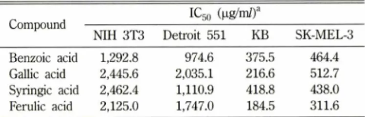
Table II - The cytotoxic activity of phenolic acid by the XTT method
Compound IC50 (^ig/m/)a
NIH 3T3 Detroit 551 KB SK-MEL-3
Benzoic acid 1,292.8 974.6 375.5 464.4
Gallic acid 2,445.6 2,035.1 216.6 512.7 Syringic acid 2,462.4 1,110.9 418.8 438.0 Ferulic acid 2,125.0 1,747.0 184.5 311.6 aIC50 value of compound against each cancer cell line which was defined as a concentration (|ig/m/) that caused 50% inhibition of cell growth in vitro. Compounds were examined in four concentrations in triplicate experiments. NIH 3T3: NIH 3T3 fibroblasts. Detroit 551: Human skin fibroblasts. KB: Human oral epithelioid carcinoma cells. SK-MEL-3: Human skin melanoma cells.
서로감소되어 관찰되었다. 일반적으로 XTT 정량분석법에서 인
체피부섬유모세포는 NIH 3T3 섬유모세포보다는세포부착능이
강하게관찰되었다.
인체구강유상피암종세포(KB)에대한세포부착능은 다른세 포와다르게가장유효한억제효능이있는것으로관찰되었으며,
ferulic acid의세포부착능은 benzoic acid의벤젠고리에 치환된
phenolic acid보다세포부착능에 대한활성이 강하게 나타났다.
Gallic acid의세포부착능이 benzoic acid보다강한활성이 나타 났으며 , gallic acid의벤젠고리에 3, 5번위치에 methoxy 그룹으 로치환된 syringic acid에서약한세포부착능이관찰되었다. MTT 정량분석법으로세포독성을측정한인체구강유상피암종세포에 대한활성과는다른효능을 관찰되었다. 이런 현상은분자구조 에따른세포활성에기인하는것으로생각된다. 일반적으로 MTT 정량분석법에서측정한 세포에대한활성과는다르게 강한효능 을관찰되었다. 따라서 인체구강유상피암종세포에 대한민감성 도 ferulic acid > gallic acid > benzoic acid > syringic acid 순서로 감소되어 관찰되었다.
인체피부흑색종세포(312 nM~464 nM)에대한세포부착능은 일반적으로인체구강유상피암종세포(185pM~419nM)에대한 세포부착능보다 약한활성이 관찰되었다. 인체구강유상피암종 세포에대한세포부착능과같이 ferulic acid에서강한활성이관 찰되었으며,gallic acid는 benzoic acid보다 약한 세포부착능이 나타났으나,syringic acid는활성이약간강하게즉정되었다. 인 체피부흑색종세포에 대한민감성도 ferulic acid > syringic acid
> benzoic acid>gallic acid 순서로감소되어관찰되었다(Table II).
결 론
이상의 연구결피를종합해보면 , 비색분석법으로 Phenolic acid 에대한세포의활성을 측정한결과는분자구조에 따라활성이 다르게 관찰되었으며,일반적으로 XTT 정량분석법으로측정한
/. Pharm. Soc. Korea
Ferulic Acid 와 관련 페놀화합물의 암세포주에 대한 독성억제효과 369
활성은 MTT 정량분석법으로측정한활성보다강한 생물검색이
관찰되었으며,XTT 정량분석법으로측정한세포중에서 인체구 강유상피암종세포에 대한세포부착능으로 ferulic acid에서가장
강한활성과 MTT 정량분석법으로측정한세포독성도인체 피
부흑색종세포에서 가장강한활성이 검색되었다. 이와같은 현상 은 phenolic acid에서 electron-withdrawing group과 side chain 이증가하면서, conjugated system을소유한분자구조의 페놀유 도체가세포에 대한활성이 클것으로생각된다.
감사의 글
이논문은 2004년도원광대학교의 교비지원에 의하여수행되
었으며,이에 감사드립니다.
참고문헌
1) Cao, Y., Lou, C.,Fang, Y. and Ye, J. : Determination of active ingredients of Rhododendron dauricum L. by capillary electrophoresis with electrochemical detection. J Chromatogr.
943,153 (2002).
2) Xu, L., Liu, J., Min, D.,Wang, S., Zhang, Z., Guo, D. and Zheng, K. : Chemical constituent. Zhongguo Zhong Yao Za Zhi. 23, 293 (1998).
3) Kong, L. D., Abliz, Z., Zhou, C. X.’ Li, L. J., Cheng, C. H. and Tan, R. X. : Glycosides and xanthine oxidase inhibitors from Conyza bonariensis. Phytochemistry 58,645 (2001).
4) De Heredia, J. B.,Torregrosa, J., Dominguez, J. R. and Peres, J. A. : Kinetic model for phenolic compound oxidation by Fenton's reagent. Chemosphere. 45,85 (2001).
5) Andrade, R B.,Oliveira, B. M., Seabra, R. M.,Ferreira, M. A., Ferreres, E and Garcia-Viguera, C. : Analysis of phenolic
compounds in Spanish Albrarino and Portuguese Alvarinho and Loureiro wines by capillary zone electrophoresis and high- performance liquid chromatography. Electrophoresis 22,1568 (2001).
6) Dawidar, A. M .,Ezmiriy,S. T., Abdel-Mogib, M .,el-Dessouki, Y. and Angawi, R. F. : New stilbene carboxylic acid from Convolvulus hystrix. Pharmazie. 55,848 (2000).
7) Heilmann, J., Calis, I., Kirmizibekmez, H., Schuhly, W., Harput, S. and Sticher, 0. : Radical scavenger activity of phenylethanoid glycosides in FMLP stimulated human poly
morphonuclear leukocytes: structure-activity relationships.
Planta Med. 66,746 (2000).
8) Hirota, A., Taki, S., Kawaii, S., Yano, M. and Abe, N.: 1,1- Diphenyl-2-picrylhy-drazyl radical-scavenging compounds from soybean miso and antiproliferative activity of isoflavones from soybean miso toward the cancer cell lines. Biosci Biotechnol Biochem. 64,1038 (2000).
9) Li, Z.,Inou, M .,Nose, M.,Kojima, K.,Sakaguchi,N.,
Isuzugawa, K.,Takeda, T. and Ogihara, Y. : Metabolic fate of gallic acid orally administered to rats. Biol. Pharm. B ull. 22, 326 (1999).
10) Sakaguchi, N.,Inoue, M. and Ogihara, Y. : Reactive oxygen species and intracellular Ca2+, common signals for apotosis induced by gallic acid. Biochem. Pharmacol. 55, 1973 (1998).
11) Mosmann, T. : Rapid colorimetric assays for cellular growth and survival: Application to proliferation and cytotoxicity assays. J. Im m unol Methods 65,55 (1983).
12) 채영암,구자옥,서학수,이영만 : 기초생물학,향문사,p. 179 (1991).
13) Inoue, M .,Suzuki, R., Sakaguchi, N., Li, Z., Takeda, T, Ogihara, Y.,Jiang, B. Y. and Chen, Y. : Selective induction of cell death in cancer cells by gallic acid. B iol Pharm. B ull. 18, 1526 (1995).