Copyrights © 2015 The Korean Society of Radiology
343
Case Report
pISSN 1738-2637 / eISSN 2288-2928 J Korean Soc Radiol 2015;73(5):343-346 http://dx.doi.org/10.3348/jksr.2015.73.5.343
INTRODUCTION
Multicystic dysplastic kidney is the most common form of cystic kidney disease in infants and children. It can arise from failure of the ureteric bud to integrate and branch appropriately into the metanephros during development (1). The involvement is usually unilateral, although bilateral disease can also occur, and in some patients there is segmental involvement of one kidney. Most multicystic dysplastic kidneys are benign and do not require surgical resection (2). Reported complications asso- ciated with multicystic dysplastic kidney include pain, infec- tion, hypertension, and neoplasia (3). However, renal cell carci- nomas are extremely rare in multicystic dysplastic kidneys. We found only three cases of renal cell carcinoma in previously published reports (4-6). Little is known regarding the imaging
characteristics of this condition. We report the sonographic and CT features of a histopathologically proven cystic papillary re- nal cell carcinoma arising from an involutional multicystic dys- plastic kidney. Institutional Review Board approval with waived informed consent (number 2014-02-009) was obtained for this case report.
CASE REPORT
A 33-year-old male with no significant past medical history was referred for evaluation of a left retroperitoneal mass inci- dentally detected on ultrasonography performed at another medical institution. Findings from a physical examination were unremarkable. Laboratory examinations, including urine cytol- ogy, yielded results within normal limits. Ultrasonography of
Cystic Papillary Renal Cell Carcinoma Arising from an Involutional Multicystic Dysplastic Kidney
퇴화된 다낭이형성신 환자에서 발생한 낭성 유두상 콩팥 세포암종
Jeong Jae Kim, MD
1, Bong Soo Kim, MD
1*, Jung-Sik Huh, MD
2, Kyung-Gi Park, MD
2, Guk Myung Choi, MD
1, Seung Hyoung Kim, MD
1, Young-Hee Maeng, MD
3Departments of 1Diagnostic Radiology, 2Urology, 3Pathology, Jeju National University School of Medicine, Jeju National University Hospital, Jeju, Korea
Multicystic dysplastic kidney is a common cystic renal disease that often occurs in infancy. Recent studies demonstrate the possibility for spontaneous involution of a dysplastic kidney. In such cases, the prognosis is generally excellent and there is a very low incidence of complications. Complications associated with multicystic dys- plastic kidney include pain, infection, hypertension, and neoplasia. Renal cell carci- nomas are extremely rare in multicystic dysplastic kidneys. To our knowledge, no case report has described a radiologic finding of renal cell carcinoma arising from an involutional multicystic dysplastic kidney. We report a case of histopathologically validated cystic papillary renal cell carcinoma arising from an involutional multicys- tic dysplastic kidney and describe its sonographic and CT features.
Index terms
Kidney Diseases, Cystic Carcinoma, Renal Cell Multicystic Dysplastic Kidney
Received April 14, 2015 Revised June 14, 2015 Accepted July 27, 2015
*Corresponding author: Bong Soo Kim, MD
Department of Radiology, Jeju National University Hospi- tal, Jeju National University School of Medicine, 15 Aran 13-gil, Jeju 63241, Korea.
Tel. 82-64-717-1371 Fax. 82-64-717-1372 E-mail: [email protected]
This is an Open Access article distributed under the terms of the Creative Commons Attribution Non-Commercial License (http://creativecommons.org/licenses/by-nc/3.0) which permits unrestricted non-commercial use, distri- bution, and reproduction in any medium, provided the original work is properly cited.
344
Cystic Papillary Renal Cell Carcinoma Arising from an Involutional Multicystic Dysplastic Kidney
jksronline.org
J Korean Soc Radiol 2015;73(5):343-346 the abdomen showed a unilocular cystic mass 7 cm in diameter
with multiple, mural nodules located in the left renal fossa (Fig.
1A). The left kidney was not visible. The patient underwent CT examination of the abdomen and pelvis (Somatom Sensation 16, Siemens Medical Solutions, Forchheim, Germany) before and after administration of IV contrast material (Ultravist 370, Bayer-Schering, Berlin, Germany). The mass had a primarily cystic nature with a discrete calcification in the wall which could be seen on the unenhanced image (Fig. 1B). Contrast- enhanced CT images showed peripheral soft-tissue mural nod- ules with mild enhancement ranging from a value of 21 Houn- sfield units (HU) on the unenhanced CT image to 50 HU on the nephrographic phase obtained 90 seconds after contrast dye administration (Fig. 1C-E). There was neither lymphadenopa- thy nor renal vein involvement.
Because this lesion had enhancing soft-tissue mural nodules, we could not immediately rule out renal malignancy in an in- volutional multicystic dysplastic kidney or a cystic retroperito-
neal tumor in a patient with left renal agenesis. Therefore, our patient underwent surgical exploration. Radical nephrectomy was performed without complications. Subsequent CT scan- ning and ultrasonography of the patient’s abdomen and urine cytology were normal at a follow-up examination performed two years post-treatment.
The excised cystic mass measured 8.7 × 7 × 6.5 cm. The outer surface was smooth and showed an area of fibrous thickening.
On cross-section, the cystic cavity was unilocular and filled with clear, serous fluid. The inner surface showed multiple foci consisting of tan, papillary excrescences (Fig. 2A). Papillary tu- mor tissue, visualized microscopically, consisted of low-grade, eosinophilic renal carcinoma cells (Fig. 2B). There were also immature tubule-like structures lined by flattened or cuboidal epithelial cells scattered in the thickened areas of the wall, which suggested dysplastic kidney (Fig. 2C).
A
D
B
E
C
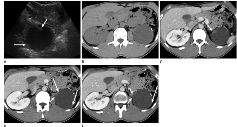
Fig. 1. 33-year-old man with a left retroperitoneal mass.
A. Ultrasonogram of the left intercostal space shows a cystic mass with multiple, discrete nodularites (arrows) in the left renal fossa. However, the left kidney was not clearly visible.
B. Unenhanced CT image shows a cystic mass with a linear calcification.
C-E. Contrast-enhanced CT images obtained during the nephrographic phase show several focal nodular enhancements (long arrows) in the cyst wall. Note the triangular, regressed, multicystic dysplastic kidney (short arrows) between the adrenal gland and the cystic mass. The normal pa- renchyma of the left kidney is not visible.
345
Jeong Jae Kim, et al
jksronline.org J Korean Soc Radiol 2015;73(5):343-346
DISCUSSION
Multicystic dysplastic kidney disease is relatively common and is estimated to occur in one in 4300 live births (7). In gen- eral, patients with multicystic dysplastic kidney disease present with a unilateral abdominal mass, and as only one kidney is in- volved, overall renal function is normal. In general, most multi- cystic dysplastic kidneys spontaneously involute or decrease in size over time and have low rates of complications and malig- nant degeneration. Therefore, such kidneys are for the most part currently left untreated, while in the past they were routinely removed (7).
Reported complications associated with multicystic dysplas- tic kidney include pain, infection, hypertension, and neoplasia (6). Until now, only three reports have been published that de- scribe renal cell carcinomas arising from multicystic dysplastic kidneys (4-6). In one case, the renal cell carcinoma was a papil- lary adenocarcinoma; histologic subtype of the other two cases is undocumented. Two of the three patients were young (15 and 26 years old). Two had mainly cystic tumors with solid compo- nents. However, these patients had aggressive tumors with early metastases involving lymph nodes, bone, and lung. Our patient is unique in that he had a cystic renal mass containing mildly enhancing mural nodules in an involutional multicystic dysplas- tic kidney, which replaced the normal kidney, and was asymp- tomatic. To our knowledge, our case is the first radiology imag- ing report of a renal cell carcinoma in an involutional multicystic dysplastic kidney.
Papillary renal cell carcinoma is the second most common histological subtype, accounting for 10–15% of all renal cell carcinomas (8). As a papillary renal cell carcinoma is common- ly a low-stage tumor, it is associated with a very good prognosis.
The most important prognostic factors for patients with renal cell carcinoma are disease stage at the time of diagnosis and nu- clear grade. A tumor is more likely to be cured by surgical re- section when it is small and low grade. Therefore, early detec- tion of renal cell carcinoma is crucial in order to improve patient survival rate. Cystic papillary renal cell carcinomas, such as that in our patient, demonstrate peripheral soft-tissue mural nod- ules that are enhanced by administration of contrast dye. They typically appear hypovascular and homogenous on contrast- enhanced CT.
Our current understanding of multicystic dysplastic kidney is incomplete. The incidence of malignant degeneration is also unknown. The majority of multicystic dysplastic kidneys are clinically benign; some cysts spontaneously decrease in size and subsequently regress. However, as shown in previous studies, dysplastic tissue may not completely disappear despite cyst shrinkage and fluid reabsorption (6), and the malignant poten- tial of even small amounts of residual tissue is unknown. Our case demonstrates that there is a potential risk of malignant de- generation even in an involutional multicystic dysplastic kid- ney. Although the incidence of malignant degeneration is low, careful consideration of elective nephrectomy is advisable in such cases.
A B C
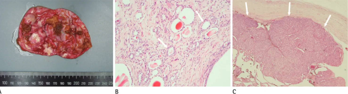
Fig. 2. Histopathologic findings of a surgically resected, left retroperitoneal mass.
A. The cut surface of the surgical specimen shows tan papillary tumor tissue on the inner surface of the wall.
B. Photomicrograph showing immature, tubule-like structures scattered in the fibrous cystic wall (arrows), which is a dysplastic kidney (hema- toxylin and eosin stain, × 100).
C. Photomicrograph showing low-grade renal cell carcinoma with papillary arrangement (arrows) (hematoxylin and eosin stain, × 40).
346
Cystic Papillary Renal Cell Carcinoma Arising from an Involutional Multicystic Dysplastic Kidney
jksronline.org
J Korean Soc Radiol 2015;73(5):343-346 Acknowledgments
We thank Bonnie Hami, MA (USA) for her editorial assis- tance in preparing the manuscript.
REFERENCES
1. Matsell DG, Bennett T, Goodyer P, Goodyer C, Han VK. The pathogenesis of multicystic dysplastic kidney disease: in- sights from the study of fetal kidneys. Lab Invest 1996;74:
883-893
2. Weinstein A, Goodman TR, Iragorri S. Simple multicystic dysplastic kidney disease: end points for subspecialty fol- low-up. Pediatr Nephrol 2008;23:111-116
3. Cambio AJ, Evans CP. Outcomes and quality of life issues in the pharmacological management of benign prostatic hyperplasia (BPH). Ther Clin Risk Manag 2007;3:181-196
4. Barrett DM, Wineland RE. Renal cell carcinoma in multi- cystic dysplastic kidney. Urology 1980;15:152-154
5. Birken G, King D, Vane D, Lloyd T. Renal cell carcinoma arising in a multicystic dysplastic kidney. J Pediatr Surg 1985;20:619-621
6. Rackley RR, Angermeier KW, Levin H, Pontes JE, Kay R. Re- nal cell carcinoma arising in a regressed multicystic dys- plastic kidney. J Urol 1994;152(5 Pt 1):1543-1545
7. Cambio AJ, Evans CP, Kurzrock EA. Non-surgical manage- ment of multicystic dysplastic kidney. BJU Int 2008;101:
804-808
8. Prasad SR, Humphrey PA, Catena JR, Narra VR, Srigley JR, Cortez AD, et al. Common and uncommon histologic sub- types of renal cell carcinoma: imaging spectrum with patho- logic correlation. Radiographics 2006;26:1795-1806; discus- sion 1806-1810
퇴화된 다낭이형성신 환자에서 발생한 낭성 유두상 콩팥 세포암종
김정재
1· 김봉수
1* · 허정식
2· 박경기
2· 최국명
1· 김승형
1· 맹영희
3다낭이형성신은 영아기에 발생하는 흔한 낭성 콩팥 질환 중 하나이다. 이 질환은 일반적으로 좋은 예후를 보이고 합병증의 빈도가 아주 적을 뿐 아니라 이형성된 콩팥이 자연적으로 퇴화되는 것으로 최근 받아들여지고 있다. 콩팥 세포암종은 이 다낭이형성신의 아주 드문 합병증이다. 퇴화된 다낭이형성신 환자에게서 발생된 콩팥 세포암종의 영상의학적 소견은 현재 까지 보고된 바가 없었다. 이 증례 보고에서는 퇴화된 다낭이형성신 환자에게서 발생한 낭성 유두상 콩팥 세포암종에 대 한 초음파와 컴퓨터전산단층촬영 소견을 보고하고자 한다.
제주대학교 의과대학 제주대학교병원 1영상의학과, 2비뇨기과, 3병리과