INTRODUCTION
Air pollutants are believed to be a factor in the increasing prevalence of asthma and to exacerbate respiratory diseases.1 Diesel exhaust particles (DEPs) as a major component of fine particulate matter (PM2.5) in the atmosphere of urban areas can induce airway hyperresponsiveness (AHR) and inflamma- tion by amplifying the T-helper 2 (Th2) immune response2-5 and are associated with cardiorespiratory mortality and the ag- gravation of disease.6-8 DEPs can induce acute AHR and acute asthma exacerbations independent of their effects on allergic sensitization.9
Long-term exposure to air pollution causes chronic respirato- ry disease,10 and chronic inhalation of DEPs leads to the devel- opment of cough and sputum. In addition, chronic bronchitis may be partially responsible for some of the exacerbations of asthma.11 The biological response to inhaled particles is aggra-
vated during chronic exposure to DEPs in a dose-dependent manner. In a previous study, inflammation and overproduction of mucus and surfactant components reached a plateau at 12 or 18 months of exposure during a 24-month experimental pe- riod, suggesting that DEPs play an important role in the devel- opment of chronic lung injury.12 Airway remodeling in asthma is defined by several structural changes, including epithelial cell mucus metaplasia, an increase in peribronchial smooth mus- cle mass, subepithelial fibrosis, and angiogenesis. Cytokines,
Long-Term Effects of Diesel Exhaust Particles on Airway Inflammation and Remodeling in a Mouse Model
Byeong-Gon Kim,
1Pureun-Haneul Lee,
1Shin-Hwa Lee,
1Young-En Kim,
1Mee-Yong Shin,
2Yena Kang,
1Seong-Hwan Bae,
1Min-Jung Kim,
1TaiYoun Rhim,
3Choon-Sik Park,
1An-Soo Jang
1*
1Department of Internal Medicine, Soonchunhyang University Bucheon Hospital, Bucheon, Korea
2Department of Pediatrics, Soonchunhyang University Bucheon Hospital, Bucheon, Korea
3Department of Bioengineering, College of Engineering, Hanyang University, Seoul, Korea
This is an Open Access article distributed under the terms of the Creative Commons Attribution Non-Commercial License (http://creativecommons.org/licenses/by-nc/3.0/) which permits unrestricted non-commercial use, distribution, and reproduction in any medium, provided the original work is properly cited.
Purpose: Diesel exhaust particles (DEPs) can induce and trigger airway hyperresponsiveness (AHR) and inflammation. The aim of this study was to investigate the effect of long-term DEP exposure on AHR, inflammation, lung fibrosis, and goblet cell hyperplasia in a mouse model. Methods:
BALB/c mice were exposed to DEPs 1 hour a day for 5 days a week for 3 months in a closed-system chamber attached to a ultrasonic nebulizer (low dose: 100 µg/m3 DEPs, high dose: 3 mg/m3 DEPs). The control group was exposed to saline. Enhanced pause was measured as an indicator of AHR.
Animals were subjected to whole-body plethysmography and then sacrificed to determine the performance of bronchoalveolar lavage and histology.
Results: AHR was higher in the DEP group than in the control group, and higher in the high-dose DEP than in the low-dose DEP groups at 4, 8, and 12 weeks. The numbers of neutrophils and lymphocytes were higher in the high-dose DEP group than in the low-dose DEP group and control group at 4, 8, and 12 weeks. The levels of interleukin (IL)-5, IL-13, and interferon-γ were higher in the low-dose DEP group than in the control group at 12 weeks.
The level of IL-10 was higher in the high-dose DEP group than in the control group at 12 weeks. The level of vascular endothelial growth factor was higher in the low-dose and high-dose DEP groups than in the control group at 12 weeks. The level of IL-6 was higher in the low-dose DEP group than in the control group at 12 weeks. The level of transforming growth factor-β was higher in the high-dose DEP group than in the control group at 4, 8, and 12 weeks. The collagen content and lung fibrosis in lung tissue was higher in the high-dose DEP group at 8 and 12 weeks. Conclusions: These results suggest that long-term DEP exposure may increase AHR, inflammation, lung fibrosis, and goblet cell hyperplasia in a mouse model.
Key Words: Chronic; diesel exhaust particles; airway remodeling
Correspondence to: An Soo Jang, MD, PhD, Department of Internal Medicine, Soonchunhyang University Bucheon Hospital,170 Jomaru-ro, Wonmi-gu, Bucheon 14584, Korea.
Tel: +32-621-5143; Fax: +32-621-6950; E-mail: [email protected] Received: April 27, 2015; Revised: August 28, 2015;
Accepted: September 8, 2015
•There are no financial or other issues that might lead to conflict of interest.
Allergy Asthma Immunol Res. 2016 May;8(3):246-256.
http://dx.doi.org/10.4168/aair.2016.8.3.246 pISSN 2092-7355 • eISSN 2092-7363
chemokines, and growth factors released from inflammatory and structural cells in the airway are believed to play a pivotal role in the development of remodeling.13-16 Chronic inflamma- tion is thought to initiate and perpetuate the cycles of tissue in- jury and repair in asthma, although remodeling may also occur in parallel with inflammation.17
Although long-term, repeated exposure to DEPs in air pollu- tion increases the risk for chronic respiratory diseases and car- diorespiratory mortality,18 the biological mechanism and re- modeling remain poorly understood during chronic exposure to DEPs. Therefore, we developed a mouse model of exposure to DEPs, and the effect of long-term exposure to DEPs as one of the air pollutants on airway inflammation and responsiveness, lung fibrosis, and goblet cell hyperplasia was examined.
MATERIALS AND METHODS DEP sources and preparation
We obtained and used DEPs from Hajime Takizawa. DEPs were collected using the following method. The engine used for preparation of DEPs was a 4JB1 type (Isuzu Automatic Co., To- kyo, Japan), light-duty (2,740-cc), 4-cylinder diesel engine. The engine was connected to an EDGY dynamometer (Meiden-Sya, Tokyo, Japan) and operated using standard diesel fuel with a speed of 1,500 rpm under a load of 10 torque (kg/m). The ex- haust was introduced into a stainless steel dilution tunnel (300×
8,400 mm) in a constant-volume sampler system equipped to the end of the dilution tunnel. The temperature at the sampling point was below 50°C. The diameter of the particles was mea- sured using an Anderson Air Sampler of the low-pressure type,19 and the mean diameter was 0.4 µm. Most of the shapes ana- lyzed using a scanning electron microscope were globular. De- tails on the DEPs used were previously described.20,21
Animal sensitization and exposure conditions
DEPs were sterilized by autoclaving and suspended in serum- free media after coating with BSA to minimize particle aggrega- tion and hydrophobicity.22 After sonication, the endotoxin con- centration of the DEP suspension was <0.064 ng/mL (0.32 EU/
mL) using the Limulus Amebocyte Lysate assay (QCL-1000;
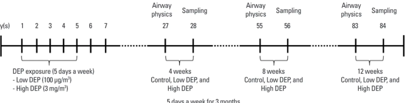
BioWhittaker Inc., Walkersville, MD, USA).23 Female Balb/c mice, 5 to 6 weeks of age and free of mice-specific pathogens, were obtained from Orient Co, Ltd (Charles River Laboratories, Seoul, Korea). The mice were housed throughout the experi- ments in a laminar flow cabinet and maintained on standard laboratory chow ad libitum. All experimental animals used in this study were treated according to guidelines approved by the Institutional Animal Care and Use Committee of the Soonc- hunhyang University Medical School. DEPs were resuspended in saline solution for 30 minutes before administration. The mice (n=8 in each group) were exposed to 100 µg/m3 and 3 mg/m3 DEPs for 1 hour a day for 5 days a week from 4 to 12 weeks (Fig. 1) in a closed-system chamber attached to an ultra- sonic nebulizer (NE-UO7; Omron Corporation, Tokyo, Japan) with an output of 1 mL/min and 1- to 5-µm particle size.
The control mice were administered and exposed to saline solution alone. Mice were sacrificed with an overdose of pento- barbital sodium (65 mg/kg body weight, administered intraper- itoneally). The chest cavity was exposed, and the catheter was carefully inserted into the trachea and secured with ligatures.
Bronchoalveolar lavage (BAL) was performed by 4 instillations of 1 mL of normal saline and gentle retrieval. Cell numbers were measured using a hemocytometer, and differential cell counts were performed on slides prepared by cyto-centrifuga- tion and Diff-Quik staining (Scientific Products, Gibbstowne, NJ, USA). Supernatants were separated by centrifugation (500 g, 5 minutes) and maintained at -70°C until use. After ligation of the right main bronchus, the left lung was fixed with 4% para- formaldehyde in phosphate-buffered saline and paraffin-em- bedded. The right lung was excised and immersed in TRI re- agent (guanidinium thiocyanate-phenol mixture; Molecular Research Center Inc., Cincinnati, OH, USA), and immediately frozen in liquid nitrogen.
Determination of airway responsiveness to methacholine Airway responsiveness was measured in unrestrained, con- scious mice 1 day after the last challenge, as previously de- scribed.24 Mice were placed in a barometric plethysmographic
Day(s) 27
Airway physics
55 Airway physics
83 Airway physics 28
Sampling
56 Sampling
84 Sampling 1 2 3 4 5 6 7
DEP exposure (5 days a week) - Low DEP (100 μg/m3) - High DEP (3 mg/m3)
4 weeks Control, Low DEP, and
High DEP
8 weeks Control, Low DEP, and
High DEP
12 weeks Control, Low DEP, and
High DEP
Fig. 1. Schematic diagram of the experimental protocol. Mice were exposed to DEPs 5 days a week for 3 months and either 100 µg/m3 or 3 mg/m3 DEPs (or saline as a control) using an ultrasonic nebulizer.
5 days a week for 3 months
chamber (All Medicus Co., Seoul, Korea), and baseline read- ings were taken and averaged for 3 minutes. Aerosolized methacholine in increasing concentrations (from 2.5 to 50 mg/
mL was nebulized through an inlet of the main chamber for 3 minutes. Readings were taken and averaged for 3 minutes after each nebulization, at which time the enhanced pause (Penh) was determined. Penh, calculated as (expiratory time/relax- ation time – 1)×(peak expiratory flow/peak inspiratory flow) according to the manufacturers’ protocol, is a dimensionless value that represents the proportion of maximal expiratory to maximal inspiratory box pressure signals and the timing of ex- piration. Penh is used as a measure of airway responsiveness to methacholine. The results are expressed as the percentage in- crease of Penh following challenge with each concentration of methacholine, where the baseline Penh (after saline challenge) is expressed as 100%. Penh values averaged for 3 minutes after each nebulization were evaluated.
Preparation of lung tissues for histology and immunohistochemistry
Trachea and lung tissues were removed from the rats. Four percent paraformaldehyde fixing solution was infused into the lungs via the trachea. The specimens were dehydrated and em- bedded in paraffin. For histological examination, 4-μm sections of embedded tissue were cut on a rotary microtome, placed on glass slides, deparaffinized, and stained sequentially with he- matoxylin and eosin.
Measurement of cytokine levels in BAL fluids
The cytokine levels were quantified in BAL fluid using a sand- wich enzyme-linked immunosorbent assay kit according to the manufacturer’s protocol (Biosource International Inc., Camaril- lo, CA, USA). Each sample was determined in triplicate. The lower limit of detection for interleukin (IL)-5, IL-13, interferon (IFN)-γ, IL-10, and vascular endothelial growth factor (VEGF) was 0.17, 30.00, 14.70, 15.60, and 2.00 pg/mL, respectively. Val- ues below these limits were considered zero for statistical anal- ysis. Inter- and intra-assay coefficients of variance were <10%.
Collagen assay
Collagen assays were performed according to the user manu- al of the Sircol collagen assay kit (Biocolor, Northern Ireland, UK). Briefly, 100 μL of the protein extract sample in lung tissue was mixed with 1 mL of Sircol dye for 30 minutes and centri- fuged at 10,000 rpm for 5 minutes to precipitate the formed col- lagen-dye complex. After decanting the suspension, droplets were dissolved in 1 mL of Sircol alkali reagent and vortexed. A total of 100 μl of the acquired solution was read at 540 nm.
Masson trichrome assay
The lung tissue slides were placed under a warmer at 60°C for 30 minutes. Next, samples were immersed in xylene for 15 min-
utes, followed by 100%, 95%, 90%, 80%, and 70% ethanol for 10 minutes each. The slides were stained in accordance with the manual of the Masson trichrome assay kit (Sigma-Aldrich, St.
Louis MO, USA). Briefly, Bouin’s solution was used as a mor- dant to intensify the color reactions. The nucleus and cyto- plasm were dyed using hematoxylin and scarlet acid. Phospho- tungstic/phosphomolybdic acid solution was used to change a positive to a negative charge. After staining with aniline blue, the samples were treated with 1% acetic acid. Prior to observa- tion, the samples were mounted with a cover slip using mount- ing media (Amresco, OH, USA). The positive trichrome-stained area was quantified using ImageJ software (National Institutes of Health, Bethesda, MD, USA). Values are reported as a per- centage of positive areas within the total sample.
Periodic acid-Schiff assay
The slides were stained in accordance with the manual of a periodic acid-Schiff assay kit (Abcam, Cambridge, MA, USA).
Briefly, lung tissue was cut into 4-μm sections and placed in pe- riodic acid solution for 5 minutes. After washing with distilled water, the specimens were immersed in Schiff’s fluid for 15 minutes. The slides were again washed with distilled water for 5 minutes and then placed in hematoxylin for 3 minutes. After the slides were exposed to running tap water for 3 minutes, blu- ing reagent was applied for 30 seconds. Before observation, the slides were mounted with a cover slip using mounting media (Amresco).
Statistical analysis
Differences between independent samples were compared using the nonparametric Kruskal-Wallis H test for continuous data. If differences were found to be significant, the Mann- Whitney U test was applied to compare differences between 2 samples. Differences were considered significant when a P val- ue was <0.05. Results are expressed as mean±standard error of the mean (SEM) unless otherwise stated.
RESULTS
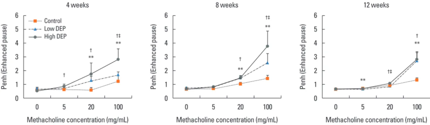
Airway responsiveness was measured 4, 8, and 12 weeks after the last exposure to DEPs in mice (Fig. 1). Airway responsiveness to aerosolized methacholine was measured in unrestrained, conscious mice. Mice were placed in the main chamber with a barometric plethysmograph and nebulized first with saline and then with increasing doses (from 2.5 to 100.0 mg/mL) of metha- choline for 3 minutes for each nebulization. Readings of breath- ing parameters were taken for 3 minutes after each nebulization, during which time Penh values were determined. AHR was higher in the low-dose and high-dose DEP groups than in the control group and higher in the high-dose DEP group than in the low-dose DEP group at 4, 8, and 12 weeks (Fig. 2).
The total cell count in the BAL fluid tended to be higher in the
low-dose DEP group than in the control group at 4, 8, and 12 weeks, and there was a significant increase in the total cell count in the high-dose DEP group than in the control group at 4 weeks (P<0.05) (Fig. 3). There were no differences in the number of macrophages among the 3 groups. The number of neutrophils was larger in the high-dose DEP group than in the control group at 4, 8, and 12 weeks (P<0.05 for each) (Fig. 3).
The number of lymphocytes was larger in the high-dose DEP group than in the control group at 4, 8, and 12 weeks (P<0.05).
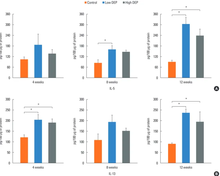
The level of IL-5 was higher in the low-dose DEP group than in the control group at 12 weeks (Fig. 4). The level of IL-13 was
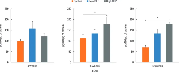
higher in the low- and high-dose DEP groups than in the con- trol group at 12 weeks (Fig. 4). The level of IFN-γ was higher in the low-dose DEP group than in the control group at 12 weeks (Fig. 5). The level of IL-10 was higher in the high-dose DEP group than in the control group at 12 weeks (Fig. 6). The level of VEGF was higher in the low- and high-dose DEP groups than in the control group at 12 weeks (Fig. 7). The level of IL-6 was higher in the low-dose DEP group than in the control group at 12 weeks (Fig. 8A). The level of transforming growth factor-β (TGF-β) was higher in the high-dose DEP group than in the control group at 4, 8, and 12 weeks (Fig. 8B).
Penh (Enhanced pause) Penh (Enhanced pause) Penh (Enhanced pause)
0 5 0 5 0 5
Methacholine concentration (mg/mL) Methacholine concentration (mg/mL) Methacholine concentration (mg/mL)
4 weeks 8 weeks 12 weeks
20 100 20 100 20 100
6 5 4 3 2 1 0
6 5 4 3 2 1 0
6 5 4 3 2 1 0
† **
**
** †‡
**
**
**
†‡
†‡
†
Fig. 2. Effect of DEP exposure on airway responsiveness in mice. **P<0.01 vs the sham group; †P<0.01 vs the sham group; ‡P<0.01 vs the low-dose DEP group.
†
† Control
Low DEP High DEP
Fig. 3. Changes in cell profiles in bronchioalveolar lavage fluid. *P<0.05 vs the control group; **P<0.01 vs the control group.
Cells in BAL fluid (×104/mL)
Total cell Macrophage Neutrophil Eosinophil Lymphocyte 60
50 40 30 20 0.6 0.4 0.2 0
24 weeks
**
*
Cells in BAL fluid (×104/mL) Cells in BAL fluid (×104/mL)
Total cell Macrophage Neutrophil Eosinophil Lymphocyte Total cell Macrophage Neutrophil Eosinophil Lymphocyte 60
50 40 30 20
60 50 40 30 20 0.6
0.4 0.2 0
0.6 0.4 0.2 0
4 weeks 8 weeks
**
***
*
* *
* *
Control Low DEP High DEP
pg/100 μg of proteinpg/100 μg of protein pg/100 μg of proteinpg/100 μg of protein pg/100 μg of proteinpg/100 μg of protein
4 weeks
4 weeks
8 weeks
8 weeks IL-5
IL-13
12 weeks
12 weeks 360
300
240
180
120
60
0
300
250
200
150
100
50
0
360
300
240
180
120
60
0
300
250
200
150
100
50
0
360
300
240
180
120
60
0
300
250
200
150
100
50
0 Control Low DEP High DEP
A
B Fig. 4. Effect of DEPs on IL-5 and IL-13 levels in BAL fluid. Bars represent mean±SEM from the 6 independent experiments. *P<0.05 vs the sham group.
*
*
*
*
*
*
*
pg/100 μg of protein pg/100 μg of protein pg/100 μg of protein
4 weeks 8 weeks
IFN-γ
12 weeks 500
400
300
200
100
0
500
400
300
200
100
0
500
400
300
200
100
0 Control Low DEP High DEP
Fig. 5. Effect of DEP on IFN-γ level in BAL fluid. Bars represent mean±SEM from the 6 independent experiments. *P<0.05 vs the sham group.
*
*
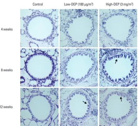
Lung fibrosis as shown by Masson trichrome staining of lung tissues was more severe in the low- and high-dose DEP groups than in the control group (Fig. 9A and B). The collagen content in lung tissues was greater in the high-dose DEP group than in the control group at 8 and 12 weeks (P<0.05) (Fig. 9C). Goblet cell hyperplasia in lung tissues was more severe in the high-dose DEP group than in the control group at 8 and 12 weeks (Fig. 10).
DISCUSSION
This study revealed that long-term exposure to DEP leads to increased airway responsiveness and inflammatory cell infiltra- tion, cytokine changes, collagen deposition, and goblet cell hy- perplasia, indicating that long-term exposure to DEPs may be associated with AHR, inflammation, and lung fibrosis.
DEPs are the particulate component of diesel exhaust, which
includes diesel soot and aerosols, such as ash particulates, me- tallic abrasion particles, sulfates, and silicates.25 Exposure to DEPs has been associated with acute short-term symptoms, such as headache, dizziness, light-headedness, nausea, cough- ing, difficult or labored breathing, chest tightness, and irritation of the eyes, nose, and throat.25 DEPs are related to allergic dis- eases, including asthma and allergic rhinitis based on extensive epidemiological studies25-27 and act as an adjuvant during aller- gen exposure and effect acute asthma exacerbations, bronchi- tis, or chronic obstructive pulmonary disease.28-38 Previous re- search has shown a strong link between particulate air pollu- tion and detrimental health effects, including cardiopulmonary morbidity and mortality.39 In this study, AHR was higher in the low- and high-dose DEP groups than in the control group and higher in the high-dose DEP group than in the low-dose DEP group at 4, 8, and 12 weeks, suggesting that DEPs can induce
pg/100 μg of protein pg/100 μg of protein pg/100 μg of protein
4 weeks 8 weeks
IL-10
12 weeks 250
200
150
100
50
0
250
200
150
100
50
0
250
200
150
100
50
0 Control Low DEP High DEP
Fig. 6. Effect of DEP on IL-10 level in BAL fluid. Bars represent mean±SEM from the 6 independent experiments. *P<0.05 vs the sham group.
*
*
pg/100 μg of protein pg/100 μg of protein pg/100 μg of protein
4 weeks 8 weeks
VEGF
12 weeks 180
150 120
90 60 30
0
180
150 120
90 60 30
0
180
150 120
90 60 30
0 Control Low DEP High DEP
Fig. 7. Effect of DEP on VEGF level in BAL fluid. Bars represent mean±SEM from the 6 independent experiments. *P<0.05 vs the sham group.
*
*
AHR in a dose-dependent manner.
DEP exposure is associated with asthma and airway cell apoptosis. DEPs may directly contribute to asthma by inducing epithelial cell death through the apoptotic pathway40 and in- ducing inflammatory responses in human airway epithelial cells.41 DEPs activate intracellular signaling pathways that cul- minate in the production of profibrotic cytokines and growth factors.38 DEPs activate T cells in asthmatics with a higher effect during exacerbations, suggesting that uncontrolled asthma is a risk factor for aggravation in individuals exposed to traffic pol- lutants.42 DEPs also induce time-dependent increases in IL-8, granulocyte-macrophage colony-stimulating factor, and IL-1β in epithelial cell lines.42 DEPs affect dendritic cells (DCs), which results in increased production of TNF, IL-6, IFN-γ, IL-12, and VEGF.41 In costimulation assays of PM-exposed DCs and allore- active CD4+ T cells, DEPs directed a Th2-like pattern of cyto- kine production (e.g., enhanced production of IL-13 and IL-18,
and suppressed of production IFN-γ).43 Pro-oxidative DEP chemicals can interfere with Th1-promoting response path- ways in a homogeneous DC population and provide a novel ex- planation for the adjuvant effect of DEPs on allergic inflamma- tion, indicative of an adjuvant effect of particulate air pollutants in allergic inflammatory disease.44 DEPs induce antigen-inde- pendent DC maturation via epithelial cell–DC interactions me- diated by the human bronchial epithelial cell-derived granulo- cyte-macrophage colony-stimulating factor.45 In our study, IL-6 and TGF-β were increased in the DEP long-term exposure group, suggesting that Th17 cytokine may be involved in airway inflammation following DEP exposure. Alveolar macrophages play an important role in particle-induced lung inflammation via direct induction of IL-13 production, suggesting that alveo- lar macrophages may act as major effectors of innate immunity to modulate immune and inflammatory responses toward a Th2-like condition via the production of IL-13, as observed in
pg/100 μg of proteinpg/100 μg of protein pg/100 μg of proteinpg/100 μg of protein pg/100 μg of proteinpg/100 μg of protein
4 weeks
4 weeks
8 weeks
8 weeks IL-6
TGF-β
12 weeks
12 weeks 360
300
240
180
120
60
0
100
80
60
40
20
0
360
300
240
180
120
60
0
100
80
60
40
20
0
360
300
240
180
120
60
0
100
80
60
40
20
0 Control Low DEP High DEP
A
B Fig. 8. Effect of DEP on IL-6 (A) and TGF-β (B) levels in BAL fluid. Bars represent mean±SEM from the 6 independent experiments. *P<0.05 vs the sham group.
* *
*
*
*
Relative trichrome-stained area (%)Collagen (μg/mL) Relative trichrome-stained area (%)Collagen (μg/mL) Relative trichrome-stained area (%)Collagen (μg/mL) 4 weeks
4 weeks
8 weeks
8 weeks
12 weeks
12 weeks 10
8
6
4
2
0
40
30
20
10
0
10
8
6
4
2
0
40
30
20
10
0
10
8
6
4
2
0
40
30
20
10
0 Control Low DEP High DEP
B
C Fig. 9. Lung fibrosis as shown by (A) Masson trichrome staining, (B) % area, and (C) collagen content. *P<0.05 vs the control group.
*
*
*
*
*
*
*
* 12 weeks
8 weeks 4 weeks
Control Low-DEP (100 μg/m3) High-DEP (3 mg/m3)
A
12 weeks 8 weeks 4 weeks
Control Low-DEP (100 μg/m3) High-DEP (3 mg/m3)
Fig. 10. Goblet cell hyperplasia in the lung based on periodic acid-Schiff staining.
the adaptive immune response.46 In our study, IL-5, IL-13, INF-γ, and IL-10 were increased in the DEP long-term exposure group, suggesting that Th2, Th1, and regulatory cytokines are involved in airway inflammation following DEP exposure. In addition, DEP exposure has been associated with the upregulation of al- lergic immune responses and airway remodeling in both ani- mal and human studies.47,48 Long-term exposure can lead to se- rious chronic health problems, such as cardiovascular disease, cardiopulmonary disease, and lung cancer.35 Fibrotic reactions are a component of these pulmonary diseases and are involved in the progressive deposition of collagen by pulmonary fibro- blasts.38 In this study, lung fibrosis, the collagen content, and VEGF in mice lungs exposed to long-term DEPs in a dose-de- pendent manner indicated that chronic exposure to DEPs can play a role in airway remodeling and angiogenesis. In addition, exposure to a higher dose of DEPs leads to increased airway in- flammation, a higher cytokine response, and more severe lung fibrosis, suggesting that environmental reduction of DEP expo- sure is important for patients with airway diseases. In conclu- sion, chronic exposure to DEPs can cause AHR, inflammation, goblet cell hyperplasia, and lung fibrosis.
ACKNOWLEDGMENTS
This research was supported by the Basic Science Research Program through the National Research Foundation of Korea funded by the Ministry of Education (2013R1A1A2005465) and
Soonchunhyang University Research Fund.
REFERENCES
1. Brandt EB, Biagini Myers JM, Acciani TH, Ryan PH, Sivaprasad U, Ruff B, et al. Exposure to allergen and diesel exhaust particles poten- tiates secondary allergen-specific memory responses, promoting asthma susceptibility. J Allergy Clin Immunol 2015;136:295-303.e7.
2. Inoue K, Tanaka M, Takano H. DEP-induced T(H)17 response in asthmatic subjects. J Allergy Clin Immunol 2014;133:1495-6, 1496.e1.
3. Folinsbee LJ. Human health effects of air pollution. Environ Health Perspect 1993;100:45-56.
4. Acciani TH, Brandt EB, Khurana Hershey GK, Le Cras TD. Diesel exhaust particle exposure increases severity of allergic asthma in young mice. Clin Exp Allergy 2013;43:1406-18.
5. Saito Y, Azuma A, Kudo S, Takizawa H, Sugawara I. Long-term in- halation of diesel exhaust affects cytokine expression in murine lung tissues: comparison between low- and high-dose diesel ex- haust exposure. Exp Lung Res 2002;28:493-506.
6. Dockery DW, Pope CA 3rd, Xu X, Spengler JD, Ware JH, Fay ME, et al. An association between air pollution and mortality in six U.S.
cities. N Engl J Med 1993;329:1753-9.
7. Samet JM, Dominici F, Curriero FC, Coursac I, Zeger SL. Fine par- ticulate air pollution and mortality in 20 U.S. cities, 1987-1994. N Engl J Med 2000;343:1742-9.
8. Hoek G, Brunekreef B, Goldbohm S, Fischer P, van den Brandt PA.
Association between mortality and indicators of traffic-related air pollution in the Netherlands: a cohort study. Lancet 2002;360:
1203-9.
9. Hao M, Comier S, Wang M, Lee JJ, Nel A. Diesel exhaust particles
exert acute effects on airway inflammation and function in murine allergen provocation models. J Allergy Clin Immunol 2003;112:
905-14.
10. Pope CA, Dockery DW, Schwartz J. Review of epidemiological evi- dence of health effects of particulate air pollution. Inhal Toxicol 1995;7:1-18.
11. Morgan WK, Reger RB, Tucker DM. Health effects of diesel emis- sions. Ann Occup Hyg 1997;41:643-58.
12. Ishihara Y, Kagawa J. Chronic diesel exhaust exposures of rats dem- onstrate concentration and time-dependent effects on pulmonary inflammation. Inhal Toxicol 2003;15:473-92.
13. Ghio AJ, Smith CB, Madden MC. Diesel exhaust particles and air- way inflammation. Curr Opin Pulm Med 2012;18:144-50.
14. Royce SG, Moodley Y, Samuel CS. Novel therapeutic strategies for lung disorders associated with airway remodelling and fibrosis.
Pharmacol Ther 2014;141:250-60.
15. van den Brûle S, Heymans J, Havaux X, Renauld JC, Lison D, Huaux F, et al. Profibrotic effect of IL-9 overexpression in a model of air- way remodeling. Am J Respir Cell Mol Biol 2007;37:202-9.
16. Henderson WR Jr, Chi EY, Bollinger JG, Tien YT, Ye X, Castelli L, et al. Importance of group X-secreted phospholipase A2 in allergen- induced airway inflammation and remodeling in a mouse asthma model. J Exp Med 2007;204:865-77.
17. Kunzmann S, Schmidt-Weber C, Zingg JM, Azzi A, Kramer BW, Blaser K, et al. Connective tissue growth factor expression is regu- lated by histamine in lung fibroblasts: potential role of histamine in airway remodeling. J Allergy Clin Immunol 2007;119:1398-407.
18. Sydbom A, Blomberg A, Parnia S, Stenfors N, Sandström T, Dahlén SE. Health effects of diesel exhaust emissions. Eur Respir J 2001;17:
733-46.
19. Andersen AA. A sampler for respiratory health hazard assessment.
Am Ind Hyg Assoc J 1966;27:160-5.
20. Song HM, Jang AS, Ahn MH, Takizawa H, Lee SH, Kwon JH, et al.
Ym1 and Ym2 expression in a mouse model exposed to diesel ex- haust particles. Environ Toxicol 2008;23:110-6.
21. Lee GB, Brandt EB, Xiao C, Gibson AM, Le Cras TD, Brown LA, et al. Diesel exhaust particles induce cysteine oxidation and s-gluta- thionylation in house dust mite induced murine asthma. PLoS One 2013;8:e60632.
22. Kim J, Natarajan S, Vaickus LJ, Bouchard JC, Beal D, Cruikshank WW, et al. Diesel exhaust particulates exacerbate asthma-like in- flammation by increasing CXC chemokines. Am J Pathol 2011;179:
2730-9.
23. Grünig G, Warnock M, Wakil AE, Venkayya R, Brombacher F, Ren- nick DM, et al. Requirement for IL-13 independently of IL-4 in ex- perimental asthma. Science 1998;282:2261-3.
24. Hamelmann E, Schwarze J, Takeda K, Oshiba A, Larsen GL, Irvin CG, et al. Noninvasive measurement of airway responsiveness in allergic mice using barometric plethysmography. Am J Respir Crit Care Med 1997;156:766-75.
25. Diaz-Sanchez D, Proietti L, Polosa R. Diesel fumes and the rising prevalence of atopy: an urban legend? Curr Allergy Asthma Rep 2003;3:146-52.
26. Jung DY, Leem JH, Kim HC, Kim JH, Hwang SS, Lee JY, et al. Effect of traffic-related air pollution on allergic disease: results of the chil- dren’s health and environmental research. Allergy Asthma Immu- nol Res 2015;7:359-66.
27. Lee JH, Lee HS, Park MR, Lee SW, Kim EH, Cho JB, et al. Relation- ship between indoor air pollutant levels and residential environ-
ment in children with atopic dermatitis. Allergy Asthma Immunol Res 2014;6:517-24.
28. Nel AE, Diaz-Sanchez D, Ng D, Hiura T, Saxon A. Enhancement of allergic inflammation by the interaction between diesel exhaust particles and the immune system. J Allergy Clin Immunol 1998;
102:539-54.
29. Takahashi G, Tanaka H, Wakahara K, Nasu R, Hashimoto M, Miyo- shi K, et al. Effect of diesel exhaust particles on house dust mite-in- duced airway eosinophilic inflammation and remodeling in mice.
J Pharmacol Sci 2010;112:192-202.
30. Takano H, Ichinose T, Miyabara Y, Yoshikawa T, Sagai M. Diesel ex- haust particles enhance airway responsiveness following allergen exposure in mice. Immunopharmacol Immunotoxicol 1998;20:
329-36.
31. Ohta K, Yamashita N, Tajima M, Miyasaka T, Nakano J, Nakajima M, et al. Diesel exhaust particulate induces airway hyperresponsive- ness in a murine model: essential role of GM-CSF. J Allergy Clin Im- munol 1999;104:1024-30.
32. Ichinose T, Takano H, Miyabara Y, Sadakaneo K, Sagai M, Shib- amoto T. Enhancement of antigen-induced eosinophilic inflam- mation in the airways of mast-cell deficient mice by diesel exhaust particles. Toxicology 2002;180:293-301.
33. Walters DM, Breysse PN, Wills-Karp M. Ambient urban Baltimore particulate-induced airway hyperresponsiveness and inflamma- tion in mice. Am J Respir Crit Care Med 2001;164:1438-43.
34. Miyabara Y, Ichinose T, Takano H, Lim HB, Sagai M. Effects of die- sel exhaust on allergic airway inflammation in mice. J Allergy Clin Immunol 1998;102:805-12.
35. Lim HB, Ichinose T, Miyabara Y, Takano H, Kumagai Y, Shimojyo N, et al. Involvement of superoxide and nitric oxide on airway inflam- mation and hyperresponsiveness induced by diesel exhaust parti- cles in mice. Free Radic Biol Med 1998;25:635-44.
36. Takano H, Yoshikawa T, Ichinose T, Miyabara Y, Imaoka K, Sagai M.
Diesel exhaust particles enhance antigen-induced airway inflam- mation and local cytokine expression in mice. Am J Respir Crit Care Med 1997;156:36-42.
37. Monforton C. Weight of the evidence or wait for the evidence? Pro- tecting underground miners from diesel particulate matter. Am J Public Health 2006;96:271-6.
38. Bonner JC. Lung fibrotic responses to particle exposure. Toxicol Pathol 2007;35:148-53.
39. Riedl M, Diaz-Sanchez D. Biology of diesel exhaust effects on respi- ratory function. J Allergy Clin Immunol 2005;115:221-8.
40. Ackland ML, Zou L, Freestone D, van de Waasenburg S, Michalc- zyk AA. Diesel exhaust particulate matter induces multinucleate cells and zinc transporter-dependent apoptosis in human airway cells. Immunol Cell Biol 2007;85:617-22.
41. Cao D, Bromberg PA, Samet JM. COX-2 expression induced by die- sel particles involves chromatin modification and degradation of HDAC1. Am J Respir Cell Mol Biol 2007;37:232-9.
42. Mamessier E, Nieves A, Vervloet D, Magnan A. Diesel exhaust par- ticles enhance T-cell activation in severe asthmatics. Allergy 2006;
61:581-8.
43. Porter M, Karp M, Killedar S, Bauer SM, Guo J, Williams D, et al.
Diesel-enriched particulate matter functionally activates human dendritic cells. Am J Respir Cell Mol Biol 2007;37:706-19.
44. Chan RC, Wang M, Li N, Yanagawa Y, Onoé K, Lee JJ, et al. Pro-oxi- dative diesel exhaust particle chemicals inhibit LPS-induced den- dritic cell responses involved in T-helper differentiation. J Allergy
Clin Immunol 2006;118:455-65.
45. Bleck B, Tse DB, Jaspers I, Curotto de Lafaille MA, Reibman J. Die- sel exhaust particle-exposed human bronchial epithelial cells in- duce dendritic cell maturation. J Immunol 2006;176:7431-7.
46. Kang CM, Jang AS, Ahn MH, Shin JA, Kim JH, Choi YS, et al. Inter- leukin-25 and interleukin-13 production by alveolar macrophages in response to particles. Am J Respir Cell Mol Biol 2005;33:290-6.
47. Nordling E, Berglind N, Melén E, Emenius G, Hallberg J, Nyberg F, et al. Traffic-related air pollution and childhood respiratory symp- toms, function and allergies. Epidemiology 2008;19:401-8.
48. Dai J, Xie C, Vincent R, Churg A. Air pollution particles produce air- way wall remodeling in rat tracheal explants. Am J Respir Cell Mol Biol 2003;29:352-8.