J Korean Soc Radiol 2018;78(2):81-87 https://doi.org/10.3348/jksr.2018.78.2.81
Korean Clinical Imaging Guideline for Hemoptysis 객혈 환자의 영상진단검사: 한국형 권고안
Mi-Jin Kang, MD
1, Jin Hwan Kim, MD
2, Yoon Kyung Kim, MD
3, Hyun Joo Lee, MD
4, Kyung Min Shin, MD
5, Jung Im Kim, MD
6, Hyun Ju Lee, MD
7, Kyung Hyun Do, MD
4, Hwan Seok Yong, MD
8, Sol Ji Choi, MPH
9, Miyoung Choi, RN
9, Jung Im Jung, MD
10*
1Department of Radiolgoy, InJe University Sanggye Paik Hospital, Seoul, Korea
2Department of Radiology, Chungnam National University Hospital, Chungnam National University School of Medicine, Daejeon, Korea
3Department of Radiology, Gachon University Gil Medical Center, Incheon, Korea
4Department of Radiology and Research Institute of Radiology, Asan Medical Center, University of Ulsan College of Medicine, Seoul, Korea
5Department of Radiology, Kyungpook National University Chilgok Hospital, Daegu, Korea
6Department of Radiology, Kyung Hee University Hospital at Gangdong, Kyung Hee University College of Medicine, Seoul, Korea
7Department of Radiology, Seoul National University Hospital, Seoul, Korea
8Department of Radiology, Korea University Guro Hospital, Korea University College of Medicine, Seoul, Korea
9Division of Healthcare Technology Assessment Research, National Evidence-based Healthcare Collaborating Agency, Seoul, Korea
10Department of Radiology, Seoul St. Mary’s Hospital, College of Medicine, The Catholic University of Korea, Seoul, Korea
권고 개발 배경
객혈은 호흡기 계통에서 출혈이 발생하여 입 혹은 코로 나오 는 것으로 그 양에 따라 임상적으로 중등도를 구분하여 진단하 고 치료한다. 그러나 실제 임상에서 객혈의 양을 정확하게 파악 하기가 어려울 뿐 아니라, 폐실질 내에 고여있는 혈액은 객혈의 양 평가에서 간과될 수 있다. 또한 객혈의 원인이 되는 질환은
기관지염 등의 급성 양성 질환과 기관지확장증 등의 만성 양성 질환 및 악성 종양까지 매우 다양하다. 따라서 적절한 진단과 처치를 위해 영상의학적 검사가 반드시 필요하다.
객혈 환자의 중등도 및 폐암의 위험도에 따른 진단 및 치료 의 권고안은 이미 미국영상전문의학회(American College of Radiology; 이하 ACR)에서 발표하였으나(1), ACR에서는 객혈 의 진단 및 치료에 있어 발견되지 않은 폐암의 중요성을 강조하 In 2014, the American College of Radiology announced a guideline for appropriate
diagnostic approach and treatment in patients with hemoptysis, according to severity of hemoptysis and risk of lung cancer. However, in Korea many patients have pulmo- nary fibrosis due to previous tuberculosis or have active tuberculosis. Therefore, appli- cation of this guideline is not appropriate. The Korean Society of Radiology and Korean Society of Thoracic Radiology proposed a guideline more closely matching the real state of diagnostic approach and treatment of patients with hemoptysis in Korea. The guideline was prepared in consensus by a development committee, working party, and an advisory committee. The process of the guideline proposal was based on method- ology for developing evidence-based clinical imaging guidelines: joint recommenda- tions by the Korean Society of Radiology and National Evidence-Based Healthcare Collaborating Agency. The clinical imaging guideline for adult patients with hemopty- sis is as follows. Chest radiography is an initial imaging modality to evaluate hemop- tysis. Contrast enhanced chest CT is recommended in patients with two lung cancer risks (> 40 years old and > 30 packs per year smoking history), moderate hemoptysis (> 30 cc/24 hours) or recurrent hemoptysis. Contrast enhanced chest CT is also rec- ommended for patients with massive hemoptysis (> 400 mL/24 hours) without car- diopulmonary compromise.
Index terms Guideline Hemoptysis
Radiography, Thoracic Tomography, X-ray Computed Evidence-Based Practice Republic of Korea
Received June 29, 2017 Revised August 4, 2017 Accepted August 7, 2017
*Corresponding author: Jung Im Jung, MD Department of Radiology, Seoul St. Mary’s Hospital, College of Medicine, The Catholic University of Korea, 222 Bandpo-daero, Seocho-gu, Seoul 06591, Korea.
Tel. 82-2-2258-1435 Fax. 82-2-599-6771 E-mail: [email protected]
This is an Open Access article distributed under the terms of the Creative Commons Attribution Non-Commercial License (http://creativecommons.org/licenses/by-nc/4.0) which permits unrestricted non-commercial use, distri- bution, and reproduction in any medium, provided the original work is properly cited.
고 있다. 반면 한국에서는 과거 결핵으로 인한 폐의 섬유화가 있는 경우가 많고, 현재의 활동성 결핵에 의한 객혈이 있는 경우 도 많아 ACR과는 차별되는 임상영상 권고안 개발의 필요성이 제기되었다. 따라서 개발위원회, 실무위원회 및 자문위원단이 의견을 모아 한국의 질병 상황에 적합한 권고안을 개발하였다.
진료지침 수용개작 과정
권고안 개발 절차는 개발위원회에서 개발한 근거기반 임상 영상 가이드라인 수용개작 방법론에 근거하여 이루어졌다(2).
핵심 질문
실무위원회에서 선정한 핵심 질문은 개발위원회 및 최종 사 용자로 예상되는 임상전문학회(대한결핵 및 호흡기학회)로부터 추천 받은 자문단의 검토를 거치고 수정 사항을 반영한 후 최종 확정되었다. 최종 확정된 문장형 핵심 질문은 아래와 같다.
객혈이 있는 성인 환자에서 객혈의 원인 진단을 위한 적절한 영상 검사는 무엇인가?
진료 지침 검색전략
검색 데이터베이스는 국외 주요 문헌 검색 데이터베이스 (Ovid-Medline, Ovid-Embase) 및 해외 진료 지침 관련 주요 사이트(National Guideline Clearinghouse, Guideline Interna- tional Network)와 국내 데이터베이스(KoreaMed, KMbase, KoMGI, KGC)를 모두 포함하여 검색하였다. 또한 주요 학회 사이트 및 주요 가이드라인이 누락되지 않도록 수기 검색을 통 해 보완하였다. 검색 기간은 2000년 이후부터 2015년 7월까지 로, 주요 검색어는 ‘hemoptysis’와 ‘computerized tomography’,
‘bronchoscopy’를 keyword로 하였으며, 데이터베이스별로 Medical Subject Heading (MeSH) term, EMTREE와 같은 색 인어를 함께 활용하였다. 이후 발간된 논문이나 가이드라인은 최신 검색을 통해 보완하였다.
진료 지침 선별 및 선정
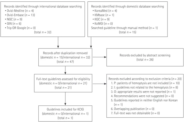
핵심 질문에 대한 문헌 검색에서 국외 검색 데이터베이스에 서 총 32건, 국내 검색 데이터베이스에서 14건, 수기 검색에서 1건의 문헌이 검색되어 총 47건이 검색되었다. 각각의 문헌에 대하여 제목과 초록의 내용을 바탕으로 직접적인 관련이 있는 문헌만을 1차 선별하여, 21건의 문헌이 선택되었다. 21건의 문
Records identified through international database searching
• Ovid-Medline (n = 4)
• Ovid-Embase (n = 13)
• NGC (n = 9)
• GIN (n = 6)
• Trip OR Google (n = 0)
(total n = 32)
Records after duplication removed (domestic n = 15/international n = 32)
(total n = 47)
Full-text guidelines assessed for eligibility (domestic n = 0/international n = 21)
(total n = 21)
Guidelines included for KCIG (domestic n = 0/international n = 1)
(total n = 1)
Records excluded by abstract screening (total n = 26)
Records excluded according to exclusion criteria (n = 20) 1. P: patients of hemoptysis are not included (n = 10) 2. I: guidelines not related to the hemoptysis (n = 8) 3. O: appropriate results were not reported (n = 1) 4. Recommendations were not suggested (n = 0) 5. Guidelines reported in neither English nor Korean
(n = 1)
6. Overlapping publication (n = 0) 7. Full-text was not obtainable (n = 0) Records identified through domestic database searching • KoreaMed (n = 4)
• KMbase (n = 1) • KGC (n = 9) • KoMGI (n = 0)
Searched guideline through manual method (n = 1) (total n = 15)
Fig. 1. Flow chart for literature selection.
GIN = Guideline International Network, I = intervention/index test, KCIG = Korean Clinical Imaging Guidelines, NGC = National Guideline Clear- inghouse, O = outcome, P = population/patient
헌에 대하여는 원문을 확보하여 개발위원회에서 선정한 지침의 요건을 충족시키지 못하는 문헌들은 배제하였으며, 2차 선정 과정을 통해 최종 1건의 문헌이 선택되었다(Fig. 1).
진료 지침의 질 평가
개발위원회에서 선택한 진료 지침에 대한 Appraisal of Guidelines for Research & Evaluation 질 평가 결과를 회신하 였고(Table 1), 실무위원회에서는 결과를 참조하여 핵심 질문에 대해 최종 1편의 지침을 선택했다.
근거 검토 및 권고안 초안 작성
최종 선택된 지침의 권고 및 권고등급을 비교하고, 권고를 지 지하는 근거 문헌을 다시 검토하여 한국 임상영상 진료지침 (Korean Clinical Imaging Guidelines)의 근거수준 결정 방법 (Tables 2, 3)에 따라 근거의 내용 및 질 평가 결과를 근거표 (evidence table) 형태로 정리했다(Supplementary Table 1 in the online-only Data Supplement). 근거표를 바탕으로 실무위 원회의 최종 논의를 거쳐 권고문 초안이 도출되었다.
권고안 합의 및 권고등급 결정
실무위원회에서 작성한 초안에 대해서는 개발위원회와의 논 의를 거쳐 최종 근거수준과 권고등급을 결정했다.
권고안의 합의
대한결핵 및 호흡기학회 등 외부 관련 학회 전문가가 포함된 자문위원회의 권고안에 대한 동의 정도를 설문조사(9점 척도) 한 결과, 핵심 질문에 대한 동의 정도는 1차에서 평균(표준편 차) 7.3-8.6 (0.5-0.9), 2차는 6.9-8.5 (0.5-0.8)이었다.
외부 검토
임상영상 가이드라인 개발에 참여하지 않은 영상의학과 전문 의와 지침의 최종사용자가 될 유관학회인 대한결핵 및 호흡기 학회에서 최종 권고문에 대해 검토하였다.
Table 2. Criteria for Evidence Level of Each Evidence Literatures
Level Content
1 Research satisfying all of criteria following three 1) Good reference standard
2) Consecutive patients study 3) Blind interpretation Systematic review of level 1
Randomized controlled trial or cross-sectional cohort study that compares index test to comparators
2 Research satisfying all of criteria following two 1) Good reference standard
2) Consecutive patients study or blind interpretation Systematic review of level 2
Observational studies that compares index test to comparators 3 Without consistently applied reference standards
4 Case-control study
Poor or non-independent reference standard 5 Expert opinion
Adated from Choi SJ et al. Korean J Radiol 2017;18:208-216 (2)
Table 3. Grades of Korean Clinical Imaging Guidelines Recommendation
Grading Content Meaning
A Recommended This intervention (examination) has enough evidence to support desired effect, and therefore, is recommended B (Conditional) recommended This intervention (examination) has intermediate to enough level of evidence to support desired effect
Provide intervention (examination) selectively, or for specific individuals based on expert’s judgment C Not recommended This intervention (examination) has enough evidence to support non-desired effect, and therefore,
is not recommended (use of this examination is not recommended)
D No recommendation This intervention (examination) does not have enough evidence to either support or reject effectiveness, and needs further research
This intervention (examination) has very low level of certainty for desired effect, and decision based on recommendation grading has no meaning
Adated from Choi SJ et al. Korean J Radiol 2017;18:208-216 (2) Table 1. AGREE II
Source of Recommendation AGREE II Score
Proposal of Develomental Committee ACR Appropriateness Criteria®
hemoptysis. 66 Recommended
Not recommended: AGREE score < 50.
ACR = American College of Radiology, AGREE = Appraisal of Guidelines for Research & Evaluation
권고
핵심 질문
객혈이 있는 성인 환자에서 객혈의 원인 진단을 위한 적절한 영상 검사는 무엇인가?
권고 1-1. 객혈이 있는 모든 환자는 초기 검사로 흉부 X선 검사를 시행할 것을 권고한다. (권고등급 A , 근거수준 II)
권고 1-2. 객혈이 있는 성인 환자에서 2가지 폐암 위험인 자가 있다면, 원인 진단을 위해 조영증강 흉부 CT를 시행할 것을 권고한다. (권고등급 A, 근거수준 II)
권고 1-3. 중등증 객혈(30-400 mL/24 hr) 또는 반복적 객혈을 보이는 성인 환자는 원인 진단을 위해 조영증강 흉부 CT를 시행할 것을 권고한다. (권고등급 A, 근거수준 II)
권고 1-4. 대량 객혈(> 400 mL/24 hr)이 있으나, 심장폐 기능은 유지되는 성인 환자는 원인 진단을 위해 조영증강 흉 부 CT를 시행할 것을 권고한다. (권고등급 A, 근거수준 III)
근거 요약
객혈은 호흡기에서 발생한 출혈을 뱉어 내는 것으로 객담에 혈흔이 섞여 있는 정도부터 대량 객혈까지 다양한 범위로 나타 난다. 객혈의 원인은 기관지확장증, 급성 및 만성 기관지염, 결 핵, 미만성폐질환, 폐혈관 기형 등의 양성 질환 및 폐종양 등이 있으며 원인 불명인 경우도 있어 적절한 선별검사 및 정밀검사 가 필요하다(3-5). 따라서 본 문헌은 객혈이 있는 성인 환자의 원인 진단을 위해 적절한 영상의학적 검사에 대한 권고안을 제 시하기 위해 작성되었다.
본 권고안에서는 객혈이 있는 모든 환자의 초기 검사로 흉부 X선 검사를 시행할 것을 권고한다. 흉부 X선 검사는 출혈 부위 를 확인하는 데 도움을 줄 뿐 아니라, 폐실질이나 흉막 등의 기 저질환 여부에 대한 선별검사이기도 하다(3, 6, 7).
객혈의 양에 따라 경증 객혈(< 30 mL/24 hr), 중등증 객혈 (30~400 mL/24 hr), 대량 객혈(> 400 mL/24 hr)로 나누어, 영상의학적인 검사 방법을 결정하였다(7-10).
흉부 X선 검사가 정상이더라도 폐암의 가능성을 배제할 수 없으므로 객혈의 양에 상관없이 폐암 위험인자가 있는 성인 환 자의 경우에는 정밀검사로 조영증강 흉부 CT를 시행할 것을 권고한다. 객혈이 있는 성인환자에서 그 원인이 종양인 경우는 논문에 따라 10~35% 정도이다(2, 3). 객혈의 원인을 발견하 지 못한 환자를 추적 관찰한 연구에서 6%의 환자에서 3년 이 내에 절제 불가능한 폐암이 발견되었으며, 폐암이 발생한 모든
환자는 40세 이상의 흡연자였다(11). 또한 객혈이 있는 환자 중 흉부 X선 검사 소견이 정상인 환자를 대상으로 시행한 CT 검사에서 9.6%의 환자에서 폐암이 발견이 되었으며, 폐암이 발견된 환자의 96%는 과거 혹은 현재의 흡연력이 있었다(12).
중등증 이상의 객혈(> 30 mL/24 hr)이나 반복적 객혈은 즉 각적인 처치를 위한 출혈 부위를 찾기보다는 객혈의 원인을 밝 혀내는 것이 더 중요하다. 문헌 고찰에 따르면, 한국에서 객혈 의 가장 흔한 원인은 기관지확장증이었으며, 활동성 폐결핵 및 폐결핵 후유증도 흔한 원인이었다(5). 그 외 폐렴, 아스페르길 루스종, 폐종양, 기관지염, 기타 원인 및 원인을 알 수 없는 경 우도 있었다(5). CT는 비침습적인 방법으로 객혈의 원인 및 출 혈 부위를 진단하는 데 있어 기관지내시경보다 진단율이 높은 장점이 있다(13, 14). 조영증강을 하지 않은 CT로도 원인 질환 인 기관지확장증, 폐결핵, 폐암 등을 진단할 수는 있다. 그러나 결핵 환자의 경우, 기관지 동맥(bronchial artery)에서 출혈이 일어나는 경우가 가장 흔하지만, 라스무센동맥류(Rasmussen aneurysm)나 비기관지 측부순환동맥(non-bronchial collateral artery)에서 출혈이 일어나는 경우도 있으므로, 정확한 출혈 부 위를 확인하기 위해 조영증강 CT를 시행할 것을 권고한다. 과 거에는 수술을 시행할 수 없는 대량 객혈이 기관지동맥색전술 의 주된 적응증이었으나, 근래에는 1주 내에 하루 100 mL 이상 의 중등도 객혈을 3회 이상하거나, 경도이지만 만성적이고 점 차 심해지는 객혈인 경우에도 기관지동맥색전술을 시행한다 (15). 조영증강 CT는 기관지동맥뿐 아니라 늑간동맥, 쇄골하 동맥, 액와동맥, 하횡격막동맥 등의 비기관지 측부순환동맥, 폐동정맥에 관한 영상해부학적 소견을 제시함으로써 기관지동 맥색전술을 가이드하고, 또 시술시간을 줄여줄 수 있다(16).
대량 객혈(> 400 mL/24 hr)이 있는 환자라도 심장폐기능이 유지되는 경우에는 조영증강 흉부 CT를 시행할 것을 권고한 다. 대량 객혈이 있으면 기관지 동맥색전술이나 수술을 시행하게 되는데, 색전술 혹은 수술 시행 전에 CT를 시행하면 출혈의 원 인이 되는 혈관을 파악하여 시술 및 수술에 도움이 된다(7, 17).
권고 고려사항
이득과 위해
객혈이 있는 환자의 초기 검사로 흉부 X선 검사를 시행하는 것은 비교적 낮은 방사선량으로 출혈 부위를 결정할 수 있고, 폐 실질 질환에 대한 선별검사로 이용할 수 있다는 점에서 매우 유 용하다.
2가지의 폐암 위험인자(40세 이상, 30갑년 이상 흡연)를 가 지고 있는 환자는 객혈의 양에 상관없이 흉부 CT를 시행할 것
을 권고하고 있는데, 이때 흉부 X선 검사에서 잘 보이지 않은 폐암을 발견할 수 있다는 장점이 있으나, CT 시행으로 인한 방 사선 피폭의 위해가 있다.
중등증 이상의 객혈이 있거나 반복적 객혈이 있는 환자에서 CT를 시행하면 객혈의 원인 및 출혈 부위를 한번에 알아낼 수 있다는 장점이 있다. 한국에서는 객혈이 활동성 결핵이나 폐결 핵의 후유증 등 염증성 질환과 관련된 경우가 많은데, 이런 환 자에서는 흉부 X선 검사만으로는 출혈 병소를 평가하기가 힘 들다. 이런 경우 CT를 시행하면 출혈 병소도 정확히 평가하고, 원인이 되는 질환도 알 수 있어 매우 유용하게 사용할 수 있다.
대량 객혈이 있는 환자나 중등증 이상의 객혈 혹은 반복적 객 혈이 있는 환자에서는 치료의 목적으로 기관지동맥색전술을 시 행할 수 있다. 색전술 시행 전에 CT를 시행하면 출혈의 위치나 기관지동맥 또는 폐동맥에 관한 영상해부학적 소견을 제시함 으로써 시술을 가이드하고, 또 시술시간을 줄여줄 수 있어 방사 선 피폭의 위해보다는 이득이 더 크다.
조영증강 CT를 시행함으로써 요오드화 조영제에 의한 위해 가 있을 수 있다. 조영제를 사용하여 얻을 수 있는 효과와 환자 에게 발생할 수 있는 위험성을 고려하여 조영제 사용 유무를 결 정해야 하며 조영제 진료지침에 따른다.
검사별 방사선량
흉부 X선 검사의 방사선량의 상대적 수준은 1 mSv 이하이고 흉부 CT 검사의 방사선량의 상대적 수준은 > 5~10 mSv이다 (Table 4).
Acknowledgments
This study was co-supported by the National Evidence-
based Collaborating Agency (NECA-C-15-003) and the Ko- rean Society of Radiology (NECA-S-15-002).
Supplementary Materials
The online-only Data Supplement is available with this arti- cle at https://doi.org/10.3348/jksr.2018.78.2.81.
RefeRences
1. ACR Appropriateness Criteria® hemoptysis. Available at:
https://acsearch.acr.org/docs/69449/Narrative/. Published Aug, 2010. Accessed Jan 31, 2017
2. Choi SJ, Jeong WK, Jo AJ, Choi JA, Kim MJ, Lee M, et al.
Methodology for developing evidence-based clinical imag- ing guidelines: joint recommendations by Korean Society of Radiology and National Evidence-Based Healthcare Col- laborating Agency. Korean J Radiol 2017;18:208-216 3. Tsoumakidou M, Chrysofakis G, Tsiligianni I, Maltezakis G,
Siafakas NM, Tzanakis N. A prospective analysis of 184 he- moptysis cases: diagnostic impact of chest X-ray, computed tomography, bronchoscopy. Respiration 2006;73:808-814 4. Fidan A, Ozdog˘an S, Oruç O, Salepçi B, Ocal Z, Cag˘layan B.
Hemoptysis: a retrospective analysis of 108 cases. Respir Med 2002;96:677-680
5. Bruzzi JF, Rémy-Jardin M, Delhaye D, Teisseire A, Khalil C, Rémy J. Multi-detector row CT of hemoptysis. Radiograph- ics 2006;26:3-22
6. Lee SJ, Rho JY, Yoo SM, Kim MD, Lee JH, Kim EK, et al. Use- Table 4. Korean Relative Radiation Level
Symbol RRL Example
0 0 Sonography, MRI
< 1 mSv Chest PA, Plain radiography, Mammography
1–5 mSv IVU, UGIS, Low dose chest CT, Brain CT, Brain CTA
> 5–10 mSv Routine Chest CT, Abdominal CT, Coronary CT
> 10 mSv 3 Phase dynamic CT (abdomen) Adated from Choi SJ et al. Korean J Radiol 2017;18:208-216 (2).
CTA = computed tomography angiography, IVU = intravenous urography, MRI = magnetic resonance imaging, RRL = relative radiation level, UGIS = upper gastrointestinal series
fulness of multi-detector computed tomography before bronchoscopy and/or bronchial arterial embolization for hemoptysis. Tuberc Respir Dis 2010;68:80-86
7. Revel MP, Fournier LS, Hennebicque AS, Cuenod CA, Meyer G, Reynaud P, et al. Can CT replace bronchoscopy in the de- tection of the site and cause of bleeding in patients with large or massive hemoptysis? AJR Am J Roentgenol 2002;
179:1217-1224
8. Delage A, Tillie-Leblond I, Cavestri B, Wallaert B, Marquette CH. Cryptogenic hemoptysis in chronic obstructive pulmo- nary disease: characteristics and outcome. Respiration 2010;
80:387-392
9. Menchini L, Remy-Jardin M, Faivre JB, Copin MC, Ramon P, Matran R, et al. Cryptogenic haemoptysis in smokers: an- giography and results of embolisation in 35 patients. Eur Respir J 2009;34:1031-1039
10. Poe RH, Israel RH, Marin MG, Ortiz CR, Dale RC, Wahl GW, et al. Utility of fiberoptic bronchoscopy in patients with he- moptysis and a nonlocalizing chest roentgenogram. Chest 1988;93:70-75
11. Herth F, Ernst A, Becker HD. Long-term outcome and lung cancer incidence in patients with hemoptysis of unknown
origin. Chest 2001;120:1592-1594
12. Thirumaran M, Sundar R, Sutcliffe IM, Currie DC. Is investi- gation of patients with haemoptysis and normal chest ra- diograph justified? Thorax 2009;64:854-856
13. McGuinness G, Beacher JR, Harkin TJ, Garay SM, Rom WN, Naidich DP. Hemoptysis: prospective high-resolution CT/
bronchoscopic correlation. Chest 1994;105:1155-1162 14. Kim HB. Bronchial artery embolization. In Korean Society
of Interventional Radiology, ed. Interventional radiology, 2nd ed. Seoul: Ilchokak 2014:321-325
15. Millar AB, Boothroyd AE, Edwards D, Hetzel MR. The role of computed tomography (CT) in the investigation of unex- plained haemoptysis. Respir Med 1992;86:39-44
16. Khalil A, Fartoukh M, Parrot A, Bazelly B, Marsault C, Carette MF. Impact of MDCT angiography on the management of patients with hemoptysis. AJR Am J Roentgenol 2010;195:
772-778
17. Hsiao EI, Kirsch CM, Kagawa FT, Wehner JH, Jensen WA, Baxter RB. Utility of fiberoptic bronchoscopy before bron- chial artery embolization for massive hemoptysis. AJR Am J Roentgenol 2001;177:861-867
객혈 환자의 영상진단검사: 한국형 권고안
강미진
1· 김진환
2· 김윤경
3· 이현주
4· 신경민
5· 김정임
6· 이현주
7도경현
4· 용환석
8· 최솔지
9· 최미영
9· 정정임
10*
2014년 미국영상전문의학회(American College of Radiology)에서 객혈로 내원한 환자에 대하여 객혈의 중등도 및 폐암 의 위험도 정도에 따른 진단 및 치료에 대한 권고안을 발표하였다. 그러나 한국에서는 과거 결핵으로 인한 폐의 섬유화가 많고, 현재의 활동성 결핵에 의한 객혈이 있는 경우도 많아 미국의 권고안을 그대로 사용하기는 어려운 실정이다. 따라서 대한영상의학회 및 대한흉부영상의학회에서는 한국의 실정에 맞는 권고안을 개발하기로 결정하였고, 개발위원회, 실무위 원회 및 자문위원단이 의견을 모아 한국의 질병 상황에 적합한 권고안을 개발하였다. 권고안 개발 절차는 개발위원회에서 개발한 근거기반 임상영상 가이드라인 수용개작 방법론에 근거하여 이루어졌다. 객혈이 있는 성인 환자에서 객혈의 원인을 진단하기 위한 영상의학적 검사에 대한 권고 내용은 다음과 같다. 객혈이 있는 모든 환자에 대한 초기 검사로 흉부 X선 검 사를 권고한다. 객혈이 있는 환자가 2가지 폐암의 위험인자(40세 이상, 30갑년 이상 흡연)가 있거나, 중등증 객혈(24시간 이내 30 mL 이상의 객혈) 또는 반복적 객혈이 있을 경우에는 조영증강 흉부 CT를 권고한다. 대량 객혈(24시간 이내 400 mL 이상의 객혈)이 있으나 심폐기능이 유지되어 있는 경우에도 조영증강 흉부 CT를 권고한다.
1인제대학교 상계백병원 영상의학과, 2충남대학교 의학전문대학원 충남대학교병원 영상의학과, 3가천대학교 길병원 영상의학과,
4울산대학교 의과대학 서울아산병원 영상의학과, 영상의학연구소, 5경북대학교 의과대학 영상의학교실,
6경희대학교 의과대학 강동경희대학교병원 영상의학과, 7서울대학교병원 영상의학과,
8고려대학교 의과대학 구로병원 영상의학과, 9한국보건의료연구원 보건의료근거연구본부,
10가톨릭대학교 의과대학 서울성모병원 영상의학과
Literatures Research Type Enrolled Patients
Evidence Level
Tsoumakidou M, Chrysofakis G, Tsiligianni I, Maltezakis G, Siafakas NM, Tzanakis N.
A prospective analysis of 184 hemoptysis cases: diagnostic impact of chest X-ray, computed tomography, bronchoscopy. Respiration 2006;73(6):808-814.
Observational (retrospective)_Dx 184 2
Fidan A, Ozdog˘an S, Oruç O, Salepçi B, Ocal Z, Cag˘layan B. Hemoptysis:
a retrospective analysis of 108 cases. Respir Med 2002;96(9):677-680.
Observational (prospective)_Dx 108 2
Bruzzi JF, Rémy-Jardin M, Delhaye D, Teisseire A, Khalil C, Rémy J.
Multi-detector row CT of hemoptysis. Radiographics 2006;26(1):3-22.
Review/other-Dx N/A 2
Ketai LH, Mohammed TL, Kirsch J, et al. ACR appropriateness criteria® hemoptysis.
J Thorac Imaging 2014;29(3):W19-W22.
Review/other-Dx N/A 2
Lee SJ, Rho JY, Yoo SM, et al. Usefulness of multi-detector computed tomography before bronchoscopy and/or bronchial arterial embolization for hemoptysis.
Tuberc Respir Dis 2010;68(2):80-86.
Observational (prospective)_Dx 125 2
Revel MP, Fournier LS, Hennebicque AS, et al. Can CT replace bronchoscopy in the detection of the site and cause of bleeding in patients with large or massive hemoptysis? AJR Am J Roentgenol 2002;179(5):1217-1224.
Observational-Dx 80 3
Delage A, Tillie-Leblond I, Cavestri B, Wallaert B, Marquette CH. Cryptogenic hemoptysis in chronic obstructive pulmonary disease: characteristics and outcome. Respiration 2010;80(5):387-392.
Observational-Dx 39 3
Menchini L, Remy-Jardin M, Faivre JB, et al. Cryptogenic haemoptysis in smokers:
angiography and results of embolisation in 35 patients. Eur Respir J 2009;34(5):
1031-1039.
Observational-Dx 35 3
Poe RH, Israel RH, Marin MG, et al. Utility of fiberoptic bronchoscopy in patients with hemoptysis and a nonlocalizing chest roentgenogram. Chest 1988;93(1):
70-75.
Observational-Dx 196 4
Herth F, Ernst A, Becker HD. Long-term outcome and lung cancer incidence in patients with hemoptysis of unknown origin. Chest 2001;120(5):1592-1594.
Review/other-Dx 722 4
Thirumaran M, Sundar R, Sutcliffe IM, Currie DC. Is investigation of patients with haemoptysis and normal chest radiograph justified? Thorax 2009;64(10):854-856.
Observational-Dx 270 2
McGuinness G, Beacher JR, Harkin TJ, Garay SM, Rom WN, Naidich DP.
Hemoptysis: prospective high-resolution CT/bronchoscopic correlation.
Chest 1994;105(4):1155-1162.
Observational-Dx 57 2
Millar AB, Boothroyd AE, Edwards D, Hetzel MR. The role of computed tomography (CT) in the investigation of unexplained haemoptysis. Respir Med 1992;86(1):39-44.
Review/other-Dx 40 4
Khalil A, Fartoukh M, Parrot A, Bazelly B, Marsault C, Carette MF. Impact of MDCT angiography on the management of patients with hemoptysis. AJR Am J Roentgenol 2010;195(3):772-778.
Observational-Dx 400 3
Hsiao EI, Kirsch CM, Kagawa FT, Wehner JH, Jensen WA, Baxter RB.
Utility of fiberoptic bronchoscopy before bronchial artery embolization for massive hemoptysis. AJR Am J Roentgenol 2001;177(4):861-867.
Review/other-Dx 28 3
Dx = diagnosis