ABSTRACT
Vertebral hemangiomas are common, benign, and asymptomatic tumors that rarely extend into the epidural space or involve the posterior elements. Surgery is recommended for aggressive vertebral hemangiomas if symptoms are severe or evolve rapidly. A 57-year-old male patient was admitted to our department for several months of back pain. A computed tomography (CT) scan and magnetic resonance imaging (MRI) were suggestive of T12 hemangioma without the involvement of the spinal canal or posterior elements. Despite aggressive conservative treatments, such as medications or nerve blocks, the back pain worsened. The CT and MRI 2 months later revealed a lesion involving the vertebral body and posterior elements with extension into the epidural space and with spinal cord compression.
The patient underwent surgery for bone cement-augmented percutaneous screw fixation followed by low-dose radiotherapy. Histological examination confirmed the diagnosis of atypical hemangioma, specifically an epithelioid hemangioendothelioma.
Keywords: Hemangioma; Vertebrae; Spinal cord compression
INTRODUCTION
Vertebral hemangiomas are relatively common, benign, and asymptomatic lesions of the spine that are most often an incidental finding.14,15) Typically, hemangiomas involve the vertebral body and rarely extend to the posterior elements, such as the laminae, pedicles, or spinous process. Hemangiomas account for approximately 2–3% of all spinal tumors, and only 0.9–1.2% are symptomatic cases, classifying them as atypical aggressive hemangiomas.2,4)
In most cases, the vertebral hemangioma has a benign course, and surgery is not
recommended. However, in rare cases, there is an extension of the lesion into the epidural space with consequent spinal cord compression and requiring surgical intervention.
We report a rare case of a rapidly progressing aggressive hemangioma with epidural extension at the T12 level. The case was managed by intraoperative vertebroplasty and percutaneous screw fixation, rather than with aggressive surgical intervention.
Case Report
Received: May 12, 2020 Revised: Jul 1, 2020 Accepted: Jul 10, 2020 Address for correspondence:
Seok Won Kim
Department of Neurosurgery, College of Medicine, Chosun University, 365 Pilmun- daero, Dong-gu, Gwangju 61453, Korea.
E-mail: [email protected]
Copyright © 2020 Korean Neurotraumatology Society
This is an Open Access article distributed under the terms of the Creative Commons Attribution Non-Commercial License (https://
creativecommons.org/licenses/by-nc/4.0/) which permits unrestricted non-commercial use, distribution, and reproduction in any medium, provided the original work is properly cited.
ORCID iDs Chi Ho Kim
https://orcid.org/0000-0002-9955-5107 Seok Won Kim
https://orcid.org/0000-0002-1910-0242 Conflict of Interest
The authors declare no conflict of interest.
Informed consent was obtained from the patient. This article presents an individual case without the need for the approval of an ethical research committee.
Chi Ho Kim and Seok Won Kim
Department of Neurosurgery, College of Medicine, Chosun University, Gwangju, Korea
Rapidly Progressive Atypical Vertebral
Hemangioma: A Case Report
CASE REPORT
A 57-year-old male patient was admitted to our department with continuous back pain without any traumatic episode. The pain persisted for several months but progressively worsened with activity or while in the standing position with relief when lying down. A computed tomography (CT) scan and magnetic resonance imaging (MRI) was performed to rule out a compression fracture. The CT revealed a characteristic ‘polka dot’ pattern of the trabecular thickening involving the whole T12 vertebral body. The MRI showed the mass was high signal intensity on the T2 weighted images and low signal intensity on the T1 weighted images without epidural extension (FIGURE 1). These findings resulted in the diagnosis of a vertebral hemangioma. Conservative treatment, including analgesic and anti-inflammatory drugs, physiotherapy, and epidural steroid injections, was performed. However, the patient complained of progressively worsening back pain over the following 2 months and re-visited our institute.
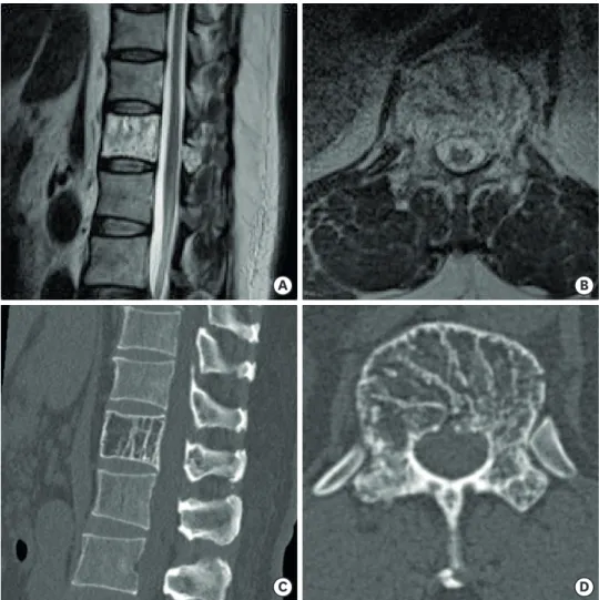
The CT and MRI took 2 months later, demonstrated an expansile mass involving the vertebral body and the posterior element with epidural extension and cord compression at
A B
C D
FIGURE 1. (A, B) T2-weighted sagittal and axial magnetic resonance images demonstrate a hyperintense signal on T12 body, with thickened trabeculae indicating the presence of typical vertebral hemangioma not compressing the spinal cord. (C, D) Sagittal and axial computed tomography scans show a “polka-dotted” appearance due to
the T12 (FIGURE 2). A preoperative embolization and vertebrectomy with reconstruction are recommended for aggressive vertebral hemangiomas. However, given the patient's intact motor power, a less invasive procedure was performed. The patient underwent intraoperative vertebroplasty and percutaneous screw fixation.
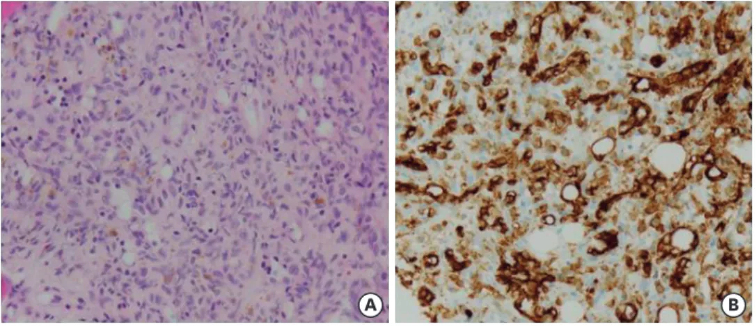
The patient received about 4 mL's of polymethylmethacrylate injected into the T12 vertebral body, followed by percutaneous screw fixation to prevent tumor growth and to enhance stability (FIGURE 3). The pathological diagnosis was consistent with epithelioid hemangioendothelioma, which is an aggressive vertebral hemangioma (FIGURE 4). After surgery, the patient's back pain improved dramatically, and he was transferred to the hemato- oncological department for low-dose radiotherapy. No further tumor growth was observed over 4 years after the procedure.
A B
C D
FIGURE 2. (A, B) Sagittal and axial T2-weighted magnetic resonance images took 2 months later demonstrate hypointense signal change on the posterior part of the T12 body, indicating the presence of bone marrow edema.
There is an epidural extension with cord compression when compared with the previous image. (C, D) Sagittal and axial computed tomography scans show more advanced fatty attenuation within the T12 vertebral body with multiple punctate thickened trabeculae.
DISCUSSION
Vertebral hemangiomas are benign vasoformative neoplasms of endothelial cells. Hence, they are generally considered neoplasms. Hemangiomas grow within marrow spaces in the bone and encase bony trabeculae. Because of the lack of aggressive histopathological features, some authors refer to hemangiomas as vascular malformations.6,7)
In most cases, hemangiomas are asymptomatic and do not require any surveillance or treatment. However, in 0.9–1.2% of patients, hemangiomas can cause symptoms because of the neural compression as a result of the bone expansion, bone erosion through cortex, hematoma, or fracture.12,13) The symptomatology classifies them as “aggressive vertebral hemangiomas,” making them more likely than their asymptomatic counterparts to involve the entire vertebral body, extend into the posterior elements, and have an irregular honeycomb pattern with lytic areas on radiological evaluation.10,11)
A B
FIGURE 3. Postoperative simple radiographs of the patient.
A B
FIGURE 4. Histopathologic images obtained during surgery. (A) The images demonstrate the proliferation of epithelioid cells and well-formed capillary-sized blood vessels. (B) The neoplastic endothelial cells have abundant cytoplasm forming intracytoplasmic lumens that express the endothelial marker CD31, meaning that these cells are immunoreactive for this marker.
The symptoms presented are dependent on the location of the tumor, the degree of the neural compression, and the involvement of the bone. The diagnosis includes a CT scan, where the typical “polka-dot-sign” is observed due to the trabecular aspect of the involved vertebra. In addition, the diagnosis may include an MRI, where the lesion appears hyperintense on both the T1 and the T2 weighted images with contrast enhancement.
Aggressive atypical hemangiomas, like epithelioid hemangiomas or hemangioendotheliomas, may demonstrate a mixed pattern, with heterogeneous signal intensity on T1 and T2
weighted MRIs that reflect the presence of inflammatory infiltration and the absence of fat.
Hemangiomas are more likely to show aggressiveness when its stroma contains less fat and is more vascular. The radiologic features demonstrating the aggressiveness of the disease include the presence of a low signal on T1 weighted images and a high signal on T2 weighted images on the MRI. In addition, avid contrast enhancement may reveal the presence of cortical erosion, soft tissue stroma between the osseous trabeculae on the CT, the presence of extradural soft tissue, expansion to the posterior elements, invasion of the spinal canal, and encroachment of the spinal cord.9)
Treatment for vertebral hemangiomas is usually only necessary when symptoms present with neurological deficits or disabling pain. The technical approach for atypical aggressive vertebral hemangiomas has been controversial in neurosurgery for centuries. Multiple therapeutic options have been described with varying degrees of success, including
radiation, percutaneous vertebroplasty or ethanol injections, embolization, surgery, and any combination of these therapies.1,3)
Among them, vertebroplasty is one of the most commonly performed treatment options for painful vertebral hemangiomas. With cement polymerisation, the exothermic reaction causes tumor necrosis, mechanical stability and prevention of vertebral collapse.
The surgical approach has been widely debated, with some authors recommending decompression and radiotherapy alone, while others support a gross total resection with 360-degree reconstruction to prevent late recurrence.8) Despite this, after extensive literature review, we propose that, for patients with atypical vertebral hemangiomas with mild or slowly progressive neurological symptoms, it is reasonable to attempt non-operative management with embolization, vertebroplasty, or radiotherapy. This treatment is recommended that provided symptoms are not due to spinal compression by a focal bony prominence, which is unlikely to resolve without surgery. However, for patients with severe or rapidly progressive symptoms, surgery should be considered. Aggressive vertebral hemangiomas are benign lesions that do not have metastatic potential and are not associated with mortality.5)
In the present case, the patient complained of severe back pain without prominent neurologic deficits. The vertebral hemangioma was treated with intraoperative vertebroplasty and percutaneous screw fixation followed by radiotherapy rather than preoperative embolization and en bloc vertebrectomy and 360-degree reconstruction. Bone cement augmented percutaneous screw fixation ensures mechanical stability and prevents vertebral collapse, and bone cement itself has the exothermic reaction causing tumor necrosis. After surgery, the disabling back pain improved dramatically, and radiological studies showed no local recurrence of the tumor. Nevertheless, regular clinical and radiologic follow-up is necessary to check for tumor recurrence.
CONCLUSION
Although most vertebral hemangioma diagnoses are often incidental imaging findings, they can display aggressive features and rapid progression requiring surgical treatment.
Diagnostic imaging awareness and an excellent understanding of the pathology is paramount to rule out other lesions that mimic aggressive hemangiomas.
REFERENCES
1. Acosta FL Jr, Dowd CF, Chin C, Tihan T, Ames CP, Weinstein PR. Current treatment strategies and outcomes in the management of symptomatic vertebral hemangiomas. Neurosurgery 58:287-295, 2006 PUBMED | CROSSREF
2. Blankstein A, Spiegelmann R, Shacked I, Schinder E, Chechick A. Hemangioma of the thoracic spine involving multiple adjacent levels: case report. Paraplegia 26:186-191, 1988
PUBMED
3. Bremnes RM, Hauge HN, Sagsveen R. Radiotherapy in the treatment of symptomatic vertebral hemangiomas: technical case report. Neurosurgery 39:1054-1058, 1996
PUBMED | CROSSREF
4. Chung SK, Nam TK, Park SW, Hwang SN. Capillary hemangioma of the thoracic spinal cord. J Korean Neurosurg Soc 48:272-275, 2010
PUBMED | CROSSREF
5. Goldstein CL, Varga PP, Gokaslan ZL, Boriani S, Luzzati A, Rhines L, et al. Spinal hemangiomas: results of surgical management for local recurrence and mortality in a multicenter study. Spine (Phila Pa 1976) 40:656-664, 2015
PUBMED | CROSSREF
6. Harrison MJ, Eisenberg MB, Ullman JS, Oppenheim JS, Camins MB, Post KD. Symptomatic cavernous malformations affecting the spine and spinal cord. Neurosurgery 37:195-204, 1995
PUBMED | CROSSREF
7. Jiang L, Liu XG, Yuan HS, Yang SM, Li J, Wei F, et al. Diagnosis and treatment of vertebral hemangiomas with neurologic deficit: a report of 29 cases and literature review. Spine J 14:944-954, 2014
PUBMED | CROSSREF
8. Karaeminogullari O, Tuncay C, Demirors H, Akin K, Sahin O, Ozyurek A, et al. Multilevel vertebral hemangiomas: two episodes of spinal cord compression at separate levels 10 years apart. Eur Spine J 14:706-710, 2005
PUBMED | CROSSREF
9. Laredo JD, Assouline E, Gelbert F, Wybier M, Merland JJ, Tubiana JM. Vertebral hemangiomas: fat content as a sign of aggressiveness. Radiology 177:467-472, 1990
PUBMED | CROSSREF
10. Laredo JD, Reizine D, Bard M, Merland JJ. Vertebral hemangiomas: radiologic evaluation. Radiology 161:183-189, 1986
PUBMED | CROSSREF
11. Lee SE, Moon KY, Jahng TA, Chung CK, Kim HJ. A case of an epidural extension of vertebral hemangioma treated by intraoperative vertebroplasty and laminectomy. Korean J Spine 6:192-196, 2009
12. Manning HJ. Symptomatic hemangioma of the spine. Radiology 56:58-65, 1951 PUBMED | CROSSREF
13. McAllister VL, Kendall BE, Bull JW. Symptomatic vertebral haemangiomas. Brain 98:71-80, 1975 PUBMED | CROSSREF
14. Nguyen JP, Djindjian M, Gaston A, Gherardi R, Benhaiem N, Caron JP, et al. Vertebral hemangiomas presenting with neurologic symptoms. Surg Neurol 27:391-397, 1987
PUBMED | CROSSREF
15. Song JW, Youn SM, Lee SH, Rhee CH, Lee SH, Lee JH, et al. Vertebral hemangioma causing cord compression: a case report. J Korean Neurosurg Soc 26:287-291, 1997