Neurocysticercosis is a relatively common parasitic disease involving the central nervous system.
Neurocysticercosis is characterized by multiple calcified lesions in the brain, like as occurs in patients with tuber- culosis. Neurocysticercosis is thought not to undergo re- activation. We report here on the first case of twice-reac- tivated neurocysticercosis.
Case Report
A 59-year-old man sought evaluation at our hospital due to perioral paresthesias and dysarthria on 25 April 2009. In 1997, he was admitted to our hospital due to headaches and seizures of 1 week duration. He had ex- perienced intermittent seizures since 1991. On the brain magnetic resonance imaging (MRI), there were multiple dark signal intensity lesions that were suggestive of cal- cifications in both cerebral hemispheres and peripheral-
enhancing cystic lesions in both frontal lobes. A dot-like, high signal intensity on the T1-weighted image and a low signal intensity on T2-weighted image were noted in the right frontal lobe, suggesting a scolex of cysticer- cosis (images not shown). Positive results were reported on the serum and cerebrospinal fluid (CSF) cysticercosis enzyme-linked immunosorbant assay (ELISA) tests (0.32 and 0.68, respectively). He was diagnosed with neuro- cysticercosis and treated with an anti-helminthic (biltri- cide). He recovered completely and was discharged.
In 2001, the patient had complaints of headaches, dizziness and numbness in both arm and legs. On brain CT, there were multiple nodular calcifications in both cerebral hemispheres and no abnormal enhancing le- sions (Figs. 1A, B), and this was all compatible with end- stage neurocysticercosis. He was hospitalized for several days and he improved. In 2002, he was admitted to our hospital with dysarthria and focal seizures of the lip sev- eral times during 1 day. There were no significant re- sults on the serum and CSF cysticercosis ELISA tests.
Thick bands of peripheral enhancement of multiple nodular calcified lesions and surrounding edema were noted on the brain MRI (Figs. 1C, D). The patient ap- peared to have reactivated neurocysticercosis and he was treated with the anti-helmintic praziquantel. His
J Korean Soc Radiol 2010;63:15-18
─ 15 ─
Second Reactivation of Neurocysticercosis: A Case Report1
Young Sup Shim, M.D., Hee Young Hwang, M.D., Hye-Young Choi, M.D., Jee Eun Kim, M.D., Hyung-Sik Kim, M.D.
1Department of Radiology, Gachon University, Gil Hospital, Incheon, Korea
Received November 10, 2009 ; Accepted February 16, 2010
Address reprint requests to : Hee Young Hwang, M.D., Department of Radiology, Gachon University, Gil Hospital, Incheon, Korea, 1198, Guwol-dong, Namdong-gu, Incheon 405-760, Korea.
Tel. 82-32-460-3060 Fax. 82-32-460-3065 E-mail: [email protected]
This report describes the first case involving a second reactivation of neurocysticer- cosis. There was peripheral enhancement and surrounding edema at multiple calcified lesions in both cerebral hemispheres on the brain MRI. One must be aware of the pos- sibility of reactivation of neurocysticercosis to make the correct diagnosis.
Index words :Neurocysticercosis Brain Edema
Magnetic Resonance Imaging Brain Diseases
symptoms improved after treatment and he was pre- scribed carbamazepine for controlling his seizures. He was healthy without any neurologic symptoms for 7 years, except for the present admission (Figs. 1E, F). He had no other primary symptoms and no remarkable findings on the physical examination. The laboratory findings, including the blood cell count, electrolytes and serum glucose level, were within the normal limits. He then underwent brain MRI with gadolinium-enhance-
ment. On brain MRI, there were several dark signal in- tensity lesions suggestive of calcifications in both cere- bral hemispheres, which were unchanged since the pre- vious brain MRI in 2004. There was edema around the calcified lesions on the axial T1- and T2-weight images, and this looked like the previous reactivation of neuro- cysticercosis in 2002 (Fig. 1G). On the gadolinium-en- hancement T1-weighted image, there were no changes in the diffuse thin bands of enhancement in the periph-
Young Sup Shim, et al: Second Reactivation of Neurocysticercosis
─ 16 ─
A B C
D E F
Fig. 1. A 59-year-old man with known neurocysticercosis.
A, B. The pre- & post-contrast brain CT that was performed for routine follow-up before the first reactivation of neurocysticercosis.
Multifocal dense nodular calcifications are detected in both cerebral hemispheres, and especially in the left frontal lobe (A). There are no abnormal enhancing lesions around the nodular calcifications on post-contrast brain CT (B).
C, D. The brain MRI with gadolinium enhancement that was performed in 2002 at the first reactivation of neurocysticercosis. A dark signal intensity lesion is shown in the left frontal lobe on the T2-weighted magnetic resonance image (arrow on C). This lesion is compatible with the dense nodular calcifications seen on the previous brain CT (A & B). There is edema around the lesion. A dif- fuse thick band of enhancement is noted in the peripheral portion of the lesion on the contrast-enhanced T1-weighted magnetic resonance image (arrow on D).
E, F. The brain MRI with gadolinium enhancement that was performed in 2004 during routine follow-up and the patient had neu- rologic symptoms. An unchanged dark signal intensity lesion is shown in the left frontal lobe on the T2-weighted magnetic reso- nance image (E). Edema around the lesion, which was detected on the previous brain MRI, is not shown. A diffuse thin band of en- hancement is noted at the peripheral portion of the lesion on the contrast-enhanced T1-weighted magnetic resonance image (F).
eral portion of the lesions (Fig. 1H). On a CSF study, he was negative for cysticercus IgG. It was thought that there was a second reactivation of neurocysticercosis and so he was treated with albendazole. The symptoms were resolved and the patient was discharged.
Discussion
Neurocysticercosis is an infection of the human cen- tral nervous system with Taenia solium, the pork tape- worm. Neurocysticercosis is a common parasitic disease in Latin America, South Africa, China, India and the United States (1). The seropositive rate for neurocys- ticercosis is 4.0% in epilepsy patients and 2.1% in other- wise healthy persons in Korea (2). Neurocysticercosis is a common cause of epilepsy in these endemic areas.
Brain imaging modalities, such as CT and MRI, are nec- essary for detecting or differentiating neurocysticercosis in epilepsy patients.
The natural course of neurocysticercosis is divided in- to four pathologic stages according to the Escobar classi- fication (3). The typical image findings on CT and MRI at each of the stages are vesicular, colloid vesicular, granular nodular and nodular calcified lesions, respec- tively (4, 5). Neurocysticercosis appears as cystic lesions without enhancement or peripheral edema in the vesic- ular stage, but in the colloid vesicular stage, surrounding edema and ring enhancement are noted as aggravated immune reactions. In the granular nodular stage, the cystic fluid is absorbed and the lesions are seen as solid nodules. Calcifications are detected in the nodular calci- fied stage, which is the terminal stage of neurocysticer- cosis, and this indicates that the larvae are dead. In our case, there were multiple cysts and calcifications on
MRI (image not shown) when the patient was initially admitted and diagnosed with neurocysticercosis in 1997; this appeared to be a mixture of the colloid vesicu- lar, granular nodular and nodular calcified stages. There were multiple calcified nodules without enhancement and no cystic lesions on the brain CT performed in 2001 (Figs. 1A, B), which appeared to be the nodular calcified stage of neurocysticercosis and accordingly death of the larvae. On the brain MRI performed in 2004, a persis- tent thin ring of enhancement was shown in the calci- fied nodules, which did not represent reactivation of neurocysticercosis, but rather, inflammation in the vicinity of the lesion (Figs. 1E, F) (6).
Peripheral thick bands of enhancement and peripher- al edema were detected twice on the follow-up brain MRIs in 2002 and 2009 (Figs. 1C, H), which were con- sidered to be reactivation of neurocysticercosis.
Unlike tuberculosis, neurocysticercosis is thought not to reactivate. One case has been reported by Sheth et al.
(7) on the reactivation of neurocysticercosis in which there were thick bands of peripheral enhancement of multiple calcified nodules with surrounding edema on the brain MRI, as in our case. The authors suggested that antigens released from dead larvae or other causes of immunologic reactions triggered an intense immune response. However, there were neither underlying dis- eases nor clinical symptoms of induced immune reac- tions in our patient, and pathologic confirmation was not performed other than the serum and CSF ELISA tests. Thus, the basis for neurocysticercosis reactivation was not demonstrated.
This is the first case report of twice-reactivated neuro- cysticercosis. Immunologic and pathologic studies are necessary for demonstrating the mechanism of reactiva-
J Korean Soc Radiol 2010;63:15-18
─ 17 ─
G H
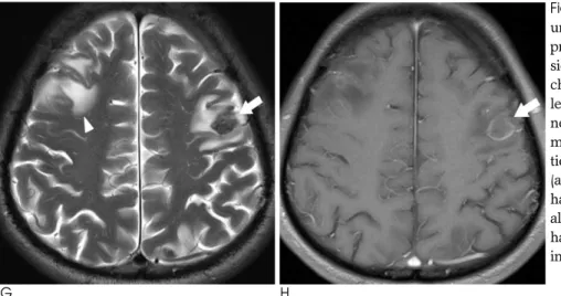
Fig. 1. G, H. The brain MRI with gadolini- um enhancement that was performed at presentation at the time of the last admis- sion. Edema is noted again around the un- changed dark signal intensity lesion in the left frontal lobe on the T2-weighted mag- netic resonance image (G). Additional ede- ma is visible around the nodular calcifica- tion (not shown) in the right frontal lobe (arrowhead). A diffuse thin band of en- hancement is not changed at the peripher- al portion of the lesion on the contrast-en- hanced T1-weighted magnetic resonance image since the previous study in 2004 (H).
tion of neurocysticercosis. Radiologists and clinicians should keep the possibility of reactivation of neurocys- ticercosis in mind, despite observing findings that are consistent with the nodular calcified stage.
References
1. White AC, Jr. Neurocysticercosis: Updates on epidemiology, pathogenesis, diagnosis, and management. Annu Rev Med 2000;51:
187-206
2. Kong Y, Cho SY, Cho MS, Kwon OS, Kang WS. Seroepidemiologi- cal observation of taenia solium cysticercosis in epileptic patients
in Korea. J Korean Med Sci 1993;8:145-152
3. Escobar A. The pathology of neurocysticercosis. In: Palacios E, Rodriguez-Carbajal J, Taveras JM. Cysticercosis of the central ner- vous system. Springfield, III: Charles C. Thomas, 1983:27-54 4. Hawk MW, Shahlaie K, Kim KD, Theis JH. Neurocysticercosis: a
review. Surg Neurol 2005;63:123-132
5. Garcia HH, Del Brutto OH. Imaging findings in neurocysticerco- sis. Acta Trop 2003;87:71-78
6. Sheth TN, Pillon L, Keystone J, Kucharczyk W. Persistent MR con- trast enhancement of calcified neurocysticercosis lesions. AJNR Am J Neuroradiol 1998;19:79-82
7. Sheth TN, Lee C, Kucharczyk W, Keystone J. Reactivation of neu- rocysticercosis: case report. Am J Trop Med Hyg 1999;60:664-667 Young Sup Shim, et al: Second Reactivation of Neurocysticercosis
─ 18 ─
대한영상의학회지 2010;63:15-18
두번째 재활성화를 보인 신경낭미충증: 증례 보고1
1가천의대 길병원 영상의학과
심영섭∙황희영∙최혜영∙김지은∙김형식
신경낭미충증은 비교적 흔한 뇌신경계 기생충감염 질환으로 재활성화를 보이지 않는 것으로 알려져 있다. 신경낭 미충증으로 진단된 환자의 추적관찰 MRI 영상에서 석회화주위 뇌실질의 조영증강과 부종을 보여 재활성화로 판단 되었다. 이에 본 저자들은 두 차례에 걸쳐 재활성화한 신경낭미충증에 대한 증례를 보고하고자 한다.