www.krspine.org
Diastematomyelia in Adult - A Case Report -
Jin-Young Lee, M.D., Joo-Sung Jo, M.D.
J Korean Soc Spine Surg 2012 Jun;19(2):68-71.
Originally published online June 30, 2012;
http://dx.doi.org/10.4184/jkss.2012.19.2.68
Korean Society of Spine Surgery
Department of Orthopedic Surgery, Inha University School of Medicine
#7-206, 3rd ST. Sinheung-Dong, Jung-Gu, Incheon, 400-711, Korea Tel: 82-32-890-3044 Fax: 82-32-890-3467
©Copyright 2011 Korean Society of Spine Surgery pISSN 2093-4378 eISSN 2093-4386
The online version of this article, along with updated information and services, is located on the World Wide Web at:
http://www.krspine.org/DOIx.php?id=10.4184/jkss.2012.19.2.68
This is an Open Access article distributed under the terms of the Creative Commons Attribution Non-Commercial License (http://
creativecommons.org/licenses/by-nc/3.0) which permits unrestricted non-commercial use, distribution, and reproduction in any medium, provided the original work is properly cited.
Journal of Korean Society of
Spine Surgery
Diastematomyelia in Adult - A Case Report -
Jin-Young Lee, M.D., Joo-Sung Jo, M.D.
Department of Orthopedic Surgery, Kangdong Sacred Heart Hospital, Hallym University School of Medicine, Seoul, Korea Study Design: Case report.
Objectives: We report an adult patient with diastematomyelia.
Summary of Literature Review: Diastematomyelia is an uncommon congenital malformation of the vertebral axis, characterized by a separation of the spinal cord with an interposed bony, cartilaginous or fibrous septum. Most of the patients present this condition in childhood. The cases in adulthood are extremely rare.
Materials and Methods: The authors experienced a 46-year old female patient with diastematomyelia presenting a gradual onset of neurologic claudication. We treated with decompressive laminectomy, septum removal and posterior instrumentation.
Results: We had satisfactory surgical results.
Conclusions: We report an extremely rare case of diastematomyelia in adulthood.
Key Words: L-spine, Diastematomyelia, Decompressive laminectomy, Septum removal, Posterior instrumentation
Received: December 10, 2010 Revised: April 30, 2012 Accepted: April 30, 2012 Published Online: June 30, 2012 Corresponding author: Jin-Young Lee M.D.
Department of Orthopedic Surgery, Kangdong Sacred Heart Hospital, 445 Gil- dong, Kangdong-gu, Seoul, 134-701, Korea
TEL: 82-2-2224-2230, FAX: 82-2-489-4391 E-mail: [email protected]
“This is an Open Access article distributed under the terms of the Creative Commons Attribution Non-Commercial License (http://
creativecommons.org/licenses/by-nc/3.0/) which permits unrestricted non-commercial use, distribution, and reproduction in any medium, provided the original work is properly cited.”
서 론
척수이분증은 척수나 마미가 골성, 연골성 혹은 섬유성 중격 에 의해 두 갈래로 분리되는 질환으로서, 신경 증상을 동반하며 이러한 임상 증상은 성장 및 병변의 위치에 따라 나타난다. 또한 종종 계류 척수(tethered cord), 유피종(dermoid)이나 표피양종 (epidermoid tumor), 척수공동증(syringomyelia), 척추측만증과 같이 척수나 척주의 다른 이상을 동반하기도 한다.1) Herren과 Edward2)에 의해서 42 예가 보고되었고 Matson3)이 이 질환의 임상증세, X-ray 소견 및 수술결과를 보고하였으며, Winter 등4) 이 26 예의 척추기형을 동반한 척수이분증을 보고한 바 있다.
척수이분증은 매우 드물게 발생하는 질환으로서, 우리나라에 서 현재까지 2 예만이 보고된 실정이다.1,3)
우리는 성인에게서 발생한 척수이분증을 치험하였기에 보고 하는 바이다.
증례 보고
현병력: 46세 여자 환자가 16년 전에 시작된 요추부 및 우측 둔부 동통 및 점진적으로 악화되는 신경학적 파행을 주소로 내 원하였다.
가족력 및 기왕력: 가족력에서는 특이사항이 없으며, 1991년 제왕절개술, 2005년에 충수돌기 절제술을 받은 바 있다.
이학적 소견: 체중 60kg, 신장 143cm이며, 우측 요추부의 측 만곡을 볼 수 있었다. 많이 걸으면 심해지는 요추부 및 우측 둔 부 통증을 호소하였으며, 우측 대퇴부 내측면 및 하퇴부 후면까 지 방사통을 호소하였고, 이로 인한 파행이 관찰되었다. 요추부
압통은 없었으며, 하지 직거상 검사도 음성이었다. 하지의 운동 및 감각은 정상이었다. 요추부 주변의 피부에 소와(dimpling), 지방종 등의 병변은 관찰 할 수 없었다.
X-선 소견: 척추 X-ray 검사에서 우측 요추부의 측만곡 및 제 2 요추부터 제 5 요추까지 척추경간 거리의 증가가 관찰되었 으며, 제 2 요추와 제 3 요추체에 걸쳐 척추체 중앙에 정상보다 경화되어 보이는 난형의 병변이 관찰되었으며, 나비형 척추, 이 분 척추, 천골 형성부전, 미골 무형성과 같은 다양한 척추 기형 이 관찰되었다(Fig. 1).
MRI: 제 1요추부 이하 척주관의 내경이 커져있었으며 제 2 요추에 골 및 연골의 신호강도를 보이는 정중 중격이 존재하며, 척주관을 완전히 분리하고 있었다. 척수이분증이 제 1 요추 부 근에서 시작해 그 이하로 연장되어 있었다. 중격에서 척수가 관 찰되는 계류 척수 또한 동반되어 있었다(Fig. 2, 3).
Diastematomyelia in Adult Journal of Korean Society of Spine Surgery
www.krspine.org
69
수술 방법 및 소견: 전신 마취 하에 복와위로 환자를 위치시킨후 피부부터 시작해 제 2 요추부터 제 4 요추의 극상돌기까지 접 근하여 양측의 근육 등 연부조직을 Cobbs elevator로 분리하였 으며, 척추경간 거리가 정상보다 증가되어 있음이 관찰되었다.
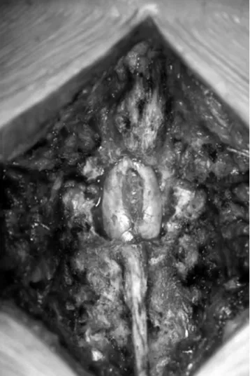
골겸자와 골정을 이용해 제 2 요추부터 제 3 요추 양측의 추궁 판을 제거한 결과 경막을 양측으로 나누고 있는 중격 및 이중척 수 소견이 관찰되었다. 중격은 매우 단단하였으며 골양 조직으 로서 2.5cm × 0.7cm 정도의 크기였고 주변에는 섬유성 조직과 지방조직이 존재하였다(Fig. 4). 중격 주변을 박리하는 과정에서 경질막과 중격의 상극 사이에 유착이 형성되어 있었으며, 경질 막을 중격으로부터 분리한 뒤 경질막이 파열되지 않도록 주의하 면서 골겸자와 골정으로 조심스럽게 중격을 제거하고 나눠져 있 던 경질막을 봉합하였다.
이후 제 2 요추체와 제 3 요추체에 척추경 나사못 고정기기로 고정한 뒤, 안정성을 확인하였다.
세척 및 지혈 후 배액관을 넣고 상처 부위를 봉합하였다. 수
술 후 1년 2개월 째 추시 관찰 중이다.
고 찰
척수이분증은 하나 혹은 여러 척추에 걸쳐 존재할 수 있으며 대부분 요추 부위에 나타나고, 그 외 흉추와 경추 부위에 발생한 경우도 보고되어 있다.4) 본 환자의 경우 제 1 요추 부위에서 시 작되어 그 이하로 연장되어 여러 부위에 걸쳐 나타난 것이 관찰 되었다.
척수이분증의 원인은 명확히 알려져 있지 않지만 발생학적 기 전에 기인한다는 주장이 인정받고 있으며, Bremer5)가 척수이분 증의 원인에 대해 부신경장관(accessory neuroenteric canal)이나 등쪽 장루(dorsal intestinal fistula)의 발현으로 설명한 이후, 다른 저자들도 발생학적 기전이 중요한 원인임을 밝히고 있다. Paul Porensky1)은 융합되지 않은 신경판(neural plate)의 뼈돌기에 의 한 1차적인 골의 이상으로, Herren과 Edward2)는 주변 척추를
Fig. 1. Preoperative anteroposterior and lateral X-ray show right scoliosis with multi-level spina bifida, hypoplastic sacrum and sclerotic bony septum at L2 vertebral body. Interpedicular distance widening is evident
형성하는 골분절의 2차적인 기형으로 척수이분증을 설명하였 다. 또한 Bentley와 Smith6)는 척삭의 비정상적 분리에 의한 내배 엽의 등쪽 이탈로 설명하였고, Gardener7)는 배아 신경관의 전후 파열로 설명하였다.
하지 근력 약화나 감각 결손, 요실금, 그리고 반사변화 등 신 경학적 이상이 종종 동반되어 5~20%의 환자에서 보고되고 있 다. 선천성 척추 기형에서 신경 조직의 기형이 동반되는 빈도는
Fig. 3. Preoperative T2 MRI shows complete division of the spinal cord into two hemicords and the lower lying conus
Fig. 4. An intraoperative gross image after bilateral total laminectomy shows bisecting bony septum and two hemicords.
Fig. 2. Preoperative coronal T1 MRI shows low signal septum dividing the spinal canal and the two hemicords
Diastematomyelia in Adult Journal of Korean Society of Spine Surgery
www.krspine.org
71
매우 높아 21~40%로 보고되고 있다.8) Winter4)에 의하면 선천성 척추측만증 환자의 4.9%에서 척수이분증을 동반한다고 하였 으며, 이와 동반될 수 있는 국소 증상으로 피부 반점, 털, 지방종 및 소와가 있다. 이 환자에서는 신경증상 및 척추측만증은 관찰 되었지만 그 밖의 국소 증상들은 보이지 않았다.
척수이분증에 의한 신경증상은 척추 성장과 척수 성장의 불균 형, 즉 정상적인 성장에 따른 척수의 근위부 이동의 장애에 기인 한다. 대부분 어린 시절 발견되며 성인에서 증상이 발현하는 경 우는 극히 드물며7), 성인에서 발현하는 경우, 외상 등의 유발인 자가 급성 신경학적 증상을 일으키는 경우가 대부분이다. 현재 까지 국내에서는 한 등9)에 의한 ‘내번족을 동반한 척수이분증의 치험 1예 보고’와 이 등10)에 의한 ‘척수 이분증을 동반한 선천성 측만증의 치험’, 이 2 예만이 보고되었으며, 9세와 14세로 모두 소아에 해당하여 국내에서 성인에 증상이 발현된 척수이분증 보 고는 본 증례가 최초이다.
성인에 척수이분증의 수술 적응증은 척추관 협착증이나 계류 척수가 신경학적 이상을 일으키는 경우이며, 본 환자도 여기에 해당된다. 또한 교정이 필요한 척추 측만증이 있을 때 측만증 교 정술에 앞서서 중격을 제거하여 측만증 교정 후에 나타날 수 있 는 계류 척수에 의한 신경장애를 미리 예방한다. 반면 척추 측만 증이 없으며 무증상인 성인에게 예방적으로 중격를 제거해주는 것은 추천되지 않는다.
결 론
저자들은 현재까지 국내 보고가 2 예 뿐인 척수이분증 1 예를 발견하고 치험하였으며 현재 1년 2개월째 추시 관찰중으로 본 질환에 대한 치료 결과 평가에 대해서는 지속적인 관찰이 필요
할 것으로 생각되기에 보고하는 바이다.
REFERENCES
1. Porensky P, Muro K, Ganju A. Adult presentation of spinal dysraphism and tandem diastematomyelia. The spine jour- nal. 2007;7:622-6.
2. Herren Y, Edwards J. Diplomyelia, Arch Patho.
1940;30:1203-12.
3. Matson DD, Woods RP. Diastematomyelia. Diagnosis and treatment. Pediatrics. 1950;6:98-112
4. Winter RB, Moe JH. Diastematomyelia and congenital spine deformity. J Bone and Joint Surg. 1974;56:27-39.
5. Bremer JL. Dorsal intestinal fistula : accessory neurenteric canal : Diastermatomyelia. Arch Pathol. 1952;54:132-8.
6. Bremer JL, Smith Jr. Development posterior enteric remnants and spinal malformations. Arch Dis Child.
1960;35:76-86.
7. Garner WJ. Embryologic origin of spinal malformation.
Acta Radiol Diagn. 1966;5:1013-23.
8. Lee CK. Study of other anatomical structure anomaly ac- companied by congenital spine anomaly. Korean society of spine surgery. The 10th symposium. 1993.
9. Kim YM, Han MS, Han SH. Diastematomyelia associ- ated with clubfoot(Case Report). J Korean Orthop Assoc.
1976;11:380-761.
10. Seo KY, Lee YK, Lee SI. Congenital Scoliosis Associ- ated with Diastematomyelia. J Korean Orthop Assoc.
1980;15:566-71.
성인에서 발견된 척수이분증 - 증례 보고 -
이진영 • 조주성
한림대학교 의과대학 강동성심병원 정형외과학교실
연구 계획: 증례보고
목적: 본 논문의 목적은 성인에서 발견된 척수이분증에 대해 보고하고자 함이다.
선행문헌의 요약: 척수이분증은 척수가 골성, 연골성 혹은 섬유성 조직 등의 격막에 의해 2가닥으로 분리되는 선천성 기형으로 대부분 소아기에 증상이 발현하며 성인기에 발현하는 경우는 극히 드물다.
대상 및 방법: 저자들은 46세 여자 환자에서 점진적으로 발현된 신경학적 파행이 동반된 척수 이분증을 감압적 척추궁 절제술, 중격 제거술 및 후방기 기 고정술로 치료하였다.
결과: 수술적인 치료로 만족할 만한 결과를 얻을 수 있었다.
결론: 척수이분증은 성인에서는 드문 질환으로 수술적 치료로 만족할 만한 결과를 얻었다.
색인단어: 요추, 척수이분증, 감압적 척추궁 절제술, 중격 제거술, 후방기기 고정술 약칭제목: 요추부 척수이분증