www.krspine.org
Osteoid Osteoma of the Sacrum - A Case Report -
Chang-Rack Lim, M.D., Ji-Hyun Ryu, M.D., Zin-Ouk Hwang, M.D., Ki-Won Kim, M.D.
J Korean Soc Spine Surg 2019 Dec;26(4):160-165.
Originally published online December 31, 2019;
https://doi.org/10.4184/jkss.2019.26.4.160
Korean Society of Spine Surgery
SMG-SNU Boramae Medical Center, 20, Boramae-ro 5-gil, Dongjak-gu, Seoul 07061, Korea Tel: +82-2-831-3413 Fax: +82-2-831-3414
©Copyright 2017 Korean Society of Spine Surgery pISSN 2093-4378 eISSN 2093-4386
The online version of this article, along with updated information and services, is located on the World Wide Web at:
http://www.krspine.org/DOIx.php?id=10.4184/jkss.2019.26.4.160
This is an Open Access article distributed under the terms of the Creative Commons Attribution Non-Commercial License (http://
creativecommons.org/licenses/by-nc/4.0) which permits unrestricted non-commercial use, distribution, and reproduction in any medium, provided the original work is properly cited.
Journal of Korean Society of
Spine Surgery
Osteoid Osteoma of the Sacrum - A Case Report -
Chang-Rack Lim, M.D., Ji-Hyun Ryu, M.D., Zin-Ouk Hwang, M.D., Ki-Won Kim, M.D.
Department of Orthopaedic Surgery, Yeouido St. Mary’s Hospital, College of Medicine, The Catholic University of Korea, Seoul, Korea Study Design: Case report.
Objectives: To report a case of osteoid osteoma arising in the sacrum in a 29-year-old male patient.
Summary of Literature Review: Osteoid osteoma is a benign osteoblastic tumor that usually arises in the long bones. Osteoid osteoma involving the sacrum is extremely rare.
Materials and Methods: A 29-year-old male patient presented with pain localized in his sacral area for 10 months. His pain was worse at night, relieved by non-steroidal anti-inflammatory drugs, and independent of physical activity. Bone scintigraphy showed increased uptake in the second sacral vertebra (S2). Computed tomography revealed a nidus located in the S2 spinous process. Magnetic resonance imaging showed bone and soft tissue edema around the nidus.
Results: En bloc excision including the nidus revealed a diagnosis of osteoid osteoma and provided immediate relief of the patient’s long-lasting sacral pain.
Conclusions: When a young patient presents with localized sacral pain that is worse at night, relieved by non-steroidal anti- inflammatory drugs, independent of physical activity, and lasts longer than expected, proper imaging studies should be performed to rule out osteoid osteoma. Although less invasive treatment modalities have been introduced, classical en bloc excision is currently the gold standard for managing osteoid osteoma.
Key words: Osteoid Osteoma, Sacrum, Benign bone tumor
Received: August 16, 2019 Revised: August 22, 2019 Accepted: October 8, 2019
Published Online: December 31, 2019 Corresponding author: Ki-Won Kim, M.D.
ORCID ID: Chang-rack Lim: https://orcid.org/0000-0001-5953-6596 Ji-Hyun Ryu: https://orcid.org/0000-0001-7550-6389 Zin-Ouk Hwang: https://orcid.org/0000-0002-4402-515X Ki-Won Kim: https://orcid.org/0000-0002-8249-1098 Department of Orthopedic Surgery, Yeouido St. Mary’s Hospital, College of Medicine, the Catholic University, 63-ro 10 Yeingdeungpo-gu, Seoul, 07345 Korea
TEL: +82-2-3779-1192, FAX: +82-2-783-0252 E-mail: [email protected]
유골 골종은 양성의 골형성 종괴로 10세-30세의 젊은 남성 에게서 호발하며 흔히 장골의 골간단부에서 발생한다.1) 천추의 유골 골종은 흔하지 않아서 외상이나 다른 병적 상태로 혼동되 어 진단이 지연될 수 있다.5,9) 저자들은 10개월간 진단이 지연되 었던 29세 남자 환자에서 천추에 발생한 유골 골종을 경험하였 기에 이를 보고하고자 한다.
증례 보고
29세 남자 환자가 특별한 외상 병력 없이 10개월 전부 터 천추부 통증이 발생하여 타 병원들에서 요천추부 염좌 (lumbosacral sprain) 진단 하에 보존적인 치료를 받았으나 증상의 호전이 없어 본원에 내원하였다. 천추부 통증은 신체 활동(physical activity)과는 관련이 없었고, 밤에 악화되는 양 상이었다. 제 2천추 부위에 압통이 있었고, 다른 이학적 검 사와 신경학적 검사들에서는 특이 소견은 없었다. 단순 방사 선 검사에서 특이 소견이 관찰되지 않아(Fig. 1) 우선적으로 비스테로이드성 항염증제 처방 후 외래 추시하였다. 외래 추 시 상 약물 복용 시에만 천추부 통증이 호전되고, 약물 효과 가 떨어지면 다시 통증이 나타나는 소견보였다.
젊은 환자에서 밤에 악화되고 비스테로이드성 항염증제 에 효과가 있는 유골 골종의 특징적인 임상 양상에 근거하 여 골주사(bone scintigraphy), 전산화 단층촬영(computed tomography, CT) 및 자기공명영상(magnetic resonance imaging, MRI)을 시행하였다. 골주사 검사에서 제 2천추 중 앙 부위에 열소(hot spot)가 관찰되었고(Fig. 2), 전산화 단 층촬영에서 제 2천추 극돌기에 유골 골종의 특징적인 핵
Osteoid Osteoma of the Sacrum Journal of Korean Society of Spine Surgery
www.krspine.org 161 (nidus)이 관찰되었다(Fig. 3). Gadolinium-증강 자기공명영 상에서 핵 주위의 골 조직과 연부 조직에 부종 소견이 관찰 되었다(Fig. 4). 이와 같은 영상 소견들을 종합할 때 유골 골 종이 가장 합당한 진단으로 결론 지었다. 확진과 치료를 위 하여 일괄절제술(en-bloc excision)을 계획하였다.
전신 마취 하에 제 5요추부터 제 2천추까지 박리하여 후 궁을 노출시켰다. 유골 골종이 있을 것으로 추정되는 부위와 주위의 정상 조직을 포함하여 직사각형 형태의 경계를 수술 용 버(surgical burr)로 표시 한 후 최대한 깊게 도랑(gutter) 을 만들었다. 제 2천추의 후궁을 들어 올리면서 도랑을 절단 하여 일괄절제술을 완료하였다(Fig. 5).
수술 후 촬영한 전산화 단층촬영 상 병변이 완전히 제거되 었음을 확인할 수 있었고(Fig. 6), 수술 후 1일째부터 코르셋 을 착용하고 보행을 시작하였다. 일괄절제술로 채취한 검체 의 조직검사에서 정상 골 조직과 구별이 확연한 유골 골종 으로 확진되었다(Fig. 7). 수술 후, 환자는 장시간 지속되었 던 천추부 통증 없이 수술 6일째 퇴원했으며 마지막 외래 추 시에서도 증상 호전 상태였다.
A B
Fig. 1. Plain radiographs.
(A) An anteroposterior plain radiograph is normal. (B) A lateral plain ra- diograph is normal.
Fig. 2. Bone scintigraphy shows increased uptake in the middle of the second sacral vertebra (S2) (arrows).
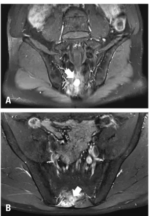
Fig. 3. Magnetic resonance imaging.
(A) A gadolinium-enhanced T1-weighted coronal magnetic resonance im- age shows a mass with high signal intensity in S2 (arrow).
(B) A gadolinium-enhanced T1-weighted axial magnetic resonance image shows a mass with non-specific surrounding edema in the bone marrow and soft tissue (arrow).
A
B
본 논문은 본원 IRB 심사면제(SC19ZESE0102)를 받은 이 후 진행되었다.
고찰
유골 골종은 1935년 Jaffe에 의해 처음 보고된 질환으로 모든 골종양의 5%, 양성 골종양의 11%가량을 차지하며, 어 Fig. 7. Histological findings (hematoxylin and eosin; original magnification
×40). The black dashed line indicates a borderline between the nidus (Ni) and surrounding reactive sclerotic bone (S). “N” represents normal bone.
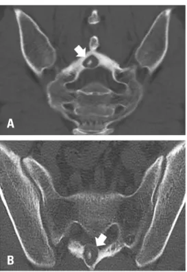
Fig. 4. Preoperative computed tomography (CT).
(A) A coronal CT image shows an oval radiolucent nidus within surround- ing sclerotic reactive bone in the spinous process of S2, and a central dot-like calcification within the nidus is also noted (arrow). (B) An axial CT image shows an osteoid osteoma bulging into the S2 spinal canal (ar- row).
A
B
Fig. 5. The surgically excised specimen shows ventral bulging of the os- teoid osteoma (arrows) as seen on the computed tomography image (Fig 4B).
Fig. 6. Postoperative computed tomography (CT).
(A) Coronal CT images show complete excision of the lesion (arrow). (B) Axial CT images show complete excision of the lesion (arrow).
A
B
Osteoid Osteoma of the Sacrum Journal of Korean Society of Spine Surgery
www.krspine.org 163 느 골격계에서나 발생할 수 있는 종양이다.1-2) 가장 호발하
는 부위는 대퇴골 근위부이며, 대퇴골이나 경골에서 50%이 상 발생한다. 척추에서는 대략 10% 정도 발생하며 이 경우 거의 대부분(70~100%) 후주(posterior column)에서 발생한 다.3) 특히 천추에서는 매우 드물게 발생하며, 지금까지 국내 에서는 1예가 보고되었다.4,8) 유골 골종의 대부분은 40세 이 하의 젊은 층에서 발생하며 남성에서 여성보다 약 2배 정도 발생 빈도가 높다.7)
유골 골종의 가장 특징적인 임상 양상은 밤에 악화되는 골 격계 통증이며 이 통증은 비스테로이성 항염증제에 의해 호 전된다는 것이다.5,7) 단순 방사선 검사에서 유골 골종의 가 장 특징적인 핵(nidus)을 관찰할수 있는 경우는 드물며 특히 천추의 경우에는 거의 대부분 정상으로 판독된다.5) 전산화 단층 촬영(computed tomography)은 특징적인 핵(nidus)을 발견하는데 가장 유용한 방법이며, 종양의 크기가 대개 작 기 때문에(2 cm 미만) 미세 절단 영상(thin-section imaging) 을 얻어야 핵(nidus)을 놓치지 않을 수 있다. 골주사(bone scintigraphy)는 가장 민감도(sensitivity)가 높은 검사 방법으 로 유골 골종을 의심하는 경우 선별 검사(screening test)로 추천된다.7) 자기공명영상(magnetic resonance imaging)은 핵 을 정확히 볼 수는 없지만 주변의 염증성 반응(inflammatory reaction)을 확인할 수 있는 장점이 있다.6)
일반적으로, 천추의 유골 골종은 매우 드물기 때문에 거의 대부분 초기 진단에 실패하며 추가적인 영상 검사들(골주 사, 전산화 단층촬영, 자기공명영상)을 통하여 유골 골종을 잠재적으로 진단할 수 있다.4,5,7,8) 본 증례에서도 마찬가지였 다. 즉, 타 병원들에서는 요천추부 염좌(lumbosacral sprain) 로 판단하여 10개월 동안 치료를 받았으나 증상 호전에 실 패하였고 위에 기술한 추가적인 영상 검사들을 통하여 유골 골종이라는 잠재적인 진단이 이루어졌다. 따라서 40세 이하 의 젊은 남자 환자에서 밤에 악화되는 천추부 통증이 있으 며 이 통증이 비스테로이성 항염증제에 의해 호전되는 임상 양상을 보인다면, 비록 드물다 하더라도, 천추의 유골 골종 을 의심해 보아야 한다는 것을 저자들은 강조하고 싶다. 또 한, 요천추부 염좌와 달리 통증 정도가 신체 활동(physical activity)의 강도와 관련이 없다는 것도 우리 환자에서는 유 골 골종을 의심케 하는 중요한 임상 소견이었다.
유골 골종은 자연 경과상 시간이 지날수록 좋아지는 경향 이 있기 때문에 비스테로이드성 항염증제를 이용한 보존적 인 치료를 하기도 한다.9-10) 하지만 장기간의 약물 복용이 필 요하고, 통증이 지속적으로 있으며 증상이 재발하는 경우가 많은 단점이 있다. 최근 경피적 고주파 응고술(percutaneous radiofrequency coagulation)이나 경피적 레이저 광응고술
(percutaneous laser photocoagulation)과 같이 비침습적인 방법들을 사용하여 성공적으로 치료한 증례도 보고되고 있
다.5-6) 주위 연부조직, 연골 및 골조직의 손상이 적다는 장
점이 있지만 핵의 불완전한 제거로 인한 재발 가능성이 있 다.3,8) 일괄절제술(en-bloc excision)은 핵을 완벽하게 제 거할 수 있어 재발이 적다. 그러므로 현재까지는 일괄절제 술이 유골 골종을 치료하는 최적의 방법(gold standard of treatment)이다.
천추의 유골 골종은 국내에서 단 1예의 증례 보고가 있을 정도로 매우 드문 질환 이다. 하지만, 특징적인 임상 양상을 보이는 젊은 환자에서는 유골 골종을 의심해 보는 것이 가 장 중요하다. 최근, 비침습적인 치료 방법들이 소개되고 있 지만 아직은 일괄절제술이 확진과 치료에 최적의 방법이라 고 판단된다.
REFERENCES
1. Jaffe HL. Osteoid-Osteoma of Bone. Radiology. 1945 Oct;45(4):319-34. DOI:10.1148/45.4.319.
2. Schajowicz F. Tumors and Tumorlike Lesions of Bone and Joints. New York: Springer; 1981.36-47.
3. Hadjipavlou AG, Lander PH, Marchesi D, et al. Minimally invasive surgery for ablation of osteoid osteoma of the spine. Spine. 2003 NOV;28(22):472-7. DOI: 10.1097/01.
BRS.0000092386.96824.DB.
4. Chung SS, Seo JS, Moon SH, et al. Osteoid os- teoma in the sacrum: a case report. J Korean Soc Spine Surg. 2006 JUN;13(2):147-51. https://doi.
org./10.4184/jkss.2006.13.2.147. DOI: 10.1097/01.
BRS.0000092386.96824.DB.
5. OZKOC G. Osteoid osteoma of the sacrum mimick- ing sacroiliitis: a case report. Turk J Rheumatol. 2013 Jan;28(1):51-3. DOI: 10.5606/tjr.2013.2863.
6. Ghanem I. The management of osteoid osteoma: update and controversied. Curr Opin Pediatr. 2006 Feb;18(1):36- 41. DOI: 10.1097/01.mop.0000193277.47119.15.
7. Etemadifar MR, Hadi A. Clinical findings and results of surgical resection in 19 cases of spinal osteoid osteoma.
Asian Spine J. 2015 Jun;9(3):386-93. DOI: 10.4184/
asj.2015.9.3.386.
8. Fukuda S, Susa M, Watanabe I, et al. Computed tomog- raphy-guided resection of osteoid osteoma of the sacrum:
a case report. Journal of Medical Case Reports. 2014
Jun;8:206. DOI: 10.1186/1752-1947-8-206.
9. Kneisl JS, Simon MA. Medical management compared with operative treatment for osteoid-osteoma. J Bone Joint Surg Am. 1992 Feb;74(2):179-85. DOI: 10.2106/00004623- 199274020-00004.
10. Feletar M, Hall S. Osteoid osteoma: a case for conserva- tive management. Rheumatology. 2002 May;41(5):585-6.
DOI: 10.1093/rheumatology/41.5.585.
© Copyright 2019 Korean Society of Spine Surgery
Journal of Korean Society of Spine Surgery. www.krspine.org. pISSN 2093-4378 eISSN 2093-4386
This is an Open Access article distributed under the terms of the Creative Commons Attribution Non-Commercial License (http://creativecommons.org/licenses/by-nc/4.0/) which permits unrestricted non-commercial use, distribution, and reproduction in any medium, provided the original work is properly cited.
165
J Korean Soc Spine Surg. 2019 Dec;26(4):160-165
Case Report
천추의 유골 골종 - 증례 보고 -
임창락 • 유지현 • 황진욱 • 김기원
가톨릭대학교 의과대학 여의도성모병원 정형외과학교실
연구 계획: 증례 보고
목적: 29세 남자 환자에서 천추에 발생한 유골 골종을 보고하고자 한다.
선행 연구문헌의 요약: 유골 골종은 양성 골형성 종괴로 천추에서는 드물게 발생한다.
대상 및 방법: 29세 남자 환자가 10개월 간 지속된 천추부 통증으로 방문하였다. 그의 통증은 야간에 악화되고, 비스테로이드성 항염증제들에 의해 호전 되며 신체 활동과 관련성이 없었다. 시행한 골주사 검사(bone scintigraphy)에서 제 2 천추 부에 열소(hot spot), 컴퓨터 단층촬영(computed tomography) 에서 제 2천추 극돌기 내에 핵(nidus)이 관찰되었다. 자기 공명 영상에서는 핵 주위의 골 조직과 연부 조직의 부종이 관찰되었다.
결과: 천추 병변 부위에 일괄절제술(en-bloc excision)을 통해 유골 골종으로 진단되었고 장시간 지속된 환자의 천추부 통증은 즉각적으로 호전되었다.
결론: 젊은 환자가 야간에 악화되고, 비스테로이드성 항염증제들에 의해 호전되고, 신체 활동과 관련성이 없고, 예상보다 장기간 지속되는 천추부 통증 을 보일 때에는 유골 골종을 배제하기 위하여 적절한 영상 검사들을 시행해야 한다. 덜 침습적인 치료 방법들이 소개되고 있지만, 현재까지는 일괄절제술 (en-bloc excision)이 유골 골종을 치료하는 최적의 방법(gold standard of treatment)이다.
색인 단어: 유골 골종, 천추, 양성 골종양 약칭 제목: 천추의 유골 골종
접수일: 2019년 8월 16일 수정일: 2019년 8월 22일 게재확정일: 2019년 10월 8일 교신저자: 김기원
서울시 영등포구 63로 10 가톨릭대학교 의과대학 여의도성모병원 정형외과학교실
TEL: 02-3779-1192 FAX: 02-783-0252 E-mail: [email protected]
![2018년도 게임그래픽 강의계획 [기초과정]](data:image/gif;base64,R0lGODlhAQABAIAAAP///wAAACH5BAEAAAAALAAAAAABAAEAAAICRAEAOw==)