Introduction
The application of cone-beam computed tomography (CBCT) in dentistry has increased rapidly since it was introduced.1 CBCT has been used as a method for creat- ing three-dimensional radiographs,2 which are one of the most sensitive imaging modalities for dental diagnostic purposes, and as a substitute for traditional CT in the assessment of pathology and dysfunction of the cranio- maxillofacial complex.3 Furthermore, CBCT has been
increasingly applied in endodontics for the diagnosis of periapical lesions, root canal observation, assessing the extent of internal and external resorption, and identifying root fractures.4
Radiographic and clinical examinations are essential for the diagnosis of vertical root fractures, and CBCT is more sensitive than periapical radiography in detecting vertical root fractures.5 Two-dimensional conventional ra- diographs with bony structure superimposition may hide the root fracture, especially when the X-ray position is not parallel to the plane of the fracture, meaning that the fracture may not be observed in the radiograph.4
One feature of vertical root fractures is a local deep pocket, the presence of a sinus tract, and halo-type lateral radiolucency on a radiograph.6 The clinical features of
The effect of metal artifacts on the identification of vertical root fractures using different fields of view in cone-beam computed tomography
Ehsan Moudi1, Sina Haghanifar1, Zahrasadat Madani2, Ali Bijani3, Zeynab Sadat Nabavi1,*
1Oral and Maxillofacial Radiology Department, Dental School, Babol University of Medical Science, Babol, Iran
2Endodontics Department, Dental School, Babol University of Medical Science, Babol, Iran
3Social Department of Health Research Center, Babol University of Medical Science, Babol, Iran
AbsTrAcT
Purpose: The aim of this study was to investigate the effects of metal artifacts on the accurate diagnosis of root fractures using cone-beam computed tomography (CBCT) images with large and small/limited fields of view (FOVs).
Materials and Methods: Forty extracted molar and premolar teeth were collected. Access canals were made in all teeth using a rotary system. In half of the teeth, fractures were created by the application of mild pressure with a hammer. The teeth were then randomly put into a wax rim on an acryl base designed in the shape of a mandible.
CBCT scans were obtained using a Newtom 5G system with FOVs of 18 cm×16 cm and 6 cm×6 cm. A metal pin was then placed into each tooth, and CBCT imaging was again performed using the same fields of view. All scans were evaluated by two oral and maxillofacial radiologists. The specificity, sensitivity, positive predictive value, negative predictive value, and likelihood ratios (positive and negative) were calculated.
result: The maximum levels of sensitivity and specificity (100% and 100%, respectively) were observed in small- volume CBCT scans of teeth without pins. The highest negative predictive value was found in the small-volume group without pins, whereas the positive predictive value was 100% in all groups except the large-volume group with pins.
conclusion: The specificity of CBCT decreased with the presence of a pin in the large-volume group, but not in the small-volume group.(Imaging Sci Dent 2015; 45: 147-51)
Key words: Cone-Beam Computed Tomography; Tooth Fractures; Artifacts; Sensitivity and Specificity
Copyright ⓒ 2015 by Korean Academy of Oral and Maxillofacial Radiology
This is an Open Access article distributed under the terms of the Creative Commons Attribution Non-Commercial License(http://creativecommons.org/licenses/by-nc/3.0) which permits unrestricted non-commercial use, distribution, and reproduction in any medium, provided the original work is properly cited.
Imaging Science in Dentistry·pISSN 2233-7822 eISSN 2233-7830 Received February 15, 2015; Revised April 17, 2015; Accepted May 10, 2015
*Correspondence to : Dr. Zeynab Sadat Nabavi
Oral and Maxillofacial Radiology Department, Dental School, Babol University of Medical Science, Ganj Afrooz Ave., Babol, Mazandaran Province, Iran
Tel) 98-911-155-5905, Fax) 98-113-229-1093, E-mail) [email protected]
root fractures can be misleading or present with a delay.
They can also be hard to distinguish from pulp and peri- apical diseases, making it challenging to identify root fractures in teeth with non-displaced parts.4 The prognosis of a tooth with a root fracture depends on various factors, such as the patient’s age, the stage of root formation, the amount of displacement of the coronal part of the tooth, the extent of looseness in the coronal part of the tooth, and the distance between the separated parts.7 Therefore, a definitive diagnosis of vertical root fracture is essential in order to avoid unnecessary tooth extraction.
Based on the field of view(FOV), CBCT systems are classified into three types: small-volume(also known as limited-volume) systems, which are usually used for scan- ning a sample of contiguous teeth or one jaw; medium- volume systems that are used to image both jaws, the max- illary sinus, and part of the nose; and large-volume sys- tems that are applied to image the entire maxillofacial area, and in some systems even the cranial vertex.8
Limited-volume systems have some advantages in terms of price, image resolution(voxel size), and radiation dose in comparison with large-volume systems. In CBCT ima- ging with a limited volume, the maxillofacial hard tissue structures located outside the imaging volume cause dis- continuity of beam information. This influences image fidelity and results in areas with variable density.1
Metal artifacts occur in all CT imaging systems. Many small and large metal items may be present in the human body, especially in the head and neck area. Metal resto- rations, metal posts located in the canal, crowns, brackets, and implants can influence the quality of the acquired CT image due to effects such as quantum noise, starvation photons, and beam hardening.9 Beam hardening results in two kinds of artifacts: the distortion of metal structures that is known as the cupping artifact because of the pat- tern of differential absorption that results, and streaks and dark bands that can appear between two dense substanc- es10 or around metal substances. The artifact may even cause a complete loss of gray values between the adjacent metal substances. As a result, the area in question is not accurately imaged, with consequences for the treatment plan.9
In 2012, Costa et al.8 investigated horizontal root frac- tures in teeth with and without metal posts using large- volume CBCT by designing a cylinder measuring 20cm×
15cm. The identification of horizontal fractures was more accurate in the group of teeth without a metal post(with or without a fracture), while weak or very weak agreement was found between two observers in the group of teeth
with a metal post. In another investigation in 2011, Costa et al.11 studied on horizontal root fractures in teeth with and without a metal post located in the canal using small- volume CBCT. They likewise found that the identifica- tion of fractures was more accurate in the group of teeth without a metal post(with or without a fracture) than in the teeth with a metal post.
The presence of prefabricated posts has little effect on the accuracy of CBCT,12 but it influences the treatment plan based on CBCT images. We hypothesized that root fractures may be more accurately observed in images with a small FOV, due to the smaller size of the pixels and the higher image resolution. Thus, the aim of this study was to investigate the effects of metal artifacts in CBCT images on the accurate diagnosis of root fractures in large and small/limited fields of view.
Materials and Methods
In this study, 40 extracted human mandibular molar and premolar teeth without fractures, periapical pathology, root resorption, or any other anomalies were collected. In order to disinfect and remove the soft cellular tissue, the teeth were placed in a 5.25% sodium hypochlorite solu- tion(Golrang, Tehran, Iran) for one hour and then placed in normal saline until the experiment was started. The col- lected teeth had not undergone any endodontic treatment, and the absence of vertical root fractures was established by examination with a stereomicroscope at 20×(Wild Photomakroskop M400, Heerbrugg, Switzerland).
Access canals in all teeth were prepared using a dia- mond bur, and the opening of both the molar distal canal and the premolar canal was accomplished with a #15 K- file. The length of the canals was visually determined with this file, and the measurement was decreased by 1mm in order to reflect functional length of the canals. The canals were prepared according to the manufacturer’s instructions using a rotary system(NSK, Fukushima, Japan), with a speed of 300rpm, controlled torque, and SX, S1, S2, F1, and F2 ProTaper files(Dentsply, Ballaigues, Switzerland).
Each canal was frequently washed with a 2% sodium hy- pochlorite solution. The prepared canals were filled with guttapercha(Pro Taper F2 & F3, Dia Dent, Burnaby, BC, Canada).
In order to create fractures in the canals, the teeth were first put in acryl(Acropars 200, Marlic Medical Industries Co., Tehran, Iran) to avoid splitting, and a thin layer of wax was added to simulate soft tissue. One screw driver- tape wedge was then placed in the canal. The fractures
were created through the application of mild pressure with a hammer in the canals of the premolar teeth and the dis- tal root of the molars. Fractures were created in half of the teeth. The teeth were evaluated again with the stereo- microscope with the same magnification in order to con- firm the absence of fractures in teeth in which a vertical root fracture was made. The teeth were then coded and randomly placed in a wax rim made of three to four wax layers on an acryl base designed in the shape of a mandi- ble. The mandibles were fixed in cardboard boxes, so that the central beam ray was perpendicular to the longitudi- nal axis of the teeth. CBCT scans were obtained using a Newtom 5G apparatus(QR s.r.1., Verona, Italy), with a potential difference of 110kV, a tube current of 9.6mA, a FOV of 18cm×16cm for the large FOV and 6cm×6 cm for the small FOV, a voxel size of 0.3mm, denture scan mode, and 4.8 seconds of exposure. The guttapercha that had been located in the canal was removed in order to place a #3 gold-plated pin(Nordin, Montreux, Switzer- land). The fracture line pattern was denoted with 1% me- thylene blue, metal pins were put in all of the teeth, and CBCT imaging was again performed using the previous specifications. The images were evaluated in the axial plane and in multiplanar image using the NNT viewer soft- ware version 3.0(QR s.r.1., Verona, Italy), with a thick- ness of 0.3mm and an interval space of 0.3mm. All scans were studied by two oral and maxillofacial radiologists in
axial and cross-sectional views(Fig. 1) in a dim room us- ing an 18.5-inch LG Flatron monitor(LG, Seoul, Korea), with a resolution of 0.3×0.3 pixel pitch. The images were randomly observed in limited numbers over consecutive days in order for the observers to identify tooth fractures.
Specificity, sensitivity, positive predictive value(PPV), and negative predictive value(NPV), and the positive and negative likelihood ratios were calculated.
results
The small-volume CBCT images of teeth without pins showed 100% sensitivity and specificity.
In the small-volume images, 100% specificity was also observed in the presence of a pin. Although the sensitivity in the small-volume images without a pin was 5% higher than in the small-volume images with a pin, no statisti- cally significant difference was found between these two groups.
The sensitivity in the large-volume images without a pin was 14% lower than in large-volume images with a pin, whereas the specificity was 11% higher in the large-vol- ume images without a pin. Overall, the sensitivity of the diagnoses of vertical root fractures made using small-vol- ume images was higher than that of the diagnoses made using large-volume images. The positive predictive value in all groups was 100%, except in large-volume images
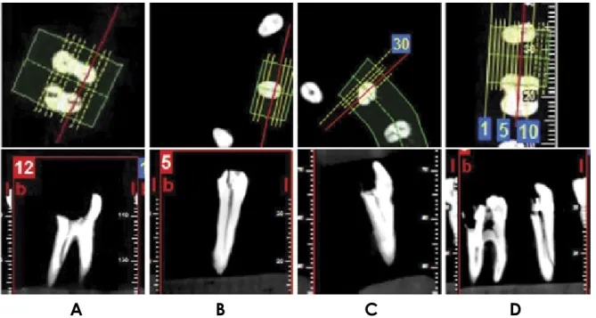
Fig. 1. Fracture lines in axial and cross-sectional cone-beam computed tomography images. A. Large volume without a pin. B. Small vol- ume without a pin. C. Large volume with a pin. D. Small volume with a pin.
A B C D
with a pin. The negative predictive value in small-volume images without a pin and in large-volume images with a pin was 100%, but was lower in small-volume images with a pin and large-volume images without a pin.
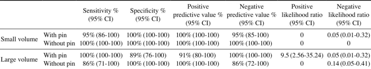
Table 1 shows the sensitivity, specificity, positive pre- dictive value, the negative predictive value, and positive and negative likelihood ratios of the various groups after CBCT was used to identify teeth root fractures, along with 95% confidence intervals.
discussion
The statistical analysis demonstrated that CBCT images of teeth without pins were highly sensitive in the diagno- sis of fracture lines in small-volume mode, in comparison with other modes. The main goal of this ex vivo study was to investigate the diagnostic accuracy of fracture lines in large and small CBCT radiographs in the presence of root canal fillings and metal pins. The sensitivity of small-vol- ume images without pins was 100%, but was reduced to 86% in the large-volume group without pins.
This is a reasonable finding in light of the decreased imaging area, the lower amount of ray dispersion, the higher signal-to-noise ratio, and the higher resolution of the radiographs. Our findings correspond to those of Costa et al.,11 who investigated horizontal fracture lines in small- volume CBCT. They are also in agreement with the find- ings of Iikubo et al.,13 who compared conventional peri- apical radiographs to CBCT images. The sensitivity of the large-volume CBCT images was 100% for the group with pins, but a lower value of specificity was reported in this group(89%). This finding is likely explicable through the beam-hardening phenomenon. Beam hardening occurs in connection with the increase of medium- energy X-rays, because lower-energy photons are absorbed by the struc- tures being imaged instead of higher-energy photons. As mentioned above, this phenomenon results in artifacts that obscure the fracture lines in the region of interest. The arti- facts that appear in the shape of dark areas or lines around endodontic materials are similar to the lines that reflect root fractures, resulting in false positives(Fig. 2).14
As seen in Figure 2A, the fracture lines and the lines
Table 1. Diagnostic parameters of different fields of view for the identification of vertical root fractures in teeth with or without pins.
Sensitivity %
(95% CI) Specificity % (95% CI)
Positive predictive value %
(95% CI)
Negative predictive value %
(95% CI)
Positive likelihood ratio
(95% CI)
Negative likelihood ratio
(95% CI) Small volume With pinWithout pin 95%(86-100)
100%(100-100) 100%(100-100)
100%(100-100) 100%(100-100)
100%(100-100) 95%(85-100)
100%(100-100) 0
0 0.05(0.01-0.32) 0 Large volume With pinWithout pin 100%(100-100)
86%(71-100) 89%(76-100)
100%(100-100) 91%(80-100)
100%(100-100) 100%(100-100)
86%(72-100) 9.5(2.56-35.24)
0 0.05(0.01-0.32) 0.14(0.05-0.41) CI: confidence interval.
Fig. 2. Axial(A) and cross-sectional(B) small-volume cone-beam computed tomography images showing fractures in teeth with a pin. The fracture and the metal atrifact are shown with a straight arrow and a curved arrow, respectively.
A B
due to metal artifacts are not distinguished easily. A study carried out by Wang et al.15 with the aim of identifying tooth fracture lines using CBCT in comparison with con- ventional radiography and to determine the effect of root canal fillings found that CBCT was more accurate in iden- tifying root fractures. The sensitivity of CBCT decreased in the presence of root canal fillings, but the specificity was not influenced by any of the factors they studied. In this study, specificity decreased with the presence of pins in the large-volume images, but not in the small-volume images. This effect might be due to the voxel size and issues involving contrast and spatial resolution.16 Large- FOV CBCT provides less contrast resolution and spatial resolution than small-FOV CBCT.6
Negative predictive value is an important functional in- dex, because when a radiologist confirms that no fracture is present in a scan, he/she estimates the possibility that a vertical root fracture is not present.4 The negative predic- tive values in the small-volume CBCT group without pins and the large-volume group with pins were 100%. The positive predictive value was 100% in all groups except the large-volume CBCT group with pins, where a slightly lower value was observed. A 100% positive predictive value means that the lines displayed as fracture lines were 100% likely to be real fracture lines.
In conclusion, small-volume CBCT scans are very accu- rate in diagnosing vertical root fractures, and the presence of pins has only a small effect on detecting root fractures.
references
1. Katsumata A, Hirukawa A, Okumura S, Naitoh M, Fujishita M, Ariji E, et al. Relationship between density variability and imaging volume size in cone-beam computerized tomographic scanning of the maxillofacial region: an in vitro study. Oral Surg Oral Med Oral Pathol Oral Radiol Endod 2009; 107:
420-5.
2. Palomo JM, Kau C, Palomo LB, Hans MG. Three-dimension- al cone beam computerized tomography in dentistry. Dent Today 2006; 25: 130-5.
3. Bechara BB, Moore WS, McMahan CA, Noujeim M. Metal artefact reduction with cone beam CT: an in vitro study. Den- tomaxillofac Radiol 2012; 41: 248-53.
4. Ferreira RI, Bahrami G, Isidor F, Wenzel A, Haiter-Neto F, Groppo FC. Detection of vertical root fractures by cone-beam computerized tomography in endodontically treated teeth with
fiber-resin and titanium posts: an in vitro study. Oral Surg Oral Med Oral Pathol Oral Radiol 2013; 115: e49-57.
5. Haghanifar S, Moudi E, Mesgarani A, Bijani A, Abbaszadeh N.
A comparative study of cone-beam computed tomography and digital periapical radiography in detecting mandibular molars root perforations. Imaging Sci Dent 2014; 44: 115-9.
6. Hassan B, Metska ME, Ozok AR, van der Stelt P, Wesselink PR. Comparison of five cone beam computed tomography systems for the detection of vertical root fractures. J Endod 2010; 36: 126-9.
7. Bornstein MM, Wölner-Hanssen AB, Sendi P, von Arx T.
Comparison of intraoral radiography and limited cone beam computed tomography for the assessment of root-fractured permanent teeth. Dent Traumatol 2009; 25: 571-7.
8. Costa FF, Gaia BF, Umetsubo OS, Pinheiro LR, Tortamano IP, Cavalcanti MG. Use of large-volume cone-beam computed tomography in identification and localization of horizontal root fracture in the presence and absence of intracanal metal- lic post. J Endod 2012; 38: 856-9.
9. Pauwels R, Stamatakis H, Bosmans H, Bogaerts R, Jacobs R, Horner K, et al. Quantification of metal artifacts on cone beam computed tomography images. Clin Oral Implants Res 2013;
24: 94-9.
10. Scarfe WC, Levin MD, Gane D, Farman AG. Use of cone beam computed tomography in endodontics. Int J Dent 2009;
2009: 634567.
11. Costa FF, Gaia BF, Umetsubo OS, Cavalcanti MG. Detection of horizontal root fracture with small-volume cone-beam com- puted tomography in the presence and absence of intracanal metallic post. J Endod 2011; 37: 1456-9.
12. Moudi E, Haghanifar S, Madani Z, Alhavaz A, Bijani A, Ba- gheri M. Assessment of vertical root fracture using cone-beam computed tomography. Imaging Sci Dent 2014; 44: 37-41.
13. Iikubo M, Kobayashi K, Mishima A, Shimoda S, Daimaruya T, Igarashi C, et al. Accuracy of intraoral radiography, multide- tector helical CT, and limited cone-beam CT for the detection of horizontal tooth root fracture. Oral Surg Oral Med Oral Pathol Oral Endod 2009; 108: e70-4.
14. Kamburoğlu K, Murat S, Yüksel SP, Cebeci AR, Horasan S.
Detection of vertical root fracture using cone-beam computer- ized tomography: an in vitro assessment. Oral Surg Oral Med Oral Pathol Oral Radiol Endod 2010; 109: e74-81.
15. Wang P, Yan XB, Lui DG, Zhang WL, Zhang Y, Ma XC. De- tection of dental root fractures by using cone-beam computed tomography. Dentomaxillofac Radiol 2011; 40: 290-8.
16. Wenzel A, Haiter-Neto F, Frydenberg M, Kirkevang LL. Vari- able-resolution cone-beam computerized tomography with en- hancement filtration compared with intraoral photostimulable phosphor radiography in detection of transverse root fractures in an in vitro model. Oral Surg Oral Med Oral Pathol Oral Ra- diol Endod 2009; 108: 939-45.