J o u r n a l o f R h e u m a t i c D i s e a s e s V o l . 2 1 , N o . 5 , O c t o b e r , 2 0 1 4
http://dx.doi.org/10.4078/jrd.2014.21.5.270 □ Case Report □
270
<Received:July 19, 2013, Revised:October 18, 2013, Accepted:October 28, 2013>
Corresponding to:Hwajeong Lee, Division of Rheumatology, Department of Internal Medicine, Catholic University of Daegu School of Medicine, 33 Duryugongwon-ro 17-gil, Nam-gu, Daegu 705-718, Korea. E-mail:[email protected]
pISSN: 2093-940X, eISSN: 2233-4718
Copyright ⓒ 2014 by The Korean College of Rheumatology
This is a Free Access article, which permits unrestricted non-commerical use, distribution, and reproduction in any medium, provided the original work is properly cited.
Renal Infarction in Antiphospholipid Antibody-negative Pediatric Systemic Lupus Erythematosus Patient
Si Hye Kim, Jung-Yoon Choe, Seong-Kyu Kim, Sung-Hoon Park, Sang Ah Baek, Hwajeong Lee
Division of Rheumatology, Department of Internal Medicine, Catholic University of Daegu School of Medicine, Daegu, Korea
Systemic lupus erythematosus (SLE) is an autoimmune disease affecting multiple organ systems, and is charac- terized by the deposition of immune complexes and a large array of autoantibodies. Thrombosis is a relatively fre- quent and serious complication of SLE; however, renal in- farction in young patients with antiphospholipid anti- body-negative lupus has rarely been reported, and no pe- diatric case has been reported in Korea. A 12-year-old fe- male patient was presented to our hospital with a 3-day history of nausea, vomiting, and right flank pain. She was diagnosed with SLE and lupus nephritis two years ago and was treated with corticosteroids and hydroxychloroquine,
azathioprine. Abdominal computed tomography (CT) showed renal infarction in the upper pole of the right kidney. Subcutaneous low molecular weight heparin was started, and warfarin was also started simultaneously and continued for 3 months. After 3 months, only minimal atrophic changes were seen on the abdominal CT. She is doing well by maintaining oral anticoagulant therapy and is being followed up regularly through outpatient clinic visits.
Key Words. Systemic lupus erythematous, Renal infarction, Antiphospholipid antibodies
Introduction
Renal infarction results from interruption of the normal blood supply to part of, or to the whole kidney. The major causes of renal infarction include emboli secondary to underlying cardiac problems, such as atrial fibrillation. According to Bourgault M et al, other causes of renal infarction include thrombi from su- prarenal aorta atheroma, endocarditis, renal artery injury, spon- taneous renal artery dissection, fibromuscular dysplasia, Ehlers-Danlos syndrome, hypercoagulable state including he- reditary thrombophilia, hyperhomocysteinemia, antiphospholipid syndrome, nephrotic syndrome and idiopathic (1). Thrombosis has been reported in approximately 10 to 26 percent of SLE patients and antiphospholipid antibodies (aPL) are important risk factors for thrombosis. Although SLE patients who test positive for antiphospholipid antibodies have a high incidence of throm- botic complications, not all patients with thrombotic events have aPL (2). Other risk factors, such as inflammation, smoking, dys-
lipidemia can also contribute to thrombosis (3,4).
Although thombosis is a relatively frequent and serious com- plication of SLE, renal infarction in antiphospholipid anti- body-negative lupus patient has rarely been reported in young patients, and no pediatric case has been reported in Korea.
Here, we report a case of an aPL-negative pediatric SLE pa- tient suffering from renal infarction.
Case Report
A 12-year-old female patient presented to the hospital with a 3-day history of right flank pain, nausea, and vomiting. She was admitted to our pediatrics department two years ago, due to intermittent low-grade fever, rash over the nose, and ar- thralgia in the left elbow. At that time, the laboratory study results were as follows: serum antinuclear antibody (ANA), 1 : 640 (normal <1 : 40); anti-RNP antibody, 1,435 U/mL (by ELISA, normal <5 U/mL); anti-SM antibody, 344 U/mL
Renal Infarction in Antiphospholipid Antibody-negative Pediatric SLE Patient 271
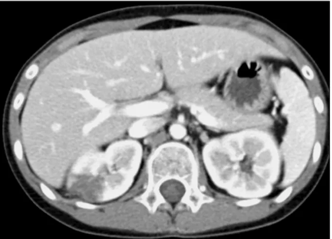
Figure 1. Abdominal CT showing a 2.2 cm sized heterogeneous hypodense lesion in the upper pole of the right kidney.
Figure 2. Focal atrophic changes in the upper pole of the right kidney.
(normal <5 U/mL); serum anti-double stranded DNA (dsDNA) antibody, 486.7 IU/mL (by ELISA, normal <5 IU/mL); anti-cardiolipin antibody IgG, equivocal; anti-car- diolipin antibody IgM, negative. Urinalysis showed 1 g of pro- tein in 24 hrs and many RBC sediments. She underwent renal biopsy, and the histologic diagnosis was lupus nephritis, mem- branous type. Due to lupus nephritis, cyclophosphamide and glucocorticoid pulse therapy was started, and she was man- aged with monthly cyclophosphamide therapy for 6 months.
After the disease activity improved, she was followed up in the outpatient clinic with deflazacort 18 mg, hydroxy- chloroquine 200 mg, azathioprine 50 mg.
On admission, her blood pressure was 120/80 mmHg, heart rate was 90 beats/min, and body temperature was 37.4oC.
Chest auscultation revealed normal breath sounds and heart sounds. There were no specific physical findings except for pain and tenderness on percussion over the right costoverte- bral angle. Initial laboratory investigations showed white blood cell (WBC) count of 7,300/mm3 (segmented neutrophils 80%, lymphocytes 10.7%), hemoglobin level of 12.3 g/dL (normal 11.5∼15.5 g/dL), platelet count of 282,000/mm3 (normal 150,000∼450,000/mm3), prothrombin time (PT) of 24 seconds (normal 11.5∼15 seconds) and activated partial thromboplastin time (aPTT) of 32.7 seconds (normal 28∼45 seconds). A serum biochemistry test showed the following: to- tal bilirubin 0.38 mg/dL (normal 0.2∼1.2 mg/dL), total pro- tein 4.6 g/dL (normal 6.5∼8.3 g/dL), albumin 2.8 g/dL (normal 3.5∼5.1 d/dL), aspartate transaminase (AST) 31 IU/L (normal 0∼35 IU/L), alanine transaminase (ALT) 20 IU/L (normal 0∼40 IU/L), blood urea nitrogen (BUN) 14.8 mg/dL (normal 8∼23 mg/dL), creatinine 0.6 mg/dL (normal 0.6∼1.5 mg/dL), erythrocyte sedimentation rate (ESR) 24 mm/hr
(normal 0∼20 mm/hr), and C-reactive protein (CRP) 76.3 mg/dL (normal 0∼5 mg/dL). The immunologic tests revealed the following: protein C activity 131% (normal 70∼130%), protein S antigen 52% (normal 60∼150%), antithrombin III 99% (normal 80∼120%), fibrinogen 608 mg/dL (normal 170
∼450 mg/dL), d-dimer 5.58 μgL/mL (normal range 0∼0.4 μgL/mL), homocysteine 12.74 μmoL/L (normal 4.7∼13.4 μmoL/L), and anti-dsDNA antibody 104 IU/mL (by ELISA, normal <5 IU/mL), C3 70.3 mg/dL (normal range 90∼180 mg/dL), C4 2.4 mg/dL (normal range 10∼40 mg/dL). The pa- tient tested negative for the following: lupus anticoagulant an- tibody 1.01 (normal <1.2), anti-beta2 glycoprotein-I antibody 1.4 U/mL (normal 0∼10 U/mL), anti-cardiolipin Ab IgG 5.0 GPL U/mL (normal <23 GPL U/mL) and IgM 1.0 MPL U/mL (normal <11 MPL U/mL). Proteinuria and hematuria were confirmed on urine analysis.
The abdominal CT revealed a 2.2 cm sized heterogeneous hypodense lesion in the upper pole of the right kidney, sug- gestive of localized right renal infarction (Figure 1). Electro- cardiography showed normal sinus rhythm and transthoracic echocardiography did not reveal intracardiac thrombosis or embolism. Although no obivous renal artery thrombosis was shown in abdominal CT, we presumed that cause of renal in- farction was thrombus formation in small branches of renal vessel, because of patient's clinical manifestations, elevated fi- brinogen and d-dimer and typical findings of focal renal in- farction on CT. Treatment with anticoagulation (enoxaparin sodium 40 mg bid) was initiated, along with hydroxy- chloroquine, azathiprine, deflazacort same dose as treated in outpatient clinic. After 4 days, oral anticoagulant (warfarin 5 mg) was combined together. Followed up laboratory tests showed PT 25.4 seconds (INR 2.37) and hematuria was no
272 Si Hye Kim et al.
more seen on urine analysis. Right flank pain and nausea gradually improved, and she was discharged on day 10 of ad- mission to follow-up in the Rheumatology outpatient clinic.
The maintenance therapy included warfarin 5mg/day, de- flazacort 16mg, hydroxychloroquine 200 mg, azathioprine 50 mg, followed by deflazacort 16 mg/day with slow tapering to 6 mg/day over 5-month period. The follow-up abdominal CT performed at three months after treatment, previously noted non-enhanced lesion of right renal upper pole is disappeared and only focal atrophic changes were seen in the infarcted area (Figure 2).
After 14 months, urinalysis showed 2 g of protein in 24 hours and the level of anti-dsDNA antibody was 104 IU/mL (by ELISA, normal <5 IU/mL). Mycophenolate mofetile (250 mg twice a day) was given for control of lupus nephritis, aza- thioprine was stopped. The maintenance therapy consisted of warfarin 5 mg, deflazacort 6 mg, hydroxychloroquine 200 mg, ramipril 2.5 mg, mycophenolate mofetil 500 mg, which was persisted over the following 5 months. And after 20 months, warfarin was stopped.
After 22 months, daily protein excretion in urine decreased to 520mg and the immunologic tests showed anti-dsDNA anti- body 66 IU/mL (by ELISA, normal <5 IU/mL), C3 101.0 mg/dL (normal range 90∼180 mg/dL) , C4 13.6 mg/dL (normal range 10∼40 mg/dL). The patient is doing well with regular follow up through outpatient clinic visits.
Discussion
Thrombosis causes substantial morbidity and mortality in SLE and occurs with greater frequency at a younger age than in the general population. Thrombotic events that affect both the arterial and venous systems are a well-known clinical entity in SLE, with a prevalence ranging from 20 to 37 percent (5).
Several environmental or genetic risk factors have been sug- gested to increase the thrombotic tendency (6-11). The pres- ence of lupus anticoagulant and anticardiolipin antibodies has been confirmed to increase the risk of thrombosis. It is known that aPL are directed against phospholipid binding proteins ex- pressed on the surface of vascular endothelial cells or plate- lets, and that they also inhibit the protein C pathway, beta 2 glycoprotein I anticoagulant activity, antithrombin activity, and displacement of annexin A5. They can also have an influ- ence by enhancing endothelial cell procoagulant activity, ex- pression of tissue factor on monocytes, dysregulation of eico- sanoids and they can initiate the formation of a thrombus (6).
Also, the other thrombophilic risk factors such as protein C, protein S, antithrombin deficiencies, factor V Leiden mutation, and G20210A prothrombin gene mutation are strongly asso-
ciated with an increased risk of thrombosis (7).
Several factors in addition to the auto-antibody may contrib- ute to the thrombotic event. The presence of inflammation af- fects blood coagulation and is associated with high risk for thrombosis. It has been shown that inflammation induces the expression of tissue factors, reduces fibrinolytic activity, and impairs the protein C pathway by downregulation of thrombo- modulin and a decrease in protein S (8). Other traditional risk factors, such as smoking, hypertension, diabetes mellitus, dys- lipidemia and hyperhomocysteinemia have also been found to be associated with thrombosis in numerous studies (3,4). The use of glucocorticoids has been found to be associated with thrombosis, probably mediated by endothelial damage and ac- celerated atherosclerosis, and abnormalities in the coagulation cascade (9).
In SLE patients, hydroxychloroquine use has been suggested to offer protection from thrombosis through multiple mecha- nisms, including inhibition of platelet aggregation and adhesion, inhibition of antiphospholipid antibody-induced GPIIb/IIIa re- ceptor expression and cholesterol-lowering mechanisms (10,11).
Our patient was diagnosed with SLE and lupus nephritis and was managed with glucocorticoids. Although she did not have antiphospholipid antibodies and lupus anticoagulants, the labo- ratory study showed proteinuria and high-titer of anti-dsDNA antibody and a low titer of C3, C4, indicating high disease activity. In our case, systemic inflammation, glucocorticoid use, dyslipidemia may lead to the occurrence of the throm- botic event.
Threre are few reports on the clinical presentation of anti- phospholipid antibody-negative SLE manifested renal artery thrombosis associated with renal infarction (2,12,13). Kang DG et al. reported 27-year-old woman with SLE and WHO classi- fication III, V lupus nephritis treated with prednisolone and an- timalarial agent (2). After discontinued taking medication, she developed left flank pain and generalized edema, and renal ar- tery thromboembolism associated with multiple renal in- farctions was shown. Similar to our case, authors presumed that systemic inflammation with high disease activity due to withdrawl medication contributed to thromboembolic event.
Membranous lupus nephritis (LN) is present in 10 to 20 per- cent of patients with lupus nephritis. Renal vessel thrombosis is most frequently reported to be associated with membranous glomerulonephritis, especially when combined with increased blood viscosity, hypercoagulation state resulting from nephrotic syndrome (14). Multiple hemostatic abnormalities have been described, including increased platelet activation, decreased levels of antithrombin and plasminogen, hyperfibrinogenemia, inhibition of plasminogen activation and the presence of
Renal Infarction in Antiphospholipid Antibody-negative Pediatric SLE Patient 273
high-molecular-weight circulating fibrinogen. Intravascular volume depletion may promote tendency to form thrombus and high dose corticosteroid use may also contributed hyper- coagulable state. The possibility of immune complex injury in the glomerulus resulting in systemic effects on clotting has also been evoked (15). Our patient was diagnosed lupus nephritis membranous type and showed nehprotic range proteinuria and treated with glucocorticoid. They also probably account for hy- percoagulable state and result in renal infarction.
It is worth noting that approximately 40 percent of patients with SLE are negative for antiphospholipid antibodies, but have developed thrombotic events. Growing evidence suggests that thrombosis is often caused due to the interaction of genet- ic defects and environmental factors that interfere with the normal hemostatic mechanism.
In conclusion, this case reveals the possibility of renal in- farction in antiphospholipid antibody negative pediatric SLE patients. Therefore, renal infarction should be suspected in cases of sudden unexplained flank or upper abdominal pain, with or without fever, nausea, and vomiting even if the patient tests negative for the antiphospholipid antibody.
Summary
Renal infarction should be suspected in cases of sudden un- explained flank or upper abdominal pain, even if the patient tests negative for the antiphospholipid antibody. Furthermore, the risk factors for thrombosis should be evaluated and man- aged in every patient with SLE.
References
1. Bourgault M, Grimbert P, Verret C, Pourrat J, Herody M, Halimi JM, et al. Acute renal infarction: a case series.
Clin J Am Soc Nephrol 2013;8:392-8.
2. Suwabe H, Moriuchi J, Hoshina Y, Iwata Y, Ichikawa Y, Arimori S. Renal and cerebral infarctions in a patient with systemic lupus erythematosus without antiphospholipid antibodies. Ryumachi 1993;33:335-40.
3. den Heijer M, Koster T, Blom HJ, Bos GM, Briet E, Reitsma PH, et al. Hyperhomocysteinemia as a risk factor for deep-vein thrombosis. N Engl J Med 1996;334:759-62.
4. Danowski A, de Azevedo MN, de Souza Papi JA, Petri M. Determinants of risk for venous and arterial thrombo- sis in primary antiphospholipid syndrome and in anti-
phospholipid syndrome with systemic lupus erythemat- osus. J Rheumatol 2009;36:1195-9.
5. Brouwer JL, Bijl M, Veeger NJ, Kluin-Nelemans HC, van der Meer J. The contribution of inherited and acquired thrombophilic defects, alone or combined with anti- phospholipid antibodies, to venous and arterial throm- boembolism in patients with systemic lupus erythema- tosus. Blood 2004;104:143-8.
6. Espinosa G, Cervera R, Font J, Reverter JC, Shoenfeld Y. Mechanisms of thrombosis in the antiphospholipid syndrome. Inmunología 2003;22:53-62.
7. De Stefano V, Martinelli I, Mannucci PM, Paciaroni K, Chiusolo P, Casorelli I, et al. The risk of recurrent deep venous thrombosis among heterozygous carriers of both factor V Leiden and the G20210A prothrombin mutation.
N Engl J Med 1999;341:801-6.
8. Esmon CT. The impact of the inflammatory response on coagulation. Thromb Res 2004;114:321-7.
9. Calvo-Alén J, Toloza SM, Fernández M, Bastian HM, Fessler BJ, Roseman JM, et al; LUMINA Study Group.
Systemic lupus erythematosus in a multiethnic US cohort (LUMINA). XXV. Smoking, older age, disease activity, lupus anticoagulant, and glucocorticoid dose as risk fac- tors for the occurrence of venous thrombosis in lupus patients. Arthritis Rheum 2005;52:2060-8.
10. Erkan D, Yazici Y, Peterson MG, Sammaritano L, Lockshin MD. A cross-sectional study of clinical thrombotic risk fac- tors and preventive treatments in antiphospholipid synd- rome. Rheumatology (Oxford) 2002;41:924-9.
11. Sisó A, Ramos-Casals M, Bové A, Brito-Zerón P, Soria N, Muñoz S, et al. Previous antimalarial therapy in pa- tients diagnosed with lupus nephritis: influence on out- comes and survival. Lupus 2008;17:281-8.
12. Kang DG, Kim BS, Jang EH, Shin SY, Kim J. Renal Artery Thrombosis in a Patient with Systemic Lupus Erythematosus without Antiphospholipid Antibody Synd- rome: A Case Study. J Korean Rheum Assoc 2009;16:
248-52.
13. Tsugawa K, Tanaka H, Kudo M, Nakahata T, Ito E. Renal artery thrombosis in a pediatric case of systemic lupus er- ythematosus without antiphospholipid antibodies. Pediatr Nephrol 2005;20:1648-50.
14. Barbour SJ, Greenwald A, Djurdjev O, Levin A, Hladunewich MA, Nachman PH, et al. Disease-specific risk of venous thromboembolic events is increased in idio- pathic glomerulonephritis. Kidney Int 2012;81:190-5.
15. Singhal R, Brimble KS. Thromboembolic complications in the nephrotic syndrome: pathophysiology and clinical management. Thromb Res 2006;118:397-407.