REVIEW ARTICLE
비알코올 지방간 질환에서 간섬유화의 진행
김문영
연세대학교 원주의과대학 내과학교실
The Progression of Liver Fibrosis in Non-alcoholic Fatty Liver Disease
Moon Young Kim
Department of Internal Medicine, Yonsei University Wonju College of Medicine, Wonju, Korea
Understanding the pathogenesis of non-alcoholic steatohepatitis (NASH) and its fibrosis progression is still evolving. Nonetheless, current evidence suggests that mechanisms involved are very complex parallel processes with multiple metabolic factors. Lipotoxicity related with excess saturated free fatty acids, obesity, and insulin resistance acts as the central driver of cellular injury via oxidative stress. Hepatocyte apoptosis and/or senescence are also contribute to the activation of inflammasome via various intra- and in- ter-cellular signaling mechanisms that lead to fibrosis. Current evidence suggests that periportal components, including ductular reaction and expansion of the hepatic progenitor cell compartment, may be involved and that the T-helper 17 cell response may medi- ate disease progression. This review aims to provide a brief overview of the pathogenesis of NASH and fibrosis progression from in- flammation to fibrosis. (Korean J Gastroenterol 2017;69:341-347)
Key Words: Non-alcoholic fatty liver disease; Fibrosis; Inflammation; Insulin resistance
Received May 25, 2017. Revised June 2, 2017. Accepted June 4, 2017.
CC This is an open access article distributed under the terms of the Creative Commons Attribution Non-Commercial License (http://creativecommons.org/licenses/
by-nc/4.0) which permits unrestricted non-commercial use, distribution, and reproduction in any medium, provided the original work is properly cited.
Copyright © 2017. Korean Society of Gastroenterology.
교신저자: 김문영, 26426, 원주시 일산로 20, 연세대학교 원주의과대학 원주세브란스기독병원 소화기내과
Correspondence to: Moon Young Kim, Division of Gastroenterology, Department of Internal Medicine, Wonju Severance Christian Hospital, Yonsei University Wonju College of Medicine, 20 Ilsan-ro, Wonju 26426, Korea. Tel: +82-33-741-1229, Fax: +82-33-741-1228, E-mail: [email protected]
Financial support: None. Conflict of interest: None.
서 론
비알코올 지방간 질환(non-alcoholic fatty liver disease, NAFLD)은 만성 간 질환으로, 제2형 당뇨병, 비만 및 대사증 후군과 밀접한 관계가 있다고 알려져 있다.1 지역마다 다소 빈도의 차이는 있으나 전 세계적으로 평균 약 20%의 환자가 NAFLD에 이환되어 있는 것으로 보고되어 있으며, 이 중 일 부에서는 지방간염 및 간섬유화의 진행을 거쳐 간경변으로 진 행하는 것으로 알려져 있다.2 NAFLD 중 단순 지방증(simple steatosis)이 비알코올 지방간염(non-alcoholic steatohepa- titis, NASH)으로 진행하는지 혹은 NASH가 단순 지방증과 는 처음부터 분명히 다른 질환인지의 여부는 확실하지 않으 나, 일반적으로 단순 지방증이 느린 조직학적 진행을 보이는 반면에 NASH는 보다 빠른 조직학적 진행을 보이며 간경변
으로 진행할 수 있음은 잘 알려져 있다.1 NASH 환자의 10-29%는 10년 이내 간경변이 발생하며, 그중 4-27%는 간암 으로 진행된다.3,4
초기에 NASH의 발병은 순차적인 자극(hit)에 의한 것으로 이해되어, 간 내 지방의 침착이 우선 이루어지고 이후 이차적 자극에 의해 간실질의 염증, 지방괴사 및 간섬유화가 일어나 는 것으로 이해되었다(two hit theory). 그러나 현재는 더 복잡 하고 다양하게 병행되는 대사성 자극들에 의해 조직 손상과 질 병의 진행이 이루어지는 것으로 이해되고 있다(multiple paral- lel theory). NASH의 발병에 있어서 지질독성(lipotoxicity)과 인슐린 저항성(insulin resistance), 산화 스트레스(oxidative stress)에 의한 간세포의 손상이 가장 중심에 있으며, 간세포 의 사멸과 이에 따른 일련의 염증 반응과 섬유화의 진행이 주요 기전으로 알려져 있다. 간섬유화는 이러한 일련의 과정
을 통한 NASH 진행의 최종 산물로, NAFLD 또는 NASH 환자에서 모든 사망 또는 간 질환 관련 사망 예후를 예측함에 있어 간섬유화의 정도가 가장 관련성이 높다는 최근의 보고는 NAFLD의 다양한 현상 중에서도 간섬유화의 중요성을 보여 주는 것이며, 향후 치료의 주요 목표가 간섬유화가 되어야 함 을 시사하는 것이라 할 수 있다.5,6 이에 본문에서는 최근에 NAFLD와 관련하여 제시되고 있는 간섬유화의 기전을 간략 히 살펴보고자 한다.
본 론
섬유화의 진행과 염증 반응은 분리해서 생각할 수 없으며 하나의 연속선상에 있는 병적과정이라 할 수 있다.7염증 반응 의 지속은 섬유화를 유발하고, 결국 간경변으로 이르게 하는 주요 기전이 된다. 만성 간 질환에서 간 내 섬유화는 원인질환 에 따라서 다소 다른 양상을 보일 수 있다. 일반적으로 어른의 NASH에서의 섬유화 초기에는 주로 간엽의 간실질을 침범하 는 ‘chicken wire’ 양상으로 불리는 peri-cellular 부위의 망 상(reticular network pattern)의 섬유화 양상을 보이게 된 다.8그러나 소아의 NASH에서는 그 양상이 달라서 문맥섬유 화(portal fibrosis)의 양상을 주로 보이게 된다.9-11 NASH에 의한 섬유화가 이와 같이 다양한 양상을 보이는 이유는 명확 하지 않지만, 섬유화 유발에 이르는 다양한 기전과 유전적 조 건 및 세포 간의 복잡한 상호작용에 의한 것으로 추정된다.11 그러나 섬유화가 진행되면 결국 문맥섬유화가 발생하게 되고 간경변에 이르게 된다. 일반적으로 문맥섬유화를 동반하지 않 는 경우에는 장기간 추적에서 간 관련 합병증의 발생이 적은 것으로 알려져 있으며,12 결국 문맥을 중심으로 한 염증의 침 윤과 섬유화의 진행이 간세포 내 지방의 침착과 간엽 내의 염증보다는 질환의 진행에 있어서 더 중요한 것으로 알려져 있다.13,14
간섬유화는 여러 자극에 의해 활성화된 근섬유아세포(myo- fibroblastic cell)가 콜라겐을 비롯한 일련의 세포 외 물질 (extra-cellular matrix)을 만들어 내고 과다하게 간 내에 침착 되는 과정이며, 간 성상세포(hepatic stellate cell)는 활성화 되어 간 내 근섬유아세포의 대부분을 이룬다. 간 성상세포는 주로 비활성화 상태로 간 내 동모양혈관(sinusoid)과 간세포 사이에 위치하여 있으며, 만성 염증에 의해 활성화되고 병변 부위로 이동하여 일련의 섬유화 작용을 일으키게 된다. 활성화 된 간 성상세포는 transforming growth factor-beta (TGFβ) 를 비롯한 여러 사이토카인(cytokines)과 케모카인(chemokines) 을 분비하여 자체의 활성을 유지하고, 동시에 다른 세포들의 활성을 증가시켜 지속적인 염증과 섬유화 반응을 일으키게 된 다. 이러한 일련의 섬유화 과정은 간 손상의 유발 원인에 따라
염증에서 섬유화로 이어지는 기전에 다소의 차이가 있을 수 있으나, 실제 섬유화가 일어나는 단계에서는 근섬유아세포를 중심으로 공통적인 기전과 양상을 보인다. 따라서 본문에서는 간 성상세포를 중심으로 하는 공통적인 간섬유화 과정보다는 NASH에서 간 성상세포의 활성을 유발하는 일련의 과정에 중점을 두고 기술하고자 한다.
1. 지질독성과 인슐린 저항성(lipotoxicity and insulin resistance)
지질독성은 NASH의 진행에 있어서 가장 중요한 기전으로 알려져 있다. 기존에 알려진 간 내 단순 중성지방 침착의 증가 나 간세포로부터 지질배출의 감소 자체는 염증을 유발하지 않
으며,15,16 오히려 일부 연구에서는 중성지방의 침착이 지질독
성을 막는 효과를 보이는 것으로 보고되고 있다.17,18지질독성 에 의한 손상은 과도한 자유 지방산(free fatty acid), 특히 포 화 자유 지방산(saturated free fatty acids)에 의해 나타나며, 이들에 의한 독성 대사산물의 생성이 질환의 진행에 있어서 중요한 역할을 한다.19식이,
de novo
지방생성(lipogenesis) 또는 인슐린 저항성에 동반된 지방질의 융해(adipose lip- olysis)에 의한 포화 자유 지방산의 증가는 결국 ceramides, diacylglycerols, lysophosphatidyl choline과 같은 지질독성 물질을 증가시키며, 이들은 산화 스트레스와 작용하여 지질독 성을 유발하게 되고, 만성적인 염증 상태를 유발하게 된다.19-21 비만은 인슐린 저항성의 발현과 toll-like receptor (TLR) 4/nuclear factor kappa B (NF-κB)를 통한 전염증성 신호체 계(pro-inflammatory signaling) 활성화에 중요한 역할을 하 는데,22 인슐린 저항성에 따른 만성적인 고인슐린혈증은 골격 근과 간 내 인슐린 신호 체계의 장애를 가져오고 간 내 지방 침착을 유발한다.23 인슐린 저항성에 의해 지방조직은 과도하 게 자유 지방산을 생성하고, 이를 통해서 간 내 지방과 독성 지질 물질을 축적시키며, 이는 다시 인슐린 저항성을 악화시 키는 악순환을 유발하게 된다.24 비만에 의한 말초 지방조직은 전염증성 tumor necrosis factor alpha (TNFα)와 interleukin- (IL-) 6의 생성 증가를 통해서도 염증과 인슐린 저항성의 증가 에 중요한 역할을 한다.25고인슐린혈증과 인슐린 저항성은 지 방조직의 c-Jun N-terminal kinases (JNK)-1 신호 체계의 증 가에 의해서도 유발되는데, 이는 말초 지방조직에 의해 만들 어지는 염증 물질이 NASH를 유발하는 주요 원인이 될 수 있음을 시사한다.26 포화 지방산은 또한 미토콘드리아의 활성 산소를 증가시키고, 이는 다시 JNK의 활성화와 세포의 인슐 린 저항성을 증가시키게 된다.27 이와 같이 비만에 의한 인슐 린 저항성과 포화 자유 지방산의 증가와 이로 인한 산화 스트 레스 및 지질독성의 증가는 간세포 손상과 NASH의 진행 및 섬유화 유발의 가장 중심에 있으며, 이후 언급하게 될 여러다른 염증 및 섬유화 유발 기전의 방아쇠와 같은 역할을 하게 된다.
2. 세포사(cell death)
지질독성은 풍선양 변화(ballooning)와 세포자멸(apoptosis) 에 의한 세포자멸체(apoptotic body) 생성 및 괴사(necrosis) 를 유발하며, 이는 간 내 염증과 섬유화를 유발하는 주요 원인 이 된다.8,28,29 세포자멸은 원래 세포사멸에 따른 세포 내 물질 의 외부 유출을 억제하고, 이를 통해 염증 발생을 최소화하는 방어적인 기전의 측면을 가지고 있으나, NASH에서 관찰되는 세포자멸의 증가는 병적 상태와 연관된다. 세포자멸의 정도는 혈중 자유 지방산의 농도와 상관성이 있다.30 자유 지방산은 세포사멸과 관련 있는 tumor necrosis factor-related apop- tosis-inducing ligand (TRAIL) 수용체의 간세포에서의 발현 을 증가시키며,31 단순 지방간에 비해서 NASH에서 세포사멸 과 관련 있는 또 다른 수용체인 Fas (cluster of differentiation 95, CD95)의 발현이 더 높게 나타난다.32,33 또한 포화 자유 지방산은 간세포 내의 endoplasmic reticulum (ER) 스트레 스를 증가시켜, ER로부터 칼슘분비를 증가시키고 활성 산소 의 증가 및 JNK 증가 등을 통해서 세포자멸을 유도한다.34,35 이러한 과도한 세포자멸은 간섬유화의 주요 원인이 되는데, 세 포자멸체는 간 성상세포를 직접적으로 활성화시키고, TGFβ 와 같은 강력한 섬유화 관련 사이토카인 생산을 증가시킨다.36
3. 선천적 면역반응(iInnate immune response)
선천적 면역반응은 NASH와 같은 간실질의 염증 반응에 있어서 매우 중요하며, 이는 중성구, 쿠퍼세포와 같은 대식세 포, 자연살해세포(natural killer cell, NK cell) 그리고 자연살 해T세포(natural killer T cell, NKT cell) 등을 통해 이루어진 다. 대식세포에서 분비 또는 발현되는 여러 인자들은 NASH 의 중증도와 상관성을 갖고, IL-6, TGFβ 등의 분비를 통해서 간 성상세포를 직접적으로 활성화시킨다.37-40 중성구는 조직 손상에 대해 최초로 염증 반응을 일으키는 세포로, IL-1수용 체를 매개로 한다.41 최근 보고에 따르면 NASH 환자에서는 말초 중성구-림프구 비율이 질환이 없는 환자에 비해서 높은 것으로 보고하고 있다.42 간에 많이 존재하는 자연살해세포는 사이토카인 등의 분비에 의한 바이러스 억제 효과 등의 선천 적 면역 방어 기능을 한다. NASH에서 그 수가 증가하며, in- terferon-γ를 통한 간 성상세포와 간세포의 세포자멸을 유도한
다.43,44 자연살해T세포는 조직 손상에 대한 후천적 면역반응
(adaptive immune response)을 담당하며 괴사염증과 관련이 있다.45
병원균이 없는 상태에서 나타나는 면역반응을 ‘sterile in- flammation’이라고 하며, 이는 간 내 염증과 NASH에서도 중
요한 역할을 한다.46-48 이러한 반응은 주로 괴사나 자멸세포 등과 같이 손상된 세포로부터 유리되는 damage associated molecular patterns (DAMPS)에 의해 유발되며, 세포질 내의
‘inflammasome’을 증가시켜 IL-1β와 같은 전염증성 사이토 카인의 합성을 증가시킨다.49-51 이 과정에서 TLR은 DAMPS 에 대한 패턴 인식 수용체(pattern recognition receptors)로 서 작용하고, 전염증성 사이토카인들과 케모카인들을 유도함 으로써 간 내 염증의 유도와 진행에 매우 중요한 역할을 한다.
TLR은 간 내 다양한 세포들에서 발현이 되는데, 쿠퍼세포는 TLR2, 3, 4와 9를 발현하며, 앞서 언급한 바와 같이 IL-1β의 분비를 통한 면역반응을 매개함으로써 sterile inflammation 의 가장 핵심적인 역할을 하는 것으로 알려져 있다.50,52-54 이 러한 TLR들은 NASH 발생에 있어서도 매우 중요한데,55-59 그 중에서도 sterile inflammation의 핵심 인자인 TLR4와 TLR9 가 간세포의 자멸과 alanine aminotransferase의 상승 및 간 섬유화와 연관성이 높은 것으로 알려져 있다.55,56,60 간섬유화 모델과 간경변증 환자들에서 세균성 lipopolysaccharide (LPS)가 증가되어 있으며,61-63 TLR4는 특히 LPS에 의해 선천 성 면역을 활성화시킴으로써, NASH 유발에 있어 매우 중요 한 역할을 한다.64-66 TLR9 또한 많은 연구를 통해서 쿠퍼세포 에서 IL-1β의 생성과 간세포 사멸에 중요한 역할을 하며, TLR9-deficient mice를 이용한 NASH 모델에서는 지방증은 물론 간 내 염증과 섬유화가 발생하지 않는 것으로 알려져 있다.56 이와 같이 inflammasome의 활성화을 통한 IL-1β 생 성 증가는 최근 NASH 발생에 대한 연구의 중심을 이루고
있다.67-71특히 포화 지방산이 간세포의 LPS에 대한 감수성을
증가시키고, 간세포의 자멸을 유발하고 DAMPS를 생성함으 로써 결국 inflammasome을 활성화하고 IL-1β를 생성한다는 점은 포화 지방산이 LPS에 의한 inflammasome의 생성과 그 를 통한 지방간염의 진행에 있어서도 중심에 있음을 다시 한 번 보여주고 있다.68,72,73
4. 후천적 면역반응(adaptive immune response)
NASH에서 문맥 주위의 만성 염증의 침윤과 진행된 섬유 화가 상관성을 보이는 것을 볼 때, 후천적 면역반응 역시 질환 의 발병 기전에서 매우 중요하다고 할 수 있다.13,74동물 실험 에서 T세포의 감소는 섬유화의 감소와 상관성을 보이며, 그 기전이 아직 명확하지 않으나 CD4+ T세포는 섬유아세포나 대식세포와 상호 반응을 보이는 반면 CD8+T세포는 직간접 적으로 간 성상세포를 활성화시킨다.75 B세포 또한 섬유화 사 이토카인인 IL-4, IL-6, IL-13을 분비하며, 이들 세포의 감소는 간세포 손상 후 섬유화를 감소시킨다.76,77
전통적인 후천적 면역반응은 주로 작용하는 T-helper (Th) 세포의 종류에 따라서 염증을 유발하는 Th1과 섬유화를 촉진
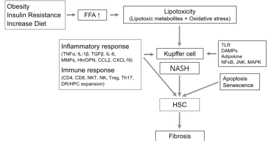
Fig. 1. Brief summary of the pathogenesis of nonalcoholic steatohepatitis with fibrosis progression. FFA, free fatty acid; TLR, toll-like receptor;
DAMPs, damage associated molecular patterns; NFκB, nuclear factor kappa-light-chain-enhancer of activated B cells; JNK, c-Jun N-terminal kinases; MAPK, mitogen-activated protein kinase; TNFα, tumor necrotizing factor-alpha; IL, interleukin; TGFβ, transforming growth factor-beta; MMPs, matrix metalloproteinases; CCL2, chemokine (C-C motif) ligand 2; CXCL16, chemokine (C-X-C motif) ligand 16; CD, cluster of differentiation; NKT, natural killer T; NK, natural killer; Treg, regulatory T cell; Th17, T helper 17 cell; Hh, hedgehog; OPN, osteopontin; DR, ductular reaction; HPC, hepatic progenitor cells; NASH, non-alcoholic steatohepatitis; HSC, hepatic stellate cell.
하는 Th2 면역반응으로 나눌 수 있다. CD4+ T-helper세포에 의한 반응에는 조절 T세포(regulatory T cells, Treg)와 Th17 세포에 의한 것이 주목을 받고 있다.78 특히, Th17세포는 IL-17을 분비하며, 병원체가 Th1이나 Th2 면역반응에 의해 적절히 제거되지 못했을 경우 작동하는데, 선천적 면역반응과 후천적 면역반응의 연결 작용을 하여 IL-17의 주요 작용 대상 인 중성구를 매개로 매우 강력히 염증 반응을 유발한다.79,80 NASH에서 Th17세포의 역할은 아직 명확히 알려져 있지 않 으나, 중성구의 침윤과 이들 세포의 유도에 중요한 IL-6, TGF β 등의 증가는 본 질환의 발생에 Th17이 관여할 수 있음을 시사한다고 할 수 있다.78,81,82
5. 노화(senescence)와 hedgehog 경로
노화는 비가역적으로 손상 받은 세포의 증식을 제한하고, 그에 따라 종양 발생을 억제하는 작용을 하는 것으로,83 NASH와 간세포암종을 비롯한 다양한 간 질환에서 확인이 되어왔으며, 아직까지 그 기전이 명확히 밝혀져 있지 않지만 NASH의 진행에 있어서 중요한 역할을 할 것으로 추정된
다.84-87임상적으로 간세포에서 노화 관련 인자 p21의 발현은
간섬유화의 단계 및 간세포암종의 발생이나 간 질환 관련 사 망 등의 임상 경과와 유의한 상관성을 갖는 것으로 보고되고 있다.86
Hedgehog (Hh)는 세포의 증식과 분화를 통한 조직 손상 회복에 중요한 역할을 하고, 그 과정에서 섬유화를 유발한다.
최근 연구에 따르면 Hh 리간드의 발현은 간세포의 손상과 유 의한 상관성을 보이며, Hh는 간 성상세포의 활성화 및 염증 세포의 침윤을 유발한다.88-90 Hh의 자극은 미성숙 담도 상피 세포로부터 Chemokine (C-X-C motif) ligand 16 (CXCL16) 의 생산을 자극하고, 이는 자연살해T세포의 침윤과 Th1, Th2 면역반응을 유도하며, hepatic progenitor cells 활성 및 ductular reaction과 관련성을 보인다. 또한 Hh와 그 표적이 되는 염증 물질인 osteopontin의 활성 정도에 따라 간섬유화 는 차이를 보이고, 이러한 소견은 NASH에서 나타나는 간섬유 화의 이질성과 다양성을 설명하는 데 도움이 될 수 있다.88,91
결 론
NASH의 병인과 간섬유화의 진행은 비만, 인슐린 저항성, 간 내 지방 침착 및 산화 스트레스에 동반된 장기간의 만성적 염증 반응에 의한 매우 복잡하고 다양한 기전에 의해 이루어 진다.
과도한 포화 자유 지방산과 그에 따른 독성 대사산물의 생 성에 의한 간세포 손상, 세포자멸 및 괴사에 의한 염증 반응과 사이토카인 증가는 간 성상세포를 자극하여 섬유화를 유발한 다. 또한 TLR4와 TLR9에 의한 무균성의 염증 반응과 그에 따른 사이토카인의 증가(TGFβ, IL-1β 등)가 염증과 섬유화를 유발한다. 최근에는 IL-1β, TGFβ, IL-6을 매개로 하는 Th17 세포에 의한 면역 반응에 의한 문맥의 섬유화가 관심을 모으
Inflammatory response
(TNFα, IL-1β, TGFβ, IL-6, MMPs, Hh/OPN, CCL2, CXCL16)
Immune response
(CD4, CD8, NKT, NK, Treg, Th17, DR/HPC expansion)
Obesity
Insulin Resistance Increase Diet
HSC
고 있으며, 이러한 만성 염증은 재생을 위한 반응을 활성화하 는데, 이를 통해서 hepatic progenitor cells 부분의 확대와 ductular reaction이 나타나고, 이는 NASH에 동반된 섬유화의 특징인 문맥 부위의 염증 침윤 및 섬유화를 반영한다(Fig. 1).
이러한 다양한 기전에 대한 이해는 결국 NASH와 그로 인한 섬유화의 치료법 개발의 근간이 되며, 향후 더 정밀한 기전에 대한 이해를 위하여 연구가 지속되어야 한다.
REFERENCES
1. Cohen JC, Horton JD, Hobbs HH. Human fatty liver disease: old questions and new insights. Science 2011;332:1519-1523.
2. Chalasani N, Younossi Z, Lavine JE, et al. The diagnosis and man- agement of non-alcoholic fatty liver disease: practice guideline by the American Association for the Study of Liver Diseases, American College of Gastroenterology, and the American Gastroenterological Association. Am J Gastroenterol 2012;107:
811-826.
3. Argo CK, Caldwell SH. Epidemiology and natural history of non-alcoholic steatohepatitis. Clin Liver Dis 2009;13:511-531.
4. Starley BQ, Calcagno CJ, Harrison SA. Nonalcoholic fatty liver disease and hepatocellular carcinoma: a weighty connection.
Hepatology 2010;51:1820-1832.
5. Ekstedt M, Hagström H, Nasr P, et al. Fibrosis stage is the stron- gest predictor for disease-specific mortality in NAFLD after up to 33 years of follow-up. Hepatology 2015;61:1547-1554.
6. Angulo P, Kleiner DE, Dam-Larsen S, et al. Liver fibrosis, but no other histologic features, is associated with long-term outcomes of patients with nonalcoholic fatty liver disease. Gastroenterology 2015;149:389-397.e10.
7. Friedman SL. Mechanisms of hepatic fibrogenesis. Gastroen- terology 2008;134:1655-1669.
8. Brunt EM. Histopathology of non-alcoholic fatty liver disease.
Clin Liver Dis 2009;13:533-544.
9. Schwimmer JB, Behling C, Newbury R, et al. Histopathology of pediatric nonalcoholic fatty liver disease. Hepatology 2005;42:
641-649.
10. Carter-Kent C, Yerian LM, Brunt EM, et al. Nonalcoholic steatohe- patitis in children: a multicenter clinicopathological study.
Hepatology 2009;50:1113-1120.
11. Skoien R, Richardson MM, Jonsson JR, et al. Heterogeneity of fib- rosis patterns in non-alcoholic fatty liver disease supports the presence of multiple fibrogenic pathways. Liver Int 2013;33:
624-632.
12. Ekstedt M, Franzén LE, Mathiesen UL, et al. Long-term follow-up of patients with NAFLD and elevated liver enzymes. Hepatology 2006;44:865-873.
13. Brunt EM, Kleiner DE, Wilson LA, et al. Portal chronic in- flammation in nonalcoholic fatty liver disease (NAFLD): a histo- logic marker of advanced NAFLD-clinicopathologic correlations from the nonalcoholic steatohepatitis clinical research network.
Hepatology 2009;49;809-820.
14. Richardson MM, Jonsson JR, Powell EE, et al. Progressive fib- rosis in nonalcoholic steatohepatitis: association with altered
regeneration and a ductular reaction. Gastroenterology 2007;
133:80-90.
15. Monetti M, Levin MC, Watt MJ, et al. Dissociation of hepatic stea- tosis and insulin resistance in mice overexpressing DGAT in the liver. Cell Metab 2007;6:69-78.
16. Liao W, Hui TY, Young SG, Davis RA. Blocking microsomal trigly- ceride transfer protein interferes with apoB secretion without causing retention or stress in the ER. J Lipid Res 2003;44:
978-985.
17. Li ZZ, Berk M, McIntyre TM, Feldstein AE. Hepatic lipid partition- ing and liver damage in nonalcoholic fatty liver disease: role of stearoyl-CoA desaturase. J Biol Chem 2009;284:5637-5644.
18. Flowers MT, Groen AK, Oler AT, et al. Cholestasis and hyper- cholesterolemia in SCD1-deficient mice fed a low-fat, high-car- bohydrate diet. J Lipid Res 2006;47:2668-2680.
19. Neuschwander-Tetri BA. Hepatic lipotoxicity and the patho- genesis of nonalcoholic steatohepatitis: the central role of non- triglyceride fatty acid metabolites. Hepatology 2010;52:774-788.
20. Han MS, Park SY, Shinzawa K, et al. Lysophosphatidylcholine as a death effector in the lipoapoptosis of hepatocytes. J Lipid Res 2008;49:84-97.
21. Tan M, Hao F, Xu X, Chisolm GM, Cui MZ. Lysophosphatidylcholine activates a novel PKD2-mediated signaling pathway that con- trols monocyte migration. Arterioscler Thromb Vasc Biol 2009;
29:1376-1382.
22. Kim F, Pham M, Luttrell I, et al. Toll-like receptor-4 mediates vas- cular inflammation and insulin resistance in diet-induced obesity. Circ Res 2007;100:1589-1596.
23. Cusi K. Role of insulin resistance and lipotoxicity in non-alcoholic steatohepatitis. Clin Liver Dis 2009;13:545-563.
24. Arner P. The adipocyte in insulin resistance: key molecules and the impact of the thiazolidinediones. Trends Endocrinol MeTab 2003;14:137-145.
25. Hotamisligil GS, Shargill NS, Spiegelman BM. Adipose ex- pression of tumor necrosis factor-alpha: direct role in obe- sity-linked insulin resistance. Science 1993;259:87-91.
26. Tilg H. Adipocytokines in nonalcoholic fatty liver disease: key players regulating steatosis, inflammation and fibrosis. Curr Pharm Des 2010;16:1893-1895.
27. Nakamura S, Takamura T, Matsuzawa-Nagata N, et al. Palmitate induces insulin resistance in H4IIEC3 hepatocytes through re- active oxygen species produced by mitochondria. J Bio Chem 2009;284:14809-14818.
28. Anderson N, Borlak J. Molecular mechanisms and therapeutic targets in steatosis and steatohepatitis. Pharmacol Rev 2008;60:311-357.
29. Tetri LH, Basaranoglu M, Brunt EM, Yerian LM, Neuschwander- Tetri BA. Severe NAFLD with hepatic necroinflammatory changes in mice fed trans fats and a high-fructose corn syrup equivalent. Am J Physiol Gastrointest Liver Physiol 2008;295:
G987-G995.
30. Bechmann LP, Gieseler RK, Sowa JP, et al. Apoptosis is asso- ciated with CD36/fatty acid translocase upregulation in non-al- coholic steatohepatitis. Liver Int 2010;30:850-859.
31. Farrell GC, Larter CZ, Hou JY, et al. Apoptosis in experimental NASH is associated with p53 activation and TRAIL receptor
expression. J Gastroenterol Hepatol 2009;24:443-452.
32. Feldstein AE, Canbay A, Angulo P, et al. Hepatocyte apoptosis and fas expression are prominent features of human non- alcoholic steatohepatitis. Gastroenterology 2003;125:437-443.
33. Ribeiro PS, Cortez-Pinto H, Solá S, et al. Hepatocyte apoptosis, expression of death receptors, and activation of NF-kappaB in the liver of nonalcoholic and alcoholic steatohepatitis patients.
Am J Gastroenterol 2004;99:1708-1717.
34. Deniaud A, Sharaf el dein O, Maillier E, et al. Endoplasmic retic- ulum stress induces calcium-dependent permeability tran- sition, mitochondrial outer membrane permeabilization and apoptosis. Oncogene 2008;27:285-299.
35. Malhi H, Kaufman RJ. Endoplasmic reticulum stress in liver disease. J Hepatol 2011;54:795-809.
36. Canbay A, Taimr P, Torok N, Higuchi H, Friedman S, Gores GJ.
Apoptotic body engulfment by a human stellate cell line is profibrogenic. Lab Invest 2003;83:655-663.
37. Holt AP, Salmon M, Buckley CD, Adams DH. Immune interactions in hepatic fibrosis. Clin Liver Dis 2008;12:861-882, x.
38. Wynn TA, Barron L. Macrophages: master regulators of in- flammation and fibrosis. Semin Liver Dis 2010;30:245-257.
39. Matsuoka M, Tsukamoto H. Stimulation of hepatic lipocyte colla- gen production by Kupffer cell-derived transforming growth fac- tor beta: implication for a pathogenetic role in alcoholic liver fibrogenesis. Hepatology 1990;11:599-605.
40. Malaguarnera L, Di Rosa M, Zambito AM, dell'Ombra N, Di Marco R, Malaguarnera M. Potential role of chitotriosidase gene in non- alcoholic fatty liver disease evolution. Am J Gastroenterol 2006;101:2060-2069.
41. Chen CJ, Kono H, Golenbock D, Reed G, Akira S, Rock KL.
Identification of a key pathway required for the sterile inflammatory response triggered by dying cells. Nat Med 2007;13:851-856.
42. Alkhouri N, Morris-Stiff G, Campbell C, et al. Neutrophil to lym- phocyte ratio: a new marker for predicting steatohepatitis and fibrosis in patients with nonalcoholic fatty liver disease. Liver Int 2012;32:297-302.
43. Zhan YT, An W. Roles of liver innate immune cells in nonalcoholic fatty liver disease. World J Gastroenterol 2010;16:4652-4660.
44. Kahraman A, Schlattjan M, Kocabayoglu P, et al. Major histo- compatibility complex class I-related chains A and B (MIC A/B):
a novel role in nonalcoholic steatohepatitis. Hepatology 2010;
51:92-102.
45. Bendelac A, Savage PB, Teyton L. The biology of NKT cells. Annu Rev Immunol 2007;25:297-336.
46. Gao B, Seki E, Brenner DA, et al. Innate immunity in alcoholic liv- er disease. Am J Physiol. Gastrointest Liver Physiol 2011;300:
G516-G525.
47. Brenner DA, Seki E, Taura K, et al. Non-alcoholic steatohepati- tis-induced fibrosis: toll-like receptors, reactive oxygen species and Jun N-terminal kinase. Hepatol Res 2011;41:683-686.
48. Maher JJ. DAMPs ramp up drug toxicity. J Clin Invest 2009;119:
246-249.
49. Matzinger P. Tolerance, danger, and the extended family. Ann Rev Immunol 1994;12:991-1045.
50. Rock KL, Latz E, Ontiveros F, Kono H. The sterile inflammatory response. Annu Rev Immunol 2010;28:321-342.
51. Davis BK, Wen H, Ting JP. The inflammasome NLRs in immunity, inflammation, and associated diseases. Annu Rev Immunol 2011;29:707-735.
52. Su GL, Klein RD, Aminlari A, et al. Kupffer cell activation by lip- opolysaccharide in rats: role for lipopolysaccharide binding pro- tein and toll-like receptor 4. Hepatology 2000;31:932-936.
53. Soehnlein O, Lindbom L. Phagocyte partnership during the on- set and resolution of inflammation. Nat Rev Immunol 2010;10:
427-439.
54. McDonald B, Pittman K, Menezes GB, et al. Intravascular danger signals guide neutrophils to sites of sterile inflammation. Science 2010;330:362-366.
55. Rivera CA, Adegboyega P, van Rooijen N, Tagalicud A, Allman M, Wallace M. Toll-like receptor-4 signaling and kupffer cells play pivotal roles in the pathogenesis of non-alcoholic steatohepatitis.
J Hepatol 2007;47:571-579.
56. Miura K, Kodama Y, Inokuchi S, et al. Toll-like receptor 9 pro- motes steatohepatitis by induction of interleukin-1beta in mice.
Gastroenterology 2010;139:323-334.e7.
57. Csak T, Velayudham A, Hritz I, et al. Deficiency in myeloid differ- entiation factor-2 and toll-like receptor 4 expression attenuates nonalcoholic steatohepatitis and fibrosis in mice. Am J Physiol Gastrointest Liver Physiol 2011;300:G433-G441.
58. Mencin A, Kluwe J, Schwabe RF. Toll-like receptors as targets in chronic liver diseases. Gut 2009;58:704-720.
59. Baffy G. Kupffer cells in non-alcoholic fatty liver disease: the emerging view. J Hepatol 2009;51:212-223.
60. Spruss A, Kanuri G, Wagnerberger S, Haub S, Bischoff SC, Bergheim I. Toll-like receptor 4 is involved in the development of fructose-induced hepatic steatosis in mice. Hepatology 2009;50:1094-1104.
61. Seki E, De Minicis S, Osterreicher CH, et al. TLR4 enhances TGF-beta signaling and hepatic fibrosis. Nat Med 2007;13:
1324-1332.
62. Nolan JP, Leibowitz AI. Endotoxins in liver disease. Gastroenterology 1978;75:765-766.
63. Grinko I, Geerts A, Wisse E. Experimental biliary fibrosis corre- lates with increased numbers of fat-storing and kupffer cells, and portal endotoxemia. J Hepatol 1995;23:449-458.
64. Akira S, Uematsu S, Takeuchi O. Pathogen recognition and in- nate immunity. Cell 2006;124:783-801.
65. Kagan JC, Medzhitov R. Phosphoinositide-mediated adaptor re- cruitment controls toll-like receptor signaling. Cell 2006;125:
943-955.
66. Wigg AJ, Roberts-Thomson IC, Dymock RB, McCarthy PJ, Grose RH, Cummins AG. The role of small intestinal bacterial over- growth, intestinal permeability, endotoxaemia, and tumour ne- crosis factor alpha in the pathogenesis of non-alcoholic steatohepatitis. Gut 2001;48:206-211.
67. Csak T, Ganz M, Pespisa J, Kodys K, Dolganiuc A, Szabo G. Fatty acid and endotoxin activate inflammasomes in mouse hep- atocytes that release danger signals to stimulate immune cells.
Hepatology 2011;54:133-144.
68. Vandanmagsar B, Youm YH, Ravussin A, et al. The NLRP3 in- flammasome instigates obesity-induced inflammation and in- sulin resistance. Nat Med 2011;17:179-188.
69. Kamari Y, Shaish A, Vax E, et al. Lack of interleukin-1α or inter- leukin-1β inhibits transformation of steatosis to steatohepatitis and liver fibrosis in hypercholesterolemic mice. J Hepatol 2011;55:1086-1094.
70. Wen H, Gris D, Lei Y, et al. Fatty acid-induced NLRP3-ASC in- flammasome activation interferes with insulin signaling. Nat Immunol 2011;12:408-415.
71. Stienstra R, Saudale F, Duval C, et al. Kupffer cells promote hep- atic steatosis via interleukin-1beta-dependent suppression of peroxisome proliferator-activated receptor alpha activity. Hepatology 2010;51:511-522.
72. Csak T, Dolganiuc A, Kodys K, et al. Mitochondrial antiviral sig- naling protein defect links impaired antiviral response and liver injury in steatohepatitis in mice. Hepatology 2011;53:1917- 1931.
73. Ji J, Zhang L, Wang P, et al. Saturated free fatty acid, palmitic acid, induces apoptosis in fetal hepatocytes in culture. Exp Toxicol Pathol 2005;56:369-376.
74. Argo CK, Northup PG, Al-Osaimi AM, Caldwell SH. Systematic re- view of risk factors for fibrosis progression in non-alcoholic steatohepatitis. J Hepatol 2009;51:371-379.
75. Safadi R, Ohta M, Alvarez CE, et al. Immune stimulation of hep- atic fibrogenesis by CD8 cells and attenuation by transgenic in- terleukin-10 from hepatocytes. Gastroenterology 2004;127:
870-882.
76. Novobrantseva TI, Majeau GR, Amatucci A, et al. Attenuated liver fibrosis in the absence of B cells. J Clin Invest 2005;115:
3072-3082.
77. Mosmann T. Complexity or coherence? Cytokine secretion by B cells. Nat Immunol 2000;1:465-466.
78. Hammerich L, Heymann F, Tacke F. Role of IL-17 and Th17 cells in liver diseases. Clin Dev Immunol 2011;2011:345803.
79. Harrington LE, Hatton RD, Mangan PR, et al. Interleukin 17-producing CD4+ effector T cells develop via a lineage distinct from the T helper type 1 and 2 lineages. Nat Immunol 2005;6:
1123-1132.
80. Miossec P, Korn T, Kuchroo VK. Interleukin-17 and type 17 helper T cells. N Engl J Med 2009;361:888-898.
81. Gadd VL, Skoien R, Powell EE, et al. The portal inflammatory in- filtrate and ductular reaction in human nonalcoholic fatty liver disease. Hepatology 2014;59:1393-1405.
82. Abiru S, Migita K, Maeda Y, et al. Serum cytokine and soluble cy- tokine receptor levels in patients with non-alcoholic steatohepatitis.
Liver Int 2006;26:39-45.
83. Campisi J, d’Adda di Fagagna F. Cellular senescence: when bad things happen to good cells. Nat Rev Mol Cell Biol 2007;8:
729-740.
84. Nakajima T, Moriguchi M, Katagishi T, et al. Premature telomere shortening and impaired regenerative response in hepatocytes of individuals with NAFLD. Liver Int 2006;26:23-31.
85. Calado RT, Brudno J, Mehta P, et al. Constitutional telomerase mutations are genetic risk factors for cirrhosis. Hepatology 2011;53:1600-1607.
86. Aravinthan A, Scarpini C, Tachtatzis P, et al. Hepatocyte sen- escence predicts progression in non-alcohol-related fatty liver disease. J Hepatol 2013;58:549-556.
87. Ikeda H, Sasaki M, Sato Y, et al. Large cell change of hepatocytes in chronic viral hepatitis represents a senescent-related lesion.
Hum Pathol 2009;40:1774-1782.
88. Syn WK, Oo YH, Pereira TA, et al. Accumulation of natural killer T cells in progressive nonalcoholic fatty liver disease. Hepatology 2010;51:1998-2007.
89. Rangwala F, Guy CD, Lu J, et al. Increased production of sonic hedgehog by ballooned hepatocytes. J Pathol 2011;224:401- 410.
90. Syn WK, Witek RP, Curbishley SM, et al. Role for hedgehog path- way in regulating growth and function of invariant NKT cells. Eur J Immunol 2009;39:1879-1892.
91. Tajiri K, Shimizu Y, Tsuneyama K, Sugiyama T. Role of liver-in- filtrating CD3+CD56+ natural killer T cells in the pathogenesis of nonalcoholic fatty liver disease. Eur J Gastroenterol Hepatol 2009;21:673-680.