Korean J Gastroenterol Vol. 69 No. 2, 151-154 https://doi.org/10.4166/kjg.2017.69.2.151 pISSN 1598-9992 eISSN 2233-6869
IMAGE OF THE MONTH
Korean J Gastroenterol, Vol. 69 No. 2, February 2017 www.kjg.or.kr
크론병에서 대나무 마디 모양의 위 내시경 소견
서광일, 문원
고신대학교 의과대학 내과학교실
Endoscopic Bamboo Joint-like Appearance of the Stomach in Crohn's Disease
Kwang Il Seo and Won Moon
Department of Internal Medicine, Kosin University College of Medicine, Busan, Korea
CC This is an open access article distributed under the terms of the Creative Commons Attribution Non-Commercial License (http://creativecommons.org/licenses/
by-nc/4.0) which permits unrestricted non-commercial use, distribution, and reproduction in any medium, provided the original work is properly cited.
Copyright © 2017. Korean Society of Gastroenterology.
교신저자: 문원, 49267, 부산시 서구 감천로 262, 고신대학교 의과대학 내과학교실
Correspondence to: Won Moon, Department of Internal Medicine, Kosin University College of Medicine, 262 Gamcheon-ro, Seo-gu, Busan 49267, Korea. Tel:
+82-51-990-5207, Fax: +82-51-990-5055, E-mail: moonone70@hanmail.net Financial support: None. Conflict of interest: None.
Case: A 23-year-old man visited the Emergency Room with epigastric abdominal pain and diarrhea for period of one week. He suffered from watery diarrhea―more than 8 times a day―without blood, accompanied by periodical epigastric pain. Two years ago, he had suffered from a severe abdomi- nal pain with fever, and he was diagnosed with Crohn's dis- ease (CD) after colonoscopy. After medical therapy, his symp- toms were resolved, showing an improvement in his follow-up clinical findings. However, he stopped the medication for 6 months before visiting our hospital.
On the initial physical examination, his blood pressure was 113/75 mmHg, pulse rate was 85 per minute, respiratory rate was 20 per minute, and body temperature was 37.2℃.
The abdomen was soft and flat. However, a mild tenderness in the epigastric and right upper quadrant area was identified. He did not have any other previous medical his- tory, including diabetes, hypertension, viral hepatitis, and tuberculosis.
Laboratory test showed white blood cell count of 10,980/uL (polymorphonuclear neutrophils, 83.5%; lymphocytes, 10.0%;
eosinophils, 0.8%), hemoglobin of 13.4 g/dL, platelet count of 335,000/uL, C-reactive protein level of 10.75 mg/dL, er- ythrocyte sedimentation rate of 38 mm/hr, and prothrombin
time of 16.6 seconds (international normalized ratio 1.30).
The renal function and electrolytes tests were both normal.
Abdominal CT scan showed diffuse enhancing wall thicken- ing in the colon, from ascending to transverse colon.
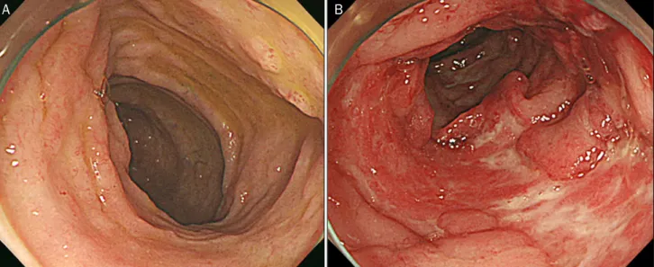
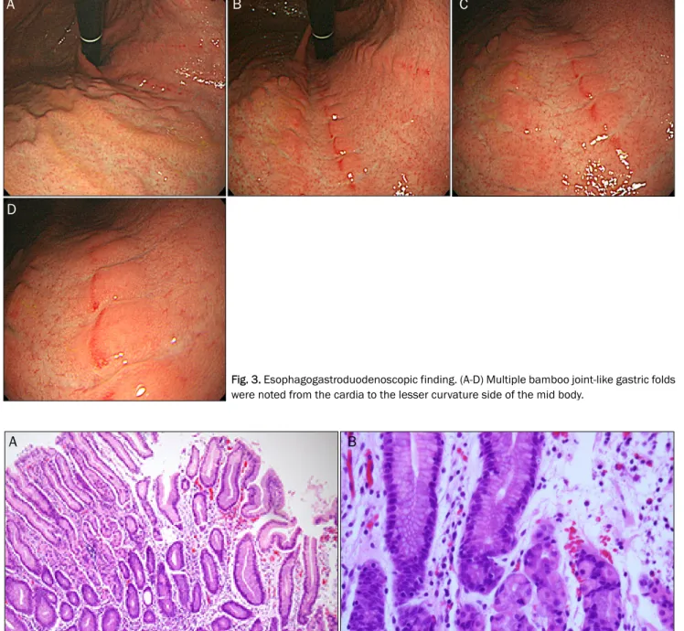
A colonoscopy showed diffuse hyperemic mucosal changes at the terminal ileum (Fig. 1A), as well as at multiple various sized longitudinal, deep active ulcers with normal looking in- tervening mucosa through the whole colon (Fig. 1B). These findings were consistent with CD. A round, slightly raised 4 mm-sized erosion was noted on the 3 o'clock direction of the perianal skin, which was located 2 cm from the anal verge (Fig. 2). Esophagogastroduodenoscopy (EGD) was per- formed for recurrent epigastric pain. Several longitudinal, hy- peremic, elevated mucosal folds transvered by multiple, reg- ularly distributed, sharp pale-colored erosive fissures were noted from the cardia to the lesser curvature of the mid-body, showing a typical bamboo joint-like gastric folds of CD (Fig. 3).
The pathologic findings of these folds showed multifocal ag- gregates of mixed inflammatory cells in the lamina propria without granuloma, consistent with chronic gastritis (Fig.
4). Therefore, the final diagnosis was CD involving the co- lon and stomach, showing a typical bamboo joint-like appearance.
152 서광일, 문원. 크론병에서 대나무 마디 모양의 위 내시경 소견
The Korean Journal of Gastroenterology Fig. 2. Perianal finding with a colonoscopy. A raised 4 mm-sized ero-
sion―round in shape―was noted in the perianal skin.
A B
Fig. 1. Colonoscopic finding. (A) In the terminal ileum, diffuse hyperemic mucosa with a few tiny erosions was noted. (B) In the whole colon, multiple longitudinal, deep active ulcers with normal looking intervening mucosa were scattered.
Diagnosis: Crohn’s disease involving the terminal ileum, the colon and the stomach
Chronic inflammation of the gastrointestinal track is a ma- jor feature of inflammatory bowel disease (IBD). The remit- ting and relapsing inflammatory changes are the natural course of this disease. The two main forms of IBD are CD and Ulcerative colitis (UC).1 Currently, the prevalence of IBD has increased in Asia, including Korea.2 Moreover, the relatively higher prevalence of intestinal tuberculosis and Behcet’s disease in Korea is a barrier for making an accurate diagnosis
of IBD.3,4 Therefore, an endoscopy is a crucial diagnostic tool for an accurate diagnosis and proper management of IBD.
Colonoscopy is commonly performed in patients with gas- trointestinal symptoms with suspicion of IBD, including stool containing mucus or blood, nocturnal diarrhea, and incontinence.1 An endoscopic examination allows for the differential diagnosis between UC and CD; it also serves to assess the disease, in- cluding activity, extent, and histology.5 Typical colonoscopic findings of CD are longitudinal ulcers, cobblestone appear- ance, and small aphthous ulcerations in a longitudinal arrangement.6 Colonoscopy findings in patients with newly diagnosed CD were aphthous ulcers (59.3%), longitudinal ul- cers (37.2%), and cobblestone appearance (23.9%).7
CD can involve the whole gastrointestinal tract, from the mouth to the anus.8 Therefore, an upper gastrointestinal in- volvement of CD is possible. However, there has not been any report on specific symptoms of CD with gastroduodenal involvement. Consequently, EGD is not routinely recom- mended in CD patients.9 Currently, the prevalence of upper gastrointestinal track involvement in IBD has been increas- ing as more patients undergo routine EGD regardless of up- per gastrointestinal symptoms.10
EGD findings in CD patients were gastric erosion, atrophic gastritis, and duodenal erosions or ulcers.11 A typical finding called bamboo joint-like appearance on EGD was found in 54% of CD patients. These lesions were found in the lesser curvature of the body and cardia, characterized by “swollen
Seo KI and Moon W. Endoscopic Bamboo Joint-like Appearance of the Stomach in Crohn's Disease 153
Vol. 69 No. 2, February 2017
A B C
D
Fig. 3. Esophagogastroduodenoscopic finding. (A-D) Multiple bamboo joint-like gastric folds were noted from the cardia to the lesser curvature side of the mid body.
A B
Fig. 4. Pathologic finding of gastric folds. The gastric mucosa showing multifocal aggregates of mixed inflammatory cells in the lamina propria without granuloma (A: H&E, ×100, B: H&E, ×400).
longitudinal folds traversed by erosive fissures or linear furrows.”12 The granuloma in pathologic finding of these gas- tric lesions was noted in 23.2%, indicating a low biopsy sensitivity.13
To date, there have been several reports regarding gastric bamboo joint-like appearance in CD patients; however, the images in these reports were insufficient to be conclusive.
Our endoscopic images of CD patient with gastric involve- ment reveals the gastric bamboo joint-like appearance very clearly, enough to remind the experts and educate the trainees. Similar to our case report, many previous studies reported that microscopic findings of gastric involvement had a low sensitivity. Therefore, pathologic findings of gastric involvement lesion have a limited diagnostic value in CD
154 서광일, 문원. 크론병에서 대나무 마디 모양의 위 내시경 소견
The Korean Journal of Gastroenterology
patients.
In conclusion, CD diagnosis can be made based on both clinical and endoscopic characteristics. Not only typical colo- noscopic findings but also gastric bamboo joint-like appear- ance may be helpful in diagnosing CD. Therefore, this case report may be useful―providing educational value―in clin- ical practice.
REFERENCES
1. Bernstein CN, Eliakim A, Fedail S, et al. World gastroenterology organisation global guidelines inflammatory bowel disease: up- date August 2015. J Clin Gastroenterol 2016;50:803-818.
2. Yang SK, Yun S, Kim JH, et al. Epidemiology of inflammatory bow- el disease in the Songpa-Kangdong district, Seoul, Korea, 1986- 2005: a KASID study. Inflamm Bowel Dis 2008;14:542-549.
3. Suzuki Kurokawa M, Suzuki N. Behcet's disease. Clin Exp Med 2004;4:10-20.
4. Kim YS, Kim YH, Lee KM, Kim JS, Park YS; IBD Study Group of the Korean Association of the Study of Intestinal Diseases. Diagnostic guideline of intestinal tuberculosis. Korean J Gastroenterol 2009;
53:177-186.
5. Moran CP, Neary B, Doherty GA. Endoscopic evaluation in diag-
nosis and management of inflammatory bowel disease. World J Gastrointest Endosc 2016;8:723-732.
6. Lee JM, Lee KM. Endoscopic diagnosis and differentiation of in- flammatory bowel disease. Clin Endosc 2016;49:370-375.
7. Park JB, Yang SK, Myung SJ, et al. Clinical characteristics at diag- nosis and course of Korean patients with Crohn's disease.
Korean J Gastroenterol 2004;43:8-17.
8. So H, Ye BD, Park YS, et al. Gastric lesions in patients with Crohn's disease in Korea: a multicenter study. Intest Res 2016;14:60-68.
9. Ye BD, Jang BI, Jeen YT, et al. Diagnostic guideline of Crohn's disease. Korean J Gastroenterol 2009;53:161-176.
10. Turner D, Griffiths AM. Esophageal, gastric, and duodenal mani- festations of IBD and the role of upper endoscopy in IBD diagnosis. Curr Gastroenterol Rep 2009;11:234-237.
11. Kang MS, Park DI, Park JH, et al. Bamboo joint-like appearance of stomach in Korean patients with Crohn's disease. Korean J Gastroenterol 2006;48:395-400.
12. Fujiya M, Sakatani A, Dokoshi T, et al. A Bamboo Joint-Like Appearance is a Characteristic Finding in the Upper Gastrointestinal Tract of Crohn's Disease Patients: A Case-Control Study. Medicine (Baltimore) 2015;94:e1500.
13. Diaz L, Hernandez-Oquet RE, Deshpande AR, Moshiree B. Upper Gastrointestinal Involvement in Crohn Disease: Histopathologic and Endoscopic Findings. South Med J 2015;108:695-700.