ISSN 0378-6471 (Print)⋅ISSN 2092-9374 (Online)
http://dx.doi.org/10.3341/jkos.2015.56.7.1117
Case Report
눈꺼풀에 발생한 일차성 전신성 아밀로이드증 1예
Primary Systemic Amyloidosis of the Eyelid: A Case Report
강연수⋅최 원⋅윤경철
Yeon Soo Kang, MD, Won Choi, MD, Kyung Chul Yoon, MD, PhD
전남대학교 의과대학 안과학교실
Department of Ophthalmology, Chonnam National University Medical School, Gwangju, Korea
Purpose: Amyloidosis involving the eyelid is a rare condition. We report a case of primary systemic amyloidosis of the eyelid.
Case summary: A 26-year-old female presented with multiple nodules on the bilateral upper and lower eyelids that had stopped growing several years prior. Multiple pearl-colored small nodular lesions were present on the upper and lower eyelid bilaterally and no clinically specific signs were observed. Surgical excision, biopsy and electrocauterization were performed. Histological examination showed amorphous and eosinophilic substances on hematoxylin & eosin (H&E) staining and orange-colored amy- loid deposits stained with Congo-red. Systemic evaluation showed amyloid nodules invading the vocal cords and external audi- tory canal, therefore the patient was diagnosed with primary systemic amylodosis. At the postoperative 6-month follow-up, re- currence or inflammation at the operation site was not observed.
Conclusions: To the best of our knowledge, this is the first case of primary systemic amyloidosis of the eyelid in Korea.
Amyloidosis should be considered in a differential diagnosis of a mass in the eyelid and can be successfully managed with surgi- cal excision and electrocauterization.
J Korean Ophthalmol Soc 2015;56(7):1117-1121
Key Words: Amyloidosis, Eyelid mass, Molluscum contagiosum
■Received: 2015. 1. 16. ■ Revised: 2015. 4. 14.
■Accepted: 2015. 6. 4.
■Address reprint requests to Kyung Chul Yoon, MD, PhD Department of Ophthalmology, Chonnam National University Hospital, #42 Jebong-ro, Dong-gu, Gwangju 501-757, Korea Tel: 82-62-220-6741, Fax: 82-62-227-1642
E-mail: [email protected]
* This study was presented as a poster at the 111th Annual Meeting of the Korean Ophthalmological Society 2014.
ⓒ2015 The Korean Ophthalmological Society
This is an Open Access article distributed under the terms of the Creative Commons Attribution Non-Commercial License (http://creativecommons.org/licenses/by-nc/3.0/) which permits unrestricted non-commercial use, distribution, and reproduction in any medium, provided the original work is properly cited.
아밀로이드증은 장기의 세포 바깥에 아밀로이드 침착물 이 쌓이는 질병군으로 정의되며,1 아밀로이드 침착물이 여 러 장기를 침범하면 전신성 아밀로이드증, 특정 부위를 침 범하면 국소성 아밀로이드증으로 구분된다.2 안와 및 주변
조직을 침범할 수 있지만 그 빈도는 낮아 두경부 아밀로이 드증의 4% 정도이고,3 대부분 국소 아밀로이드증의 양상을 보인다고 알려져 있다.1 국내에서는 Chung et al4에 의해 각 막의 아밀로이드증이 보고된 이래 각막에서 발생한 3예와 결막에서 발생한 2예, 각막 및 결막을 함께 침범한 1예, 눈 물샘에서 발생한 2예, 안와에서 발생한 2예, 유리체에서 발 생한 1예가 보고되었다.5-15 눈꺼풀에서 발생한 일차성 전신 성 아밀로이드증은 드문 질환으로 아직까지 국내에는 보고 된 바가 없다. 저자들은 눈꺼풀에 작고 진줏빛을 띠는 원형 의 결절 병변을 보여 전염성 연속종으로 의심하였다가 절 제생검 후 진단된 일차성 전신성 눈꺼풀 아밀로이드증 1예 를 경험하고 성공적으로 치료하였기에 이를 문헌 고찰과 함께 보고하고자 한다.
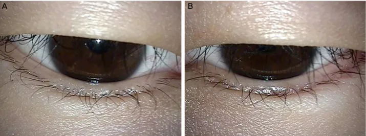
Figure 1. Clinical photograph of a 26 year-old woman with multiple, yellowish nodular lesion in the right upper (A), left upper (B),
right lower (C), left lower (D) eyelid.증례보고
26세 여자 환자가 양측 위, 아래 눈꺼풀의 종물을 주소로 내원하였다. 환자의 진술상 수 년 전부터 상기 부위에 종물 이 있었으며 크기의 변화는 없다고 하였다. 면역결핍질환 등의 전신적인 기저질환은 없었고, 이학적 검사상 양측 위, 아래 눈꺼풀에 진줏빛을 띤 여러 개의 포도송이 모양의 융 기된 병변이 관찰되었다(Fig. 1). 양안의 최대교정시력은 1.0이었고 안압, 안구운동검사, 전안부 및 안저검사상 특이 소견은 없었다.
임상소견상 작고, 진줏빛을 띠는 원형의 전염성 연속종 의 병변이 모여 있는 양상과 비슷하여, 전염성 연속종 의심 하에 수술을 계획하였다. 국소마취하에 위, 아래 눈꺼풀의 종물을 미세가위로 절제한 후 전기소작술을 시행하였다.
술 후 병리조직검사 결과 Hematoxylin & Eosin 염색에서 무정형의 호산성 물질이 관찰되었고, 면역조직화학 분석에 서는 Congo-red 염색에서 주황색으로 염색되는 아밀로이드
침착물이 관찰되었다(Fig. 2). 전신적인 검사에서 혈액학적 검사, 심전도 및 흉부 방사선검사에서는 특이소견이 보이 지 않았고, 이루 및 목쉼이 있어 시행한 이경 및 후두경검 사에서 성대 및 외이도를 침범한 아밀로이드 결절을 관찰 할 수 있었다. 이러한 소견을 종합하여 위, 아래 눈꺼풀에 서 발생한 일차성 전신성 아밀로이드증으로 진단하였다. 술 후 6개월까지 병변 부위는 재발 및 이상소견을 보이지 않았다(Fig. 3).
고 찰
아밀로이드증은 일차성 국소성 아밀로이드증, 일차성 전 신성 아밀로이드증, 만성질환에 의한 이차성 아밀로이드증, 다발성골수종에 의한 이차성 아밀로이드증, 가족형 아밀로 이드증 등 5가지로 분류된다.16 안와 및 주변조직을 침범하 는 아형은 대부분 일차성 국소성, 일차성 전신성 아밀로이 드증이며 각막, 결막, 유리체, 눈물샘, 외안근, 눈꺼풀 등 다
A B
C D
Figure 2. (A) Hematoxylin and eosin stained section (×200) shows amorphous, eosinophilic substances. (B) Congo-red stained sec-
tion (×200) reveals orange colored stained amyloid deposits.Figure 3. At the postoperative 6-month follow-up, there was no recurrence or inflammation at the operation site. (A) Right eyelid.
(B) Left eyelid.
양한 조직을 침범할 수 있다.17,18
일차성 국소성 아밀로이드증은 선행되는 질환 없이 신체 국소에 침착되는데 피부, 후두, 방광, 요도 등에 결절을 형 성하며 드물게 각막, 결막, 눈꺼풀 등에 결절을 형성하고 안검하수나 안구돌출을 보일 수 있다.19,20 일차성 전신성 아 밀로이드증은 선행 전신 및 국소 질환 없이 주로 근육, 피 부, 신경, 혈관 등을 침범하여 간, 비장, 신장, 부신 등에 침 착되어 심부전, 위장관 궤양 및 출혈 등을 일으켜 치명적인 결과를 취할 수 있다. 안구 침범은 드물지만 눈꺼풀, 유리 체, 외안근 등에 침착될 수 있어 눈꺼풀결절, 안검하수 등 을 유발할 수 있다.19,20
전염성 연속종은 폭스바이러스 감염에 의해 발생하며 피 부 및 점막조직에 다수의 결절을 형성한다. 병변은 대부분 작고, 가운데가 움푹 들어가 있는 양상을 보인다.21 전염성
연속종은 소아에서 흔하나, 최근에는 면역결핍질환이 있는 성인에서 발생빈도가 늘어나고 있다.22 눈꺼풀은 안구주위 전염성 연속종의 가장 흔한 발생 장소로 알려져 있고, 여포 각결막염을 동반하는 경우가 많다.21,23 본 증례의 종물은 작 고 원형의 진줏빛을 띠는 다수의 결절이 모여 포도송이 모 양을 띠고 있어, 전염성 연속종으로 오인할 수 있었다.
Nair et al24에 의해 보고된 바와 같이 눈꺼풀에 생긴 아밀 로이드증은 임상적으로 콩다래끼와 혼동될 수 있다. 하지 만 본 증례에서는 결막쪽에서 관찰하였을 때는 종물의 형 태가 보이지 않아 전형적인 콩다래끼의 임상소견과는 맞지 않으며, 따라서 비교적 쉽게 감별이 가능하였다.
눈꺼풀에 발생한 종물의 정확한 분석을 위해서는 면역조 직화학염색을 이용한 분석이 필요할 수 있다. 아밀로이드 증의 경우 Congo-red 염색에서 주황색으로 염색되는 아밀
A B
A B
로이드 결절을 관찰할 수 있고, 편광현미경으로 관찰했을 때 녹색의 복굴절 소견이 나타난다.25,26 본 증례의 경우 Congo-red 염색에서 주황색으로 염색되는 아밀로이드 결절 은 명확히 관찰할 수 있었지만, 편광현미경으로 관찰했을 때 녹색의 복굴절 소견은 저명하게 나타나지 않았다. 아밀 로이드증의 진단에는 환자의 증상, 기왕력, 이학적 검사, 조 직병리검사 등 종합적인 판단이 필요하기 때문에,26,27 본 증 례의 환자를 충분히 아밀로이드증으로 진단할 수 있었다.
아밀로이드증으로 진단된 경우는 국소적 질환인지 전신 적 질환인지를 감별하기 위해 전신적인 검사를 시행하는 것이 중요하다.11 혈청 내 단백질, 알부민 등을 포함한 감염 성 질환, 자가면역질환, 기타 만성 질환에 대한 혈액 및 요 검사가 필요하며 흉부 방사선검사, 심전도 검사 등을 시행 한다. 또한 내과, 피부과 등과의 협진을 통해 전신적 침범 여부를 파악해야 한다. 본 증례에서도 아밀로이드 확진 후 시행한 검진에서 전신 피부에 결절은 촉지되지 않았고 흉 부 방사선검사 및 심전도 검사에서도 정상 소견을 보였으 며 혈액 및 요검사에서 이상을 보이지 않았지만, 성대 및 외이도를 침범한 아밀로이드 결절이 발견되어 눈꺼풀을 침 범한 일차성 전신성 아밀로이드증으로 진단할 수 있었다.
국소성 또는 전신성 아밀로이드증의 치료 방법을 결정할 때에는 침범된 위치가 매우 중요하다.1 눈꺼풀 아밀로이드 증의 치료로는 단순 절제술, 침범된 눈꺼풀 조직을 포함한 광범위절제술, 침범된 결막 절제술, 방사선치료 등이 있
다.28,29 단순 절제술은 불완전한 절제 및 국소적인 재발의
위험성이 있고,16 광범위절제술은 수술 후 눈꺼풀의 구조 및 기능에 이상을 초래할 수 있어30 병변의 절제와 함께 전 기소작술, 냉동응고술, 선택적 소파술, 방사선치료 등을 함 께 이용하면 효과적으로 병변을 제거할 수 있다.31 본 증례 에서도 단순 절제술 및 전기소작술을 함께 이용하여 좋은 결과를 얻을 수 있었다. 또한 성공적인 수술적 절제 이후에 도 20-23%의 환자에서는 재발이 관찰되기 때문에31,32 지속 적인 경과관찰이 필요하다.
본 증례의 특징적 소견으로는 Congo-red 염색 후 편광현 미경으로 관찰했을 때 녹색의 복굴절 소견이 저명하게 나타 나지 않았다는 점이다. 하지만 Mollee et al26에 의하면 드문 경우에서 Congo-red 염색 후 편광현미경하에서 복굴절 소 견이 나타나지 않을 수 있고, 이런 경우 Lambda right chain 보다 kappa right chain이 축적되는 Light and heavy chain deposition disease를 의심할 수 있다고 하였다. 또한 Bozkurt et al27은 편광현미경 소견 대신 공초점현미경을 이용하여 아밀로이드 침착물을 관찰하여 아밀로이드증을 진단하였다.
본 증례에서는 편광현미경으로 관찰했을 때 복굴절 소견이 저명하지 않았고, 이에 추가적인 면역화학염색이나 공초점
현미경 검사를 시행하지 못했다는 점에 한계가 있다. 결론적으로, 눈꺼풀 아밀로이드증을 진단하고 성공적으 로 치료하였기에 이를 보고하는 바이며, 눈꺼풀 종물의 감 별진단 중 하나로 아밀로이드증도 고려하여야 할 것이다.
REFERENCES
1) Aryasit O, Preechawai P, Kayasut K. Clinical presentation, treat- ment, and prognosis of periocular and orbital amyloidosis in a uni- versity-based referral center. Clin Ophthalmol 2013;7:801-5.
2) Tyradellis C, Peponis V, Kulwin DR. Surgical management of re- current localized eyelid amyloidosis. Ophthal Plast Reconstr Surg 2006;22:308-9.
3) Gean-Marton AD, Kirsch CF, Vezina LG, Weber AL. Focal amy- loidosis of the head and neck: evaluation with CT and MR imaging.
Radiology 1991;181:521-5.
4) Chung EH, Oum BS, Lee SH, Hong SH. A case of corneal amyloidosis. J Korean Ophthalmol Soc 1983;24:383-6.
5) Kim DS, Kim CH, Kim YK. A case of corneal amyloidosis. J Korean Ophthalmol Soc 1996;37:554-8.
6) Park YH, Lee JY, Chung SK, Myung YW. Corneal amyloidosis de- veloped after penetrating keratoplasty. J Korean Ophthalmol Soc 1998;39:3093-7.
7) Nam H, Cho KR. Primary systemic amyloidosis involved the conjunctiva. J Korean Ophthalmol Soc 1991;32:604-8.
8) Song BR, Kim YK, Yoo JH, Chu YC. Primary localized amyloi- dosis of bulbar conjunctiva and cornea. J Korean Ophthalmol Soc 1993;34:352-6.
9) Lee JH, Kim YD. Primary localized orbital amyloidosis. J Korean Ophthalmol Soc 2001;42:1793-7.
10) Kim WT, Kim JS, Park HB. A case of vitreous amyloidosis. J Korean Ophthalmol Soc 2003;44:1948-53.
11) Choi HJ, Choung HK, Khwarg SI. Primary localized amyloidosis of the lacrimal gland. J Korean Ophthalmol Soc 2004;45:1567-72.
12) Kim YT, Kim JY, Kim YD. Primary localized amyloidosis of the lacrimal gland. J Korean Ophthalmol Soc 2004;45:1573-7.
13) Kim JT, Kim EY, Lee HI, et al. A report of localized corneal amy- loidosis secondary to the trichiasis. J Korean Ophthalmol Soc 2006;47:2035-40.
14) Kim BY, Song JH, Yang SW, Kim MS. A case of bilateral con- junctiva amyloidosis treated with mass excision and cryotherapy. J Korean Ophthalmol Soc 2011;52:628-32.
15) Lyu IJ, Woo KI, Kim YD. Primary orbital MALT lymphoma asso- ciated with localized amyloidosis. J Korean Ophthalmol Soc 2013;54:1109-13.
16) Caggiati A, Campanella A, Tenna S, et al. Primary amyloidosis of the eyelid: a case report. In Vivo 2010;24:575-8.
17) Brownstein MH, Elliott R, Helwig EB. Ophthalmologic aspects of amyloidosis. Am J Ophthalmol 1970;69:423-30.
18) Kosch G, Meyer-Rüsenberg HW. Primary localized amyloidosis of the eyelid and conjunctiva. Klin Monbl Augenheilkd 1993;202:
56-9.
19) George TF, John SL, Ronald ES. The cornea in systemic disease.
In: Tasman W, Jaeger EA, eds. Duane’s clinical ophthalmology, re- vised ed. Philadelphia: JB Lippincott, 1993; chap. 15.
= 국문초록 =
눈꺼풀에 발생한 일차성 전신성 아밀로이드증 1예
목적: 눈꺼풀에 발생한 아밀로이드증은 드문 질환으로 아직까지 국내에 보고된 바 없다. 저자들은 눈꺼풀에서 발생한 일차성 전신성 아밀로이드증 1예를 경험하였기에 이를 보고하고자 한다.
증례요약: 26세 여자 환자가 양측 위, 아래 눈꺼풀의 종물을 주소로 내원하였다. 환자의 진술상 수 년 전부터 상기 부위에 종물이 있었으며 크기의 변화는 없다고 하였다. 이학적 검사상 양측 위, 아래 눈꺼풀에 진줏빛을 띤 여러 개의 포도송이 모양의 융기된 병변 이 관찰되었다. 전염성 연속종 의심하에 양측 눈꺼풀의 병변에 대한 절제생검 및 전기소작술을 시행하였다. 조직병리검사 결과 Hematoxylin & Eosin 염색에서 무정형의 호산성 물질이 관찰되었고, Congo-red 염색에서 주황색으로 염색되는 아밀로이드 침착물 이 관찰되었다. 전신적인 검사에서 성대 및 외이도에 침착된 아밀로이드 결절이 있어 일차성 전신성 아밀로이드증으로 진단할 수 있었다. 수술 부위는 6개월째 염증이나 재발 없이 안정적으로 유지되었다.
결론: 눈꺼풀에 발생한 아밀로이드증은 드물지만 종물의 감별 진단 시 고려해야 하며, 절제술 및 전기소작술에 의해 성공적으로 치료 될 수 있다.
<대한안과학회지 2015;56(7):1117-1121>
20) Edward LH, Narsing AR. Basic mechanisms in pathology. In:
Spencer WH, ed. Ophthalmic pathology: an atlas and textbook, re- vised ed. Philadelphia: WB Saunders, 1990; chap. 13.
21) Schornack MM, Siemsen DW, Bradley EA, et al. Ocular manifes- tations of molluscum contagiosum. Clin Exp Optom 2006;89:
390-3.
22) Massa AF, Borges-Costa J, Soares-Almeida L, Sacramento-Marques M. Molluscum contagiosum eyelid lesions in an HIV-patient.
Dermatol Online J 2013;19:10.
23) Ingraham HJ, Schoenleber DB. Epibulbar molluscum conta- giosum. Am J Ophthalmol 1998;125:394-6.
24) Nair AG, Mukherjee B, Krishnakumar S, et al. Unilateral primary cutaneous amyloidosis of the eyelid masquerading as a chalazion.
Can J Ophthalmol 2014;49:e112-4.
25) Picken MM. Amyloidosis-where are we now and where are we heading? Arch Pathol Lab Med 2010;134:545-51.
26) Mollee P, Renaut P, Gottlieb D, Goodman H. How to diagnose amyloidosis. Intern Med J 2014;44:7-17.
27) Bozkurt B, Kiratli H, Soylemezoglu F, Irkec M. In vivo confocal microscopy in a patient with conjunctival amyloidosis. Clin Experiment Ophthalmol 2008;36:173-5.
28) Patrinely JR, Koch DD. Surgical management of advanced ocular adnexal amyloidosis. Arch Ophthalmol 1992;110:882-5.
29) Stack RR, Vote BJ, Evans JL, Elder MJ. Bilateral ptosis caused by localized superficial eyelid amyloidosis. Ophthal Plast Reconstr Surg 2003;19:239-40.
30) Iijima S. Primary systemic amyloidosis: a unique case complaining of diffuse eyelid swelling and conjunctival involvement. J Dermatol 1992;19:113-8.
31) Demirci H, Shields CL, Eagle RC Jr, Shields JA. Conjunctival amyloidosis: report of six cases and review of the literature. Surv Ophthalmol 2006;51:419-33.
32) Leibovitch I, Selva D, Goldberg RA, et al. Periocular and orbital amyloidosis: clinical characteristics, management, and outcome.
Ophthalmology 2006;113:1657-64.