Atul Goel
430 Asian Spine J 2016;10(3):430-435
C2–3 Fusion, C3–4 Cord Compression and C1–2 Posterior Facetal Instability:
An Evaluation of Treatment Strategy Based on Four Surgically Treated Cases
Atul Goel
Department of Neurosurgery, K.E.M. Hospital and Seth G.S. Medical College, Parel, India
Study Design: Four patients had C2–3 vertebral fusion and radiologically demonstrated cord compression at C3–4 level related to disc bulge with or without association of osteophytes and C1–2 posterior facetal dislocation. The outcome of treatment by atlanto- axial and subaxial facetal fixation is discussed.
Purpose: The article evaluates the significance of atlantoaxial facetal instability in cases having C2–3 vertebral fusion and cord com- pression at the level of C3–4 disc.
Overview of Literature: C2–3 vertebral fusions are frequently encountered in association with basilar invagination and chornic atlantoaxial dislocations. Even when basilar invagination and atlantoaxial dislocation are not identified by conventional parameters, atlantoaxial instability can be the nodal point of pathogenesis in cases with C2–3 vertebral fusion.
Methods: Between June 2013 and November 2014 four patients having C2–3 fusion presented with progressive symptoms of my- elopathy that were related to cord compression at the level opposite the C3–4 disc space. Further investigations revealed C1–2 pos- terior facetal dislocation.
Results: All patients were males. Ages ranged from 18 to 50 years (average, 36 years). All patients were treated by atlantoaxial facetal plate and screw, and subaxial single or multi-segmental transarticular screw fixation. Follow-up (average, 15 months) using a recently described clinical grading system and the Japanese Orthopaedic Association scoring system confirmed marked improvement of symptoms.
Conclusions: Identification and treatment of atlantoaxial facetal instability may be crucial for a successful outcome in cases having C2–3 fusion and high cervical (C3–4) disc related cord compression.
Keywords: Atlantoaxial dislocation; High cervical disc prolapse; Transarticular fixation
Copyright Ⓒ 2016 by Korean Society of Spine Surgery
This is an Open Access article distributed under the terms of the Creative Commons Attribution Non-Commercial License (http://creativecommons.org/licenses/by-nc/3.0/) which permits unrestricted non-commercial use, distribution, and reproduction in any medium, provided the original work is properly cited.
Asian Spine Journal • pISSN 1976-1902 eISSN 1976-7846 • www.asianspinejournal.org
Received Sep 16, 2015; Revised Sep 17, 2015; Accepted Sep 17, 2015 Corresponding author: Atul Goel
Department of Neurosurgery, K.E.M. Hospital and Seth G.S. Medical College, Parel, Mumbai-400012, India Tel: +91-22-24129884, Fax: +91-22-24143435, E-mail: [email protected]
ASJ A SJ
Clinical Study Asian Spine J 2016;10(3):430-435 • http://dx.doi.org/10.4184/asj.2016.10.3.430
Introduction
The author presents clinical details of four patients who had C2–3 vertebral body fusion and cord compression
opposite the C3–4 disc space. All patients had posterior atlantoaxial facetal instability [1]. There was no radio- graphic evidence of odontoid process related instability as traditionally assessed by an increase in atlantodental in-
terval or cord compression at the craniovertebral junction.
Treatment by atlantoaxial and subaxial single or multi- segmental facetal fixation resulted in remarkable recovery from symptoms of myelopathy. The treatment philosophy is discussed.
Materials and Methods
From June 2013 to November 2014, four patients present- ed with symptoms related to cervical myelopathy. Investi- gations revealed C2–3 vertebral fusion and predominant cord compression opposite C3–4 disc space. The four patients were evaluated.
Results
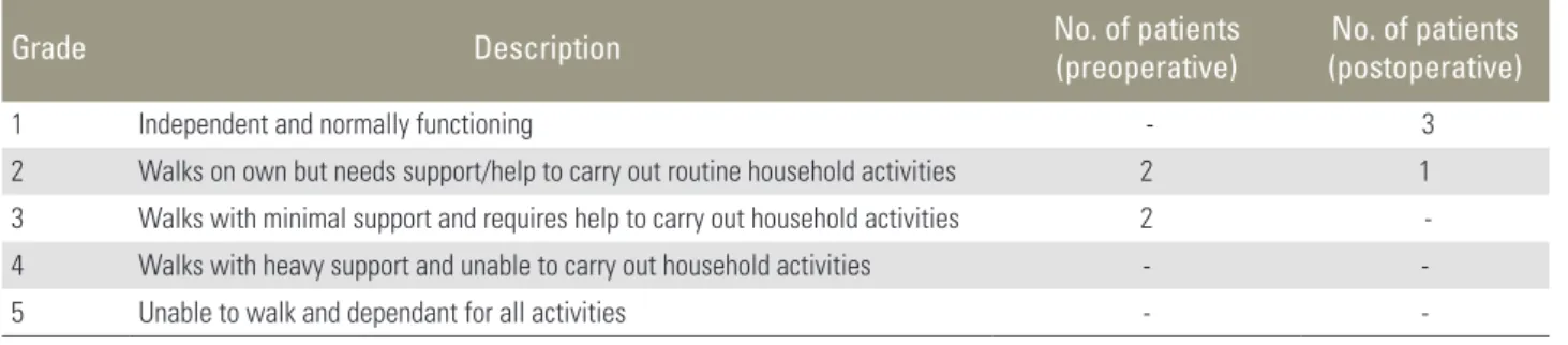
All patients were males. The demographic and radiologi- cal details are summarized in Table 1. Our recently dis- cussed clinical grading system(Table 2) [2] and Japanese Orthopaedic Association scoring system (Table 3) [3]
were used to monitor neurological status both before and after surgery. Preoperative investigations included dynam- ic computed tomography (CT) scan, magnetic resonance imaging (MRI) and plain radiological images in flexion
and extension neck position. Investigations showed no, insignificant or only marginal cord compression at the level of odontoid process (Fig. 1). The atlantodental in- terval was within the range of normal. Cervical imaging revealed C2–3 fusion and most prominent cord compres- sion opposite C3–4 disc space. Moderate cord compres- sion at C4–5 level was seen in 3 cases and opposite C5–6 level disc space in 1 case. On reviewing the CT and MRI slices focused on C1–2 facets, posterior atlantoaxial fac- etal (Type B) dislocation was evident in all cases [1] (Fig.
1). Additional radiological findings included occipitaliza- tion of the atlas in one case and presence of bifid posterior arch of atlas in one case. All patients underwent surgery Table 1. Table showing the presenting demographic and radiological features
Patient no. Sex/age
(yr) Radiological findings Posterior atlantoaxial
instability Other
anomalies Levels fixed 1. Male/39 C2–3 fusion with cord compression
at C3–4 level Present - C1–2, C3–4 and
C4–5 2. Male/18 C2–3 fusion with cord compression
at C3–4, C4–5 and C5–6 levels Present Occipitalization
of atlas C1–2, C3–4, C4–5 and C5–6 levels 3. Male/36 C2–3 fusion with cord compression
at C3–4 and C4–5 levels Present - C1–2, C3–4 and
C4–5 4. Male/50 C2–3 fusion with cord compression
at C3–4 and C4–5 levels
Present Bifid posterior arch of atlas
C1–2, C3–4 and C4–5
Table 2. Distribution as per our clinical grading system
Grade Description No. of patients
(preoperative) No. of patients (postoperative)
1 Independent and normally functioning - 3
2 Walks on own but needs support/help to carry out routine household activities 2 1
3 Walks with minimal support and requires help to carry out household activities 2 -
4 Walks with heavy support and unable to carry out household activities - -
5 Unable to walk and dependant for all activities - -
Table 3. Table showing the preoperative and postoperative clinical assessment as per JOA scoring system
JOA score Preoperative
(no. of patients) Postoperative (no. of patients)
<7 - 4
8–12 4 -
13–15 - -
16–17 - -
JOA, Japanese Orthopaedic Association.
by posterior cervical route in prone position. Cervical traction was applied after induction of anesthesia and prior to turning the patient. The head end of the table was elevated by 30 degrees. Head elevation allowed direct sur- gical access to the craniovertebral junction and reduced venous congestion in the operative field. Cervical traction was applied basically to keep the head steady during the conduct of surgery and keep the pressure off the face and eyes. Atlantoaxial fixation was done using lateral mass plate and screws with the technique described by us in 1994 [4,5]. The atlantoaxial joint was widely exposed. The
articular cartilage was denuded using power driven drills and the joint cavity was stuffed with bone graft harvested from iliac crest. The C2 ganglion was either sectioned or was elevated superiorly to have a wide exposure of the field and to conduct the instrumentation. C3–4, C4–5, and C5–6 facets were exposed and inspected by physi- cal manual handling. Instability of the facets was clearly identified during surgery at the C3–4 level. Additionally, facetal instability was observed in all four patients at the C4–5 level and in one patient at the C5–6 level. Of these cases, instability was identified and treated at the C4–5
Fig. 1. Images of a 50-year-old male patient. (A) T2-weighted magnetic resonance imaging (MRI) shows C2–3 fusion and altered cord signal opposite C3–4 disc space. There is no evidence of any cord compression at the level of tip of odontoid process. (B) Sagittal cut of MRI at the level of facets showing Goel type B atlantoaxial facetal instability. (C) Computed tomography (CT) scan showing the C2–3 fusion. (D) Three-dimensional (3D) CT scan of the cervical spine showing the fusion of C2–3 vertebrae and the facets. (E) Posterior view of the 3D CT scan showing the bifid arch of atlas. (F) Postoperative CT scan findings. (G) Postoperative scan shows fixation of C1–2 (lateral mass plate and screw technique) and C3–4 and C4–5 by transarticular method of fixation.
A B C
D
E
F G
level in one case despite the fact that radiological images did not show significant cord compression at these levels.
Transarticular screw fixation was done at all the subaxial levels by deploying recommended parameters [6]. A suit- able size osteotome was introduced into the joint cavity with its flat edge and turned by 90 degrees in a screw- ing fashion to affect denuding of the articular cartilage.
Transarticular fixation was then completed. Screws used for transarticular fixation averaged 2.7 mm in diameter and 14 mm in length. All the soft tissues and ligaments attached to the spinous process were sharply sectioned and removed. Spinous processes at the levels of surgery were sectioned at the base. Bone material of the spinous processes was meshed into thin pieces and placed over the midline and over the instrumented facets after appropri- ately preparing the host bone. Additional bone graft mate- rial was obtained from the iliac crest. Postoperatively, the patient used hard cervical collar for 8 weeks after surgery and neck movement was kept to a minimum. Postopera- tive investigations were done 3 weeks and 3 months after surgery. Subsequent imaging was done after one year. Fol- low-up for the four cases was at 6, 12, 18, and 24 months.
During follow-up, all patients improved significantly in neurological function (Tables 2, 3). There were no infec- tion- or implant-related mechanical problems.
Discussion
C2–3 vertebral bone fusion is relatively a frequent obser- vation in cases with craniovertebral instability in general and basilar invagination in particular. In 2009, we present- ed a series of 170 cases having basilar invagination and identified 44 cases having C2–3 vertebral fusion [7]. In the article, we concluded that musculoskeletal changes that included bone fusions were secondary to primary atlanto- axial instability. Increase in neck height, reversal of torti- collis, reversal of platybasia and a number of other features that were associated with postoperative craniovertebral realignment were identified after the atlantoaxial joint was fixed and stabilized. We also discussed the potential of re- versal of secondary osteophytic changes and bone fusions following atlantoaxial joint stabilization surgery.
C3–4 disc-related cord compression is frequently iden- tified in cases with basilar invagination and atlantoaxial dislocation. In 2004 and 2008, we identified the associa- tion of basilar invagination and atlantoaxial instability and C3–4 disc related cord compression [8,9]. We discussed
that craniovertebral junction instability was the primary pathology and cervical spondylotic changes in general and C3–4 disc disease in particular were secondary spinal alterations that are designed to affect shortening of the neck size. We suggested the need for atlantoaxial stabiliza- tion as the treatment for such a condition and advocated that there was no necessity of surgically dealing with the spinal spondylotic alterations [8,9].
In the present cases, C2–3 vertebral body fusion and C3–4 disc-osteophyte-related cord compression were evi- dent. There was no or insignificant basilar invagination. In one patient there was assimilation of atlas and in one case there was presence of bifid posterior arch of atlas. Both features suggested a more generalized spinal malformation that included a craniovertebral junctional anomaly. There was no evidence of cord compression or compromise of spinal canal dimensions related to odontoid process.
Also, conventional dynamic flexion-extension imaging did not reveal any abnormal movements of the odontoid process that could suggest instability. As per our recently described classification of atlantoaxial facetal dislocation and instability[10], all patients had Type B dislocation, wherein the facet of atlas was dislocated posterior to the facet of axis. In our publications related to basilar invagi- nation, Chiari malformation, syringomyelia and atlanto- axial dislocation, we identified that Type B and Type C or central or axial facetal dislocation (the facets are in align- ment, but there is atlantoaxial instability that is identified and confirmed only during surgery) are associated with more chronic forms of craniovertebral anomalies [2,11].
The symptom complex is subtle and longstanding as there is no direct compression of the cord by the odontoid pro- cess. This is in variance with Type A facetal dislocation, wherein the facet of atlas is dislocated anterior to the facet of axis, a process that results in atlantoaxial dislocation and basilar invagination and posterior migration of odon- toid process into the spinal canal and resultant cord com- pression. Despite the fact that there is no direct odontoid process related neural compression in Type B and Type C atlantoaxial facetal dislocation, the entire musculoskeletal and neural transformation that includes Chiari malforma- tion and syringomyelia is related to atlantoaxial instability [2,11]. Atlantoaxial fixation results in relatively rapid re- versal of neurological symptoms and potential or manifest reversal of all musculoskeletal and neural malformations [7]. We recently identified the presence of atlantoaxial facetal instability in cases with degenerative cervical spon-
dyltoic myelopathy. Despite the fact that there was no cord compression related to odontoid process, we recom- mended atlantoaxial fixation in such cases and concluded that missing atlantoaxial instability in such cases can be a cause of failure of surgical treatment [1].
Although there are no morphological or radiological studies that discuss the relative alignment of facet of atlas and facet of axis, it does seem that some degree of mal- alignment may be in the range of normalcy. It appears that the facetal instability should be viewed in a wider perspective and clinical interpretations should be based on association of a number of other morphological and clinical factors. Our classification of atlantoaxial instability and basilar invagination based on the type of facetal mal- alignment, rather than the conventional evaluation of the region based on odontoid process related cord compres- sion, presents an alternative format of understanding of instability at the craniovertebral junction. Drawing on our long-term experience spanning over 25 years and dealing with over 1,400 cases having instability of the atlantoaxial joint and treatment by direct manipulation of facets, we identified that there was evidence of marked instability of atlantoaxial joint in Types B and C dislocation. Stabiliza- tion of the atlantoaxial joint forms the basis of treatment.
In all four patients, there was evidence of Type B atlan- toaxial facetal dislocation. The association of C2–3 fusion and C3–4 disc disease did not appear incidental in these cases. In all four cases, facetal fixation of C3–4 was done in addition to atlantoaxial fixation. As with our experi- ence in cases with basilar invagination and high cervical discs wherein we suggested that only atlantoaxial fixa- tion is sufficient and dealing with the cervical disc related cord compression is not necessary, it is unclear if only atlantoaxial fixation would have sufficed in the presented cases. Instability was identified by direct observations and manual handling of both atlantoaxial and C3–4 facets joint [12-16]. Stabilization resulted in remarkable recov- ery of symptoms that begun in the immediate postopera- tive phase and was progressive. Although our experience is limited, it does appear that ignoring of either of the unstable joints at C1–2 or at C3–4 could have adversely affected the clinical outcome. Despite our successful clini- cal outcome, it is clear that blinded analysis is required to assess the individual roles of atlantoaxial fixation and subaxial cervical spinal fixation. However, the relative rar- ity of such cases may make adoption of research protocols difficult.
Conclusions
Identification and treatment of atlantoaxial instability despite absence of odontoid process related cord compres- sion can be a defining feature in success of surgical treat- ment in the complex clinical scenario of C2–3 bone fusion and C3–4 disc related cord compression.
Conflict of Interest
No potential conflict of interest relevant to this article was reported.
References
1. Goel A. Posterior atlantoaxial ‘facetal’ instability as- sociated with cervical spondylotic disease. J Cranio- vertebr Junction Spine 2015;6:51-5.
2. Goel A. Is atlantoaxial instability the cause of Chiari malformation? Outcome analysis of 65 patients treated by atlantoaxial fixation. J Neurosurg Spine 2015;22:
116-27.
3. Fujiwara A, Kobayashi N, Saiki K, Kitagawa T, Tamai K, Saotome K. Association of the Japanese Orthopae- dic Association score with the Oswestry Disability Index, Roland-Morris Disability Questionnaire, and short-form 36. Spine (Phila Pa 1976) 2003;28:1601-7.
4. Goel A, Desai KI, Muzumdar DP. Atlantoaxial fixa- tion using plate and screw method: a report of 160 treated patients. Neurosurgery 2002;51:1351-6.
5. Goel A, Laheri V. Plate and screw fixation for atlanto- axial subluxation. Acta Neurochir (Wien) 1994;129:
47-53.
6. Roy-Camille R, Saillant G. Surgery of the cervical spine: 2. Dislocation. Fracture of the articular pro- cesses. Nouv Presse Med 1972;1:2484-5.
7. Goel A, Shah A. Reversal of longstanding musculo- skeletal changes in basilar invagination after surgical decompression and stabilization. J Neurosurg Spine 2009;10:220-7.
8. Goel A. High cervical C3-4 ‘disc’ compression as- sociated with basilar invagination. Neurol India 2008;56:68-70.
9. Goel A, Phalke U, Cacciola F, Muzumdar D. Surgical management of high cervical disc prolapse associated with basilar invagination: two case reports. Neurol Med Chir (Tokyo) 2004;44:142-5.
10. Goel A. Goel’s classification of atlantoaxial “facetal”
dislocation. J Craniovertebr Junction Spine 2014;5:3- 8.
11. Goel A. Is Chiari malformation nature’s protective
“air-bag”? Is its presence diagnostic of atlantoaxial in- stability? J Craniovertebr Junction Spine 2014;5:107- 9.
12. Goel A. Facet distraction spacers for treatment of degenerative disease of the spine: rationale and an alternative hypothesis of spinal degeneration. J Cra- niovertebr Junction Spine 2010;1:65-6.
13. Goel A. Facet distraction-arthrodesis technique: Can it revolutionize spinal stabilization methods? J Cra-
niovertebr Junction Spine 2011;2:1-2.
14. Goel A. Only fixation for cervical spondylosis: report of early results with a preliminary experience with 6 cases. J Craniovertebr Junction Spine 2013;4:64-8.
15. Goel A, Shah A, Gupta SR. Craniovertebral instabil- ity due to degenerative osteoarthritis of the atlanto- axial joints: analysis of the management of 108 cases.
J Neurosurg Spine 2010;12:592-601.
16. Goel A, Shah A. Facetal distraction as treatment for single- and multilevel cervical spondylotic radicu- lopathy and myelopathy: a preliminary report. J Neu- rosurg Spine 2011;14:689-96.