INTRODUCTION
Coronary computed tomography angiography (CCTA) has
been widely used as a non-invasive assessment in patients with stable coronary artery disease (CAD). Its utility is attributed to intuitive visualization of the coronary artery lumen, which has been deemed highly sensitive for either detecting an obstruc- tive coronary lesion in stable CAD1 or preclusion from acute coronary syndrome in the emergency department setting.2 Advancement in imaging technology and constant clinical val- idation in recent years have justified the role of CCTA in the diagnostic approach in patients with suspected CAD.3 Al- though there are some concerns and disputes about the ap- propriate use and interpretation of CCTA, additional use of CCTA was found to play a positive role in the assessment and management of CAD.3 However, further investigation is need-
Clinical Implications of Moderate Coronary Stenosis on Coronary Computed Tomography Angiography in Patients with Stable Angina
Choongki Kim1, Sung-Jin Hong1, Chul-Min Ahn1,2, Jung-Sun Kim1,2, Byeong-Keuk Kim1,2, Young-Guk Ko1,2, Byoung-Wook Choi3,4, Donghoon Choi1,2, Yangsoo Jang1,2, and Myeong-Ki Hong1,2
Divisions of 1Cardiology and 3Cardiovascular Radiology, Severance Cardiovascular Hospital, Yonsei University College of Medicine, Seoul;
2Cardiovascular Research Institute, Yonsei University College of Medicine, Seoul;
4Department of Radiology, Yonsei University College of Medicine, Seoul, Korea.
Purpose: The present study investigated the diagnostic accuracy and clinical implications of moderate stenosis (50–69%, Coro- nary Artery Disease Reporting and Data System, grade 3) on coronary computed tomography angiography (CCTA), compared with invasive coronary angiography (ICA).
Materials and Methods: Two hundred and seventy-six patients who underwent ICA due to moderate stenosis alone on CCTA were selected from our prospective registry cohort.
Results: Diagnostic concordance between CCTA and ICA was found in only 50 (18%) patients. Among the 396 vessels and 508 segments with moderate stenosis, diagnostic concordance was found in 132 vessels (33%) and 127 segments (25%). Segments with calcified plaque had lower diagnostic concordance than those with mixed or non-calcified plaque (22% vs. 28% vs. 27%, re- spectively, p=0.001). While calcified plaque burden did not have an influence on severe stenosis (≥70%) on ICA, higher burden of non-calcified plaque was correlated with a greater incidence of ICA-based severe stenosis, which was more frequent in patients with ≥3 segments of non-calcified plaque (75%) than those without non-calcified plaque (22%, p<0.001). Typical angina and mixed or non-calcified plaque were correlated with a higher incidence of under-diagnosis, while the use of next-generation computed to- mography scanners reduced the incidence of under-diagnosis. Increased body weight, left circumflex artery involvement, and calcified plaque were independent factors that increased the risk of over-diagnosis of CCTA.
Conclusion: The diagnosis of moderate stenosis by CCTA may be limited in estimating the exact degree of ICA-based anatomical stenosis. Unlike calcific burden, non-calcific burden was positively correlated with the presence of severe stenosis on ICA.
Key Words: Coronary artery disease, coronary stenosis, computed tomography angiography
pISSN: 0513-5796 · eISSN: 1976-2437
Received: May 3, 2018 Revised: July 12, 2018 Accepted: August 2, 2018
Corresponding author: Myeong-Ki Hong, MD, PhD, Division of Cardiology, Sev- erance Cardiovascular Hospital, Yonsei University College of Medicine, 50-1 Yon- sei-ro, Seodaemun-gu, Seoul 03722, Korea.
Tel: 82-2-2228-8458, Fax: 82-2-2227-7943, E-mail: [email protected]
•The authors have no financial conflicts of interest.
© Copyright: Yonsei University College of Medicine 2018
This is an Open Access article distributed under the terms of the Creative Com- mons Attribution Non-Commercial License (https://creativecommons.org/licenses/
by-nc/4.0) which permits unrestricted non-commercial use, distribution, and repro- duction in any medium, provided the original work is properly cited.
Yonsei Med J 2018 Oct;59(8):937-944 https://doi.org/10.3349/ymj.2018.59.8.937
ed to ascertain how general physicians or cardiologists can utilize CCTA findings in decision-making in the cardiac cathe- terization laboratory. A recent consensus document drew rec- ommendations for future strategies according to degrees of maximal coronary stenosis on CCTA.4 However, these recom- mendations were not based on objective validation. In particu- lar, moderate stenosis on CCTA may be a worrisome and con- fusing finding for physicians, as it implies obstructive CAD, but not severe anatomical stenosis. Therefore, we sought to inves- tigate the diagnostic accuracy of CCTA in patients with seg- ments with moderate stenosis alone on CCTA. The CCTA re- sults were compared with the results of invasive coronary angiography (ICA), which was utilized as a confirmative study.
MATERIALS AND METHODS
Study population
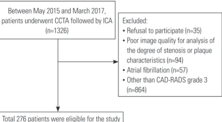
A prospective, real-world registry was used for the consecu- tive enrollment of all patients with suspicious, stable ischemic heart disease who underwent ICA for the diagnosis of CAD within 3 months after CCTA was performed. Data from 1326 patients were added to the registry between May 2015 and March 2017. We included 276 individuals who were classified as Coronary Artery Disease Reporting and Data System (CAD- RADS) grade 3. Participants had one or more segments with a maximal degree of stenosis of 50–69% in diameter in the epi- cardial arteries, excluding the left main trunk (Fig. 1).4 Pretest probability was estimated using the Duke Clinical Score.5 The study protocol was approved by the Institutional Review Board of the two institutions (1-2013-0084). Written informed con- sent was obtained from all patients.
Acquisition and assessment of coronary computed tomographic angiography
CCTA images were acquired with conventional 64-detector- row scanners (SOMATOM Sensation 64, Siemens, Forchheim, Germany; Philips Brilliance 64, Philips Medical System, Best, the Netherlands; LightSpeed VCT and CT750 HD, GE Health- care, Waukesha, WI, USA) and next generation 128 or 256-de- tector-row scanners (SOMATOM Definition Flash, Siemens;
Revolution CT, GE Healthcare). The protocol recommended by the Society of Cardiovascular Computed Tomography at the time each scan was used as the standard protocol for im- age acquisition at the two institutions.6 One to two hours be- fore the CCTA examination, oral metoprolol was administered to patients with a baseline heart rate (HR) ≥70 beats/min in the absence of any contraindications. Sublingual nitroglycerin (0.3 mg to 0.6 mg) was administered immediately before con- trast injection. Patients with a regular HR underwent CCTA scanning under prospectively electrocardiogram (ECG)-trig- gered axial mode. Spiral mode, if offered, was utilized if the HR was <60 beats/min. Retrospectively ECG-gated scanning
was applied during image acquisition if a patient had frequent irregular beats or a higher HR. A bolus of 60 mL to 80 mL of io- pamidol was injected into the antecubital vein using a triple- phase injection method, followed by 30 mL of 30% blended io- pamidol and a 20 mL saline flush at a 5 mL/s flow rate. The optimal delay times were determined using automatic evalua- tion of the enhancement of the ascending aorta.
The coronary arteries were divided into 15 segments follow- ing the defined tree model.7 Segments with a >2.0 mm diame- ter were visually evaluated at a core laboratory (Severance Cardiovascular Hospital, Seoul, Korea) by a single experienced radiologist (BWC, 17 years of experience) who was blinded to patient and coronary angiographic information. A cardiolo- gist (CK, 4 years of experience) assessed reliability and repro- ducibility in 30 randomly chosen patients (55 lesions). Any of the following available post-processed reconstructed images were used for the assessment of coronary artery stenosis: two- dimensional axial, three-dimensional maximal intensity pro- jection, multi-planar reformat, cross-sectional analysis, or the volume-rendered technique using a three-dimensional com- puted tomography (CT) workstation (Wizard, Siemens Medi- cal Solutions, Erlangen, Germany).8
The degree of stenosis was classified by a quantitative ste- nosis grading system.4 CAD-RADS recommends reporting stenosis from grade 0 (absence of atherosclerosis) to 5 (pres- ence of at least one total occlusion) based on the maximal coronary stenosis. All coronary plaques with maximal steno- sis at each segment were classified into one of three categories:
calcified (plaque with high CT attenuation compared to the contrast-enhanced lumen), mixed (non-calcified and calcified elements in a single plaque), or non-calcified plaque (plaque with lower CT attenuation compared to the contrast-enhanced lumen without any evidence of calcification).9,10 Overall imag- ing quality was also qualitatively assessed as follows: 1) opti- mal images, clear delineation without motion artifact, minor motion artifacts, or blurring, but diagnostic in quality, without phase change; 2) suboptimal images, moderate to severe arti- fact or blurring, but evaluable, using different phase or addi-
Between May 2015 and March 2017, patients underwent CCTA followed by ICA
(n=1326)
Excluded:
• Refusal to participate (n=35)
• Poor image quality for analysis of the degree of stenosis or plaque characteristics (n=94)
• Atrial fibrillation (n=57)
• Other than CAD-RADS grade 3 (n=864)
Total 276 patients were eligible for the study
Fig. 1. Study flow. CAD-RADS, Coronary Artery Disease Reporting and Data System; CCTA, coronary computed tomography angiography; ICA, invasive coronary angiography.
tional manual reconstruction; and 3) unevaluable images, which were excluded in the current study.11 We used the seg- ment involvement score (SIS), which indicates the number of segments (per patient) with any coronary plaque. We calcu- lated calcified and non-calcified SISs separately by counting the numbers of calcified and non-calcified plaques, respectively.12 Invasive coronary angiography and revascularization All eligible patients underwent ICA a median of 17 days (inter- quartile range 8–41 days) after the initial CCTA examination.
The degree of coronary stenosis at each segment was visually assessed by interventional cardiologists in the catheterization laboratory. Analysts at an independent core laboratory (Car- diovascular Research Center, Seoul, Korea), who were blinded to the findings of the initial assessment and patient informa- tion, reviewed the ICA findings. Comparing the findings of ICA and CCTA, the analysts verified stenosis of any corresponding lesion identifiable on CCTA. Discrepant results were evaluat- ed by the investigators (CK, SJH, and MKH) who reached a consensus concerning the degree of stenosis while continuing to be blinded to patient information. Quantitative coronary an- giography analysis was also performed in 30 randomly chosen patients using an offline computerized quantitative coronary angiographic system (CASS system, Pie Medical Instruments, Maastricht, the Netherlands) for assessment of agreement with visual estimation. The minimal lumen diameter and ref- erence diameters of coronary lesions were measured in the view with the narrowest lumen and the least amount of foreshort- ening by comparison to the diameter of a guidance catheter from diastolic frames in a single, matched view. We compared the CCTA- and ICA-derived diagnoses for each patient. The diagnoses were analyzed and confirmed as concordant at the patient level if the degree of stenosis was matched for all ma- jor epicardial arteries. The CCTA-derived diagnosis was de- fined as under-diagnosis if the maximal stenosis of a vessel or segment determined by ICA was ≥70% of the diameter steno- sis at the same location on CCTA. Correspondingly, the CCTA- derived diagnosis was defined as over-diagnosis if the maximal stenosis was less than 50% of the diameter stenosis determined by ICA. At the patient-level, under-diagnosis of CCTA was de- fined as greater numbers of vessels with stenosis ≥50% or the presence of stenosis ≥70% in any vessels; over-diagnosis was defined as fewer numbers of vessels with stenosis ≥50% or no stenosis ≥50% in any vessels.
Statistical analysis
Categorical data are expressed as numbers (%) and were ana- lyzed with chi-square statistics or Fisher’s exact test. Continu- ous variables are expressed as medians (interquartile range) because of a skewed distribution. To compare differences in diagnostic concordance, patients were categorized by quar- tiles of calcified or non-calcified SIS and Agatston calcium score. A generalized linear mixed model was used to deter-
mine under- or over-diagnosis of moderate stenosis found on CCTA, considering the random effect of vessel-level and pa- tient-level clustering. All variables with a p value <0.10 were entered into the multivariate model, and backward elimina- tion was applied as sequentially deleted non-significant inter- actions with the largest p values one at a time to obtain a parsi- monious final model. Cohen’s kappa13 and intraclass correlation coefficient14 were used for assessment of the agreement be- tween CCTA interpreters and between visual and quantitative coronary angiography-derived assessment of coronary steno- sis, respectively. All statistical analyses were performed using R statistical software (version 3.3.2; R Foundation for Statistical
Table 1. Baseline Characteristics
Variables Patients (n=276)
Age (yr) 67 (59–73)
Male 168 (61)
Height (cm) 163 (157–170)
Weight (kg) 65 (58–74)
Body mass index (kg/m2) 24.6 (22.8–26.4)
Previous myocardial infarction 3 (1)
Previous percutaneous coronary intervention 11 (4) Previous coronary artery bypass graft 1 (0.4) Comorbidities
Hypertension 175 (63)
Diabetes mellitus 85 (31)
Dyslipidemia 177 (64)
Chronic heart failure 11 (4)
Chronic kidney disease 12 (4)
Previous cerebrovascular attack 37 (13)
Smoking 36 (13)
Any chest pain 163 (59)
Typical angina 106 (38)
Atypical angina 57 (21)
Pretest probability
<15% 40 (14)
15–85% 176 (64)
>85% 60 (22)
Left ventricular ejection fraction (%) 66 (60–71) CT scanner
64-detector-row 70 (25)
128 or 256-detector-row 206 (75)
Agatston calcium score 182 (15–561)
Heart rate on CT scan (beats/min) 60 (56–68) Image quality
Optimal 203 (74)
Suboptimal 73 (26)
Segment involvement score 5 (4–8)
Calcified 3 (1–5)
Non-calcified 1 (0–2)
CT, computed tomography.
Data are expressed as numbers (%) or medians (interquartile range).
Computing, Vienna, Austria). All statistical analyses were con- ducted as two-sided. p values <0.05 were considered indicative of statistical significance.
RESULTS
Baseline characteristics of the 276 enrolled patients are de- scribed in Table 1. Three-fourths of the patients underwent evaluation in next generation CT scanners and obtained opti- mal quality CCTA imaging.
The intra- (κ=0.86) and inter-observer (κ=0.75) agreements for CCTA grading was good, and visual estimation of coronary stenosis was highly reliable, compared with quantitative coro- nary angiography [intraclass correlation coefficient 0.94, 95%
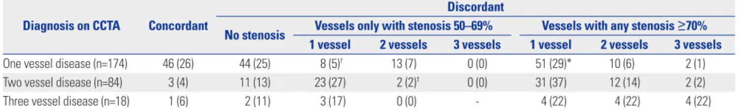
confidence interval (CI) 0.89–0.96]. Patient-level analysis re- vealed that diagnostic concordance, which indicates the cor- rect diagnosis of the presence of moderate stenosis in all three vessels, was found in only 50 (18%) patients. Among the 174 patients who were diagnosed with single-vessel disease on
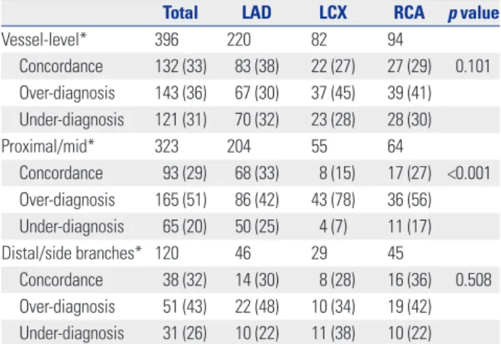
CCTA, 46 (26%) patients had a confirmed concordant diagno- sis by ICA and 44 (25%) patients had no obstructive CAD (Ta- ble 2). In 102 patients suspected to have stenosis in two or more vessels on CCTA, only 4 (4%) patients had a confirmed concordant diagnosis by ICA. Furthermore, more than half of the patients who were assumed to have multi-vessel disease by CCTA had a confirmed under-diagnosis by CCTA, compared to the ICA findings. The proportions of under-diagnosis or se- vere stenosis by ICA were significantly higher as the number of diseased vessels with CCTA-identified stenosis increased (p<0.001) (Fig. 2). Per-vessel analysis showed that one-third of the vessels that were suspected to have moderate stenosis on CCTA had a similar degree of stenosis on ICA, and 36% and 31%
of the vessels were at the risk of over-diagnosis and under-diag- nosis, respectively (Table 3). Per-segment analysis showed that the overall diagnostic concordance was 25% among segments with moderate stenosis on CCTA (Table 4). Over-diagnosis of CCTA was more frequently found in the left circumflex artery (68%) than other vessels (52% in the left anterior descending artery and 61% in the right coronary artery, p=0.049). Compared Table 2. Diagnostic Concordance of CCTA Findings in Per-Patient Analysis
Diagnosis on CCTA Concordant
Discordant
No stenosis Vessels only with stenosis 50–69% Vessels with any stenosis ≥70%
1 vessel 2 vessels 3 vessels 1 vessel 2 vessels 3 vessels
One vessel disease (n=174) 46 (26) 44 (25) 8 (5)† 13 (7) 0 (0) 51 (29)* 10 (6) 2 (1)
Two vessel disease (n=84) 3 (4) 11 (13) 23 (27) 2 (2)† 0 (0) 31 (37) 12 (14) 2 (2)
Three vessel disease (n=18) 1 (6) 2 (11) 3 (17) 0 (0) - 4 (22) 4 (22) 4 (22)
CCTA, coronary computed tomographic angiography; ICA, invasive coronary angiography.
Data are expressed as numbers (%).
*Two patients were diagnosed with isolated left main disease (≥50%) by ICA, discordant with CCTA findings, †CCTA indicates different vessels having stenosis, compared to the findings on ICA.
Fig. 2. Diagnostic correlation between CCTA and ICA per patient analysis. (A) Diagnostic accuracy. (B) Degree of maximal stenosis. CCTA, coronary com- puted tomography angiography; ICA, invasive coronary angiography; VD, vessel disease.
46 (26%) 44 (25%)
8 (5%)
44 (25%) 67 (39%)
76 (44%) 63 (36%)
45 (54%) 45 (54%)
34 (40%) 28 (33%)
11 (13%) 2 (2%)
p<0.001 p=0.013
Diagnosis by CCTA Diagnosis by CCTA
1 VD (n=174)
1 VD (n=174) 2 VD
(n=84)
2 VD (n=84) 3 VD
(n=18)
3 VD (n=18)
3 (4%) 1 (6%) 2 (11%)
5 (28%) 4 (22%)
12 (67%) 12 (67%)
Diagnostic concordance of CCTA compared to ICA Maximal stenosis on ICASimilar extent,
but wrong vessel
No obstructive stenosis Concordant
Over-diagnosis
Under-diagnosis Severe
(≥70% stenosis)
Moderate (50–69% stenosis)
A B
to mixed (28%) or non-calcified (27%) plaque, calcified plaque had lower diagnostic concordance (22%, p=0.001), which was mainly attributed to a higher incidence of over-diagnosis. Also, the degree of stenosis of non-calcified plaques (26%) was more frequently underestimated than that of calcified (12%) or mixed plaque (20%) (Table 4). A higher burden of non-calci- fied plaque, assessed by SIS, was correlated with an increased risk of severe stenosis by ICA (Fig. 3). At least one segment with severe stenosis was found by ICA in 75% of patients who had
≥3 non-calcified SIS and in 22% of patients who lacked non- calcified plaque on the coronary tree (p<0.001). While assess- ment of calcified plaque burden, such as calcified SIS or calci-
um score, did not have an influence on diagnostic accuracy.
Risk factors for under-diagnosis or over-diagnosis of CCTA at the segment of moderate stenosis on CCTA are shown in Table 5. The presence of the typical symptoms of angina [odds ratio (OR) 3.32, 95% CI 1.72–6.41, p<0.001] and mixed (OR 1.99, 95% CI 1.00–3.96, p=0.049) or non-calcified plaque (OR 2.55, 95% CI 1.27–5.11, p=0.009) were independently correlated with a higher incidence of under-diagnosis. Use of a next gen- eration CT scanner lowered the incidence of under-diagnosis (OR 0.50, 95% CI 0.25–0.99, p=0.047). Increased body weight
Table 3. Diagnostic Accuracy of Coronary Computed Tomographic An- giography Findings in Per-Vessel Analysis
Total LAD LCX RCA p value
Vessel-level* 396 220 82 94
Concordance 132 (33) 83 (38) 22 (27) 27 (29) 0.101 Over-diagnosis 143 (36) 67 (30) 37 (45) 39 (41) Under-diagnosis 121 (31) 70 (32) 23 (28) 28 (30)
Proximal/mid* 323 204 55 64
Concordance 93 (29) 68 (33) 8 (15) 17 (27) <0.001 Over-diagnosis 165 (51) 86 (42) 43 (78) 36 (56) Under-diagnosis 65 (20) 50 (25) 4 (7) 11 (17)
Distal/side branches* 120 46 29 45
Concordance 38 (32) 14 (30) 8 (28) 16 (36) 0.508 Over-diagnosis 51 (43) 22 (48) 10 (34) 19 (42) Under-diagnosis 31 (26) 10 (22) 11 (38) 10 (22)
LAD, left anterior descending artery; LCX, left circumflex artery; RCA, right coronary artery.
Data are expressed as numbers (%).
*Vessels including any segments with 50–69% stenosis were assessed.
Table 4. Diagnostic Accuracy among Segments with Moderate Stenosis on CCTA
Characteristics N Concordance Over- diagnosis
Under- diagnosis
p value
Total 508 127 (25) 291 (57) 90 (18)
Epicardial artery 0.049
LAD 296 82 (28) 155 (52) 59 (20)
LCX 94 15 (16) 64 (68) 15 (16)
RCA 118 30 (25) 72 (61) 16 (14)
Location 0.467
Proximal or mid 373 98 (26) 208 (56) 67 (18) Distal or side branch 135 29 (21) 83 (61) 23 (17)
Plaque characteristics 0.001
Calcified 238 52 (22) 158 (66) 28 (12)
Mixed 139 39 (28) 72 (52) 28 (20)
Non-calcified 131 36 (27) 61 (47) 34 (26) CCTA, coronary computed tomographic angiography; LAD, left anterior de- scending artery; LCX, left circumflex artery; RCA, right coronary artery.
At the segment of moderate stenosis on CCTA, the assessment was deter- mined as over-diagnosis or under-diagnosis if the degree of stenosis evaluat- ed by invasive coronary angiography was <50% or ≥70%, respectively. Data are expressed as numbers (%).
30 (37%) 22 (27%) 33 (41%)
31 (38%)
25 (40%)
10 (19%) 18 (22%)
28 (35%)
35 (56%)
39 (75%)
2 (3%) 0 (n=81)
Non-calcified SIS p<0.001
1 (n=81) 2 (n=62) ≥3 (n=52) 3 (6%) Severe
(≥70% stenosis)
No obstructive stenosis Moderate (50–69% stenosis)
Maximal stenosis on ICA
A
23 (26%)
23 (29%)
14 (18%) 38 (42%)
22 (40%) 16 (31%) 29 (32%)
43 (54%) 23 (42%) 25 (49%)
10 (18%)
0–1 (n=90) 2–3 (n=80) 4–5 (n=55)≥6 (n=51) Calcified SIS
p=0.159
10 (20%) Severe
(≥70% stenosis)
No obstructive stenosis Moderate (50–69% stenosis)
Maximal stenosis on ICA
B
C
19 (28%)
25 (36%)
9 (13%) 26 (38%)
27 (39%)
21 (30%) 24 (35%)
35 (51%) 27 (39%)
34 (49%)
15 (22%) 1Q (0–15)
(n=69)
2Q (16–181) (n=69)
3Q (182–560) (n=69)
4Q (≥561) (n=69) Calcium score
p=0.297
14 (20%) Severe
(≥70% stenosis)
No obstructive stenosis Moderate (50–69% stenosis)
Maximalstenosis on ICA
Fig. 3. Presence of any significant stenosis in patients according to plaque burden. Patients were categorized by quartiles of non-calcified (A) and calcified (B) SIS, and calcium score (C). CT, computed tomogra- phy; SIS, segment involvement score; ICA, invasive coronary angiography.
(OR 1.02, 95% CI 1.00–1.04, p=0.023), left circumflex artery (OR 2.00, 95% CI 1.15–3.47, p=0.014), and calcified plaque (OR 2.08, 95% CI 1.39–3.10, p<0.001) were identified as independent fac- tors affecting the possibility of over-diagnosis of CCTA in mul- tivariable analysis.
DISCUSSION
This study investigated the clinical implications of moderate stenosis on CCTA. Current guidelines recommend functional assessment for moderate stenosis in patients with stable isch- emic heart disease.4 Moreover, a CCTA diagnosis of moderate stenosis would be provisional and require further investiga- tion using another non-invasive, functional assessment before coronary catheterization. When considering cardiac catheter- ization, physicians may have difficulty interpreting CCTA find- ings. For example, there could be confusion as to whether CC- TA-identified moderate stenosis would reliably reflect moderate risk and underestimate the actual risk of severe stenosis in need of revascularization, or vice versa. This has been a gray zone in the diagnostic decision process for ischemic heart disease un- til now.
Our findings highlighted the aforementioned concerns about the indeterminacy of CCTA-identified moderate stenosis, which poorly matched with the degree of anatomical stenosis determined by ICA. Only 18% of patients had diagnostic con- cordance in regards to the degree of stenosis and extent of dis- eased vessel between CCTA and ICA. More than 50% of pa- tients had a more severe degree of stenosis or more vessels with obstructive lesions on ICA, compared with CCTA findings.
The limited capabilities of CCTA for delineation of coronary lu- men and for prediction of the need for revascularization were previously demonstrated.8,15,16
Other studies, on the other hand, have insisted that CCTA may provide sufficient specificity and sensitivity for the diag- nosis of obstructive CAD.1,17,18 These trials demonstrated posi-
tive predictive value of stenosis >50% in vessel-level analysis ranging between 51%17 and 83%,1 which was widely dispersed across to the trials. Another study using a dual-source CT scan- ner demonstrated that 39–50% of segments suspected to have moderate stenosis on CCTA had a similar degree of stenosis on ICA.19 The authors insisted that most segments were catego- rized within a 1-grade discrepancy, which contained a wide range of stenosis between 25% to 89%. In addition, their study included a small number of patients (n=84), included all se- verities of stenosis, and did not demonstrate how the diagnos- tic discordance at each segment would impact the diagnosis at the patient-level. Meanwhile, another compelling diagnostic tool, CCTA-derived fractional flow reserve has been shown to have clinical utility for diagnosis of myocardial ischemia in in- termediate stenosis without noticeably altering the sensitivity of CCTA.20
If diagnosis is not highly accurate at each segment level, the possibility of diagnostic discordance may be exponentially in- creased at the patient level as the number of plaques to be eval- uated was increased on CCTA. In the present study, only 4% of patients who were diagnosed with multi-vessel disease on CCTA had CCTA findings that were completely concordant with ICA findings. Furthermore, the presence of any severe stenosis was found more frequently as the number of vessels with moder- ate stenosis or non-calcified plaques increased.
Although the ability of CCTA to determine the exact degree of stenosis per segment is limited, CCTA may have a benefit in the estimation of overall atherosclerotic burden of the coronary tree, even when compared with functional assessment and ICA. Total plaque burden, especially non-calcified plaque, on the coronary tree may have clinical implications for risk strati- fication of patients with the possibility of severe stenosis. CC- TA-identified overall plaque burden was revealed to have inde- pendent value for prediction of future cardiac events.21 Calcium scoring is a surrogate for plaque burden and has been well es- tablished to estimate the risk of future cardiac events.22,23 Cal- cification is associated with advanced stage atherosclerosis, Table 5. Risk Factors for Misdiagnosis among Segments with Moderate Stenosis Assessed by Coronary Computed Tomographic Angiography
Univariate analysis Multivariate model
OR 95% CI p value OR 95% CI p value
Under-diagnosis
Typical angina 3.52 1.79–6.90 <0.001 3.32 1.72–6.41 <0.001
Use of next generation CT scanner 0.49 0.24–0.99 0.046 0.50 0.25–0.99 0.047
Plaque characteristics (over calcified plaque)
Mixed plaque 1.99 1.01–3.94 0.047 1.99 1.00–3.96 0.049
Non-calcified plaque 2.86 1.45–5.72 0.003 2.55 1.27–5.11 0.009
Over-diagnosis
Male 1.59 1.03–2.45 0.035 - - -
Weight, per 1 kg 1.02 1.00–1.04 0.010 1.02 1.00–1.04 0.023
Left circumflex artery (over the other vessels) 1.85 1.10–3.12 0.020 2.00 1.15–3.47 0.014
Calcified plaque (over the other plaques) 2.12 1.43–3.16 <0.001 2.08 1.39–3.10 <0.001
CI, confidence interval; OR, odds ratio; CT, computed tomopgraphy.
and it is most commonly found on fibrocalcific plaques, which are at low risk for rapid progression.24 However, non-calcified plaque burden, which was found to have more influence on the possibility of severe stenosis in our study, was proposed to be a better predictor for myocardial ischemia than calcium score or stenosis severity.25 Because CCTA tends to underestimate the size of non-calcified plaques and overestimate that of cal- cified plaques due to calcification-caused artifacts, a higher burden of non-calcified plaques may be interpreted as a high risk finding for the presence of more severely stenotic lesions.26 The CAD-RADS4 consensus mentioned only the degree of maximal coronary stenosis, but not overall plaque burden, in consideration of the diagnostic process for patients presenting with chest pain. Considering the limited reliability of CCTA- identified stenosis severity, physicians should be cautious when applying the suggested recommendations to daily clinical practice. Instead, the interpretation of CCTA findings should be individualized, and physicians should use clinical risk fac- tors or another non-invasive assessment, or proceed to cardi- ac catheterization for treatment decisions.
Our study had several limitations. First, we validated CCTA- identified moderate stenosis in comparison with the degree of anatomical stenosis assessed by ICA. Because the functional significance of coronary stenosis may not be determined solely by ICA, further functional evaluation is required for the deci- sion of revascularization in some cases. However, our study focused on the diagnostic reliability of the anatomical assess- ment of CCTA. The degree of maximal stenosis assessed by CCTA may be an insufficient evaluation to differentiate patient risk and to direct future investigation in clinical practice. An- other functional test may be required for the decision of revas- cularization. However, its actual utility in clinical practice should be addressed in another study. Second, we used visual estimation to assess the degree of stenosis in both ICA and CCTA. Visual estimation is a common and comparable method for evaluation of stenosis severity in both ICA27 and CCTA19 in clinical practice. We found that visual assessment of ICA was also reliable compared with quantitative coronary angiogra- phy in randomly selected patients. Third, the diagnostic accu- racy of CCTA may not be interpreted as the result in the over- all patients who used CCTA as a non-diagnostic assessment because our prospective registry only enrolled the patients whose ICA information was available. The clinical reasons for cardiac catheterization, such as clinical suspicion or high prob- ability of CAD, may reflect a higher prevalence of severe CAD in selected patients. However, 22% of the patients in our study had a high pretest probability, which did not impact the diag- nostic concordance.
In conclusion, CCTA-identified moderate stenosis may be limited in estimating the exact degree of anatomical stenosis assessed by ICA. CCTA-identified stenosis per se may not be appropriate for predicting exact degree of coronary stenosis, although a high burden of non-calcified plaque on a coronary
tree may increase the risk of significant coronary stenosis.
ACKNOWLEDGEMENTS
This study was supported by a grant from the Korea Health- care Technology R&D Project, Ministry for Health, Welfare &
Family Affairs, Republic of Korea (A085012, HI15C1277 and A085136), the Basic Science Research Program through the National Research Foundation of Korea funded by the Minis- try of Education (2014R1A1A2055258), and the Cardiovascu- lar Research Center, Seoul, Korea.
ORCID
Myeong-Ki Hong https://orcid.org/0000-0002-2090-2031
REFERENCES
1. Miller JM, Rochitte CE, Dewey M, Arbab-Zadeh A, Niinuma H, Gottlieb I, et al. Diagnostic performance of coronary angiography by 64-row CT. N Engl J Med 2008;359:2324-36.
2. Litt HI, Gatsonis C, Snyder B, Singh H, Miller CD, Entrikin DW, et al. CT angiography for safe discharge of patients with possible acute coronary syndromes. N Engl J Med 2012;366:1393-403.
3. SCOT-HEART investigators. CT coronary angiography in patients with suspected angina due to coronary heart disease (SCOT- HEART): an open-label, parallel-group, multicentre trial. Lancet 2015;385:2383-91.
4. Cury RC, Abbara S, Achenbach S, Agatston A, Berman DS, Budoff MJ, et al. CAD-RADS(TM) Coronary Artery Disease - Reporting and Data System. An expert consensus document of the Society of Car- diovascular Computed Tomography (SCCT), the American College of Radiology (ACR) and the North American Society for Cardiovas- cular Imaging (NASCI). Endorsed by the American College of Car- diology. J Cardiovasc Comput Tomogr 2016;10:269-81.
5. Pryor DB, Harrell FE Jr, Lee KL, Califf RM, Rosati RA. Estimating the likelihood of significant coronary artery disease. Am J Med 1983;75:771-80.
6. Taylor AJ, Cerqueira M, Hodgson JM, Mark D, Min J, O’Gara P, et al. ACCF/SCCT/ACR/AHA/ASE/ASNC/NASCI/SCAI/SCMR 2010 appropriate use criteria for cardiac computed tomography. A re- port of the American College of Cardiology Foundation Appropri- ate Use Criteria Task Force, the Society of Cardiovascular Comput- ed Tomography, the American College of Radiology, the American Heart Association, the American Society of Echocardiography, the American Society of Nuclear Cardiology, the North American So- ciety for Cardiovascular Imaging, the Society for Cardiovascular Angiography and Interventions, and the Society for Cardiovascu- lar Magnetic Resonance. J Am Coll Cardiol 2010;56:1864-94.
7. Austen WG, Edwards JE, Frye RL, Gensini GG, Gott VL, Griffith LS, et al. A reporting system on patients evaluated for coronary artery disease. Report of the Ad Hoc Committee for Grading of Coronary Artery Disease, Council on Cardiovascular Surgery, American Heart Association. Circulation 1975;51(4 Suppl):5-40.
8. Hong SJ, Her AY, Suh Y, Won H, Cho DK, Cho YH, et al. Coronary computed tomographic angiography does not accurately predict the need of coronary revascularization in patients with stable an- gina. Yonsei Med J 2016;57:1079-86.
9. Achenbach S, Raggi P. Imaging of coronary atherosclerosis by computed tomography. Eur Heart J 2010;31:1442-8.
10. Groothuis JG, Beek AM, Meijerink MR, Brinckman SL, Heymans MW, van Kuijk C, et al. Positive predictive value of computed to- mography coronary angiography in clinical practice. Int J Cardiol 2012;156:315-9.
11. Dey D, Lee CJ, Ohba M, Gutstein A, Slomka PJ, Cheng V, et al. Im- age quality and artifacts in coronary CT angiography with dual- source CT: initial clinical experience. J Cardiovasc Comput Tomogr 2008;2:105-14.
12. Øvrehus KA, Marwan M, Bøtker HE, Achenbach S, Nørgaard BL.
Reproducibility of coronary plaque detection and characterization using low radiation dose coronary computed tomographic angiog- raphy in patients with intermediate likelihood of coronary artery disease (ReSCAN study). Int J Cardiovasc Imaging 2012;28:889-99.
13. McHugh ML. Interrater reliability: the kappa statistic. Biochem Med (Zagreb) 2012;22:276-82.
14. Shrout PE, Fleiss JL. Intraclass correlations: uses in assessing rater reliability. Psychol Bull 1979;86:420-8.
15. Doh JH, Koo BK, Nam CW, Kim JH, Min JK, Nakazato R, et al. Di- agnostic value of coronary CT angiography in comparison with invasive coronary angiography and intravascular ultrasound in patients with intermediate coronary artery stenosis: results from the prospective multicentre FIGURE-OUT (Functional Imaging criteria for GUiding REview of invasive coronary angiOgraphy, in- travascular Ultrasound, and coronary computed Tomographic an- giography) study. Eur Heart J Cardiovasc Imaging 2014;15:870-7.
16. Kim C, Hong SJ, Shin DH, Kim JS, Kim BK, Ko YG, et al. Limitations of coronary computed tomographic angiography for delineating the lumen and vessel contours of coronary arteries in patients with stable angina. Eur Heart J Cardiovasc Imaging 2015;16:1358-65.
17. Budoff MJ, Dowe D, Jollis JG, Gitter M, Sutherland J, Halamert E, et al. Diagnostic performance of 64-multidetector row coronary com- puted tomographic angiography for evaluation of coronary artery stenosis in individuals without known coronary artery disease: re- sults from the prospective multicenter ACCURACY (Assessment by Coronary Computed Tomographic Angiography of Individuals Undergoing Invasive Coronary Angiography) trial. J Am Coll Car- diol 2008;52:1724-32.
18. Meijboom WB, Meijs MF, Schuijf JD, Cramer MJ, Mollet NR, van Mieghem CA, et al. Diagnostic accuracy of 64-slice computed to- mography coronary angiography: a prospective, multicenter, mul-
tivendor study. J Am Coll Cardiol 2008;52:2135-44.
19. Cheng V, Gutstein A, Wolak A, Suzuki Y, Dey D, Gransar H, et al.
Moving beyond binary grading of coronary arterial stenoses on cor- onary computed tomographic angiography: insights for the imager and referring clinician. JACC Cardiovasc Imaging 2008;1:460-71.
20. Baumann S, Renker M, Hetjens S, Fuller SR, Becher T, Loßnitzer D, et al. Comparison of coronary computed tomography angiogra- phy-derived vs invasive fractional flow reserve assessment: meta- analysis with subgroup evaluation of intermediate stenosis. Acad Radiol 2016;23:1402-11.
21. Naoum C, Berman DS, Ahmadi A, Blanke P, Gransar H, Narula J, et al. Predictive value of age- and sex-specific nomograms of global plaque burden on coronary computed tomography angiography for major cardiac events. Circ Cardiovasc Imaging 2017;10:e004896.
22. Kelkar AA, Schultz WM, Khosa F, Schulman-Marcus J, O'Hartaigh BW, Gransar H, et al. Long-term prognosis after coronary artery calcium scoring among low-intermediate risk women and men.
Circ Cardiovasc Imaging 2016;9:e003742.
23. Okwuosa TM, Greenland P, Ning H, Liu K, Bild DE, Burke GL, et al.
Distribution of coronary artery calcium scores by Framingham 10- year risk strata in the MESA (Multi-Ethnic Study of Atherosclero- sis) potential implications for coronary risk assessment. J Am Coll Cardiol 2011;57:1838-45.
24. Otsuka F, Sakakura K, Yahagi K, Joner M, Virmani R. Has our under- standing of calcification in human coronary atherosclerosis pro- gressed? Arterioscler Thromb Vasc Biol 2014;34:724-36.
25. Bauer RW, Thilo C, Chiaramida SA, Vogl TJ, Costello P, Schoepf UJ.
Noncalcified atherosclerotic plaque burden at coronary CT angi- ography: a better predictor of ischemia at stress myocardial perfu- sion imaging than calcium score and stenosis severity. AJR Am J Roentgenol 2009;193:410-8.
26. De Filippo M, Capasso R. Coronary computed tomography angi- ography (CCTA) and cardiac magnetic resonance (CMR) imaging in the assessment of patients presenting with chest pain suspected for acute coronary syndrome. Ann Transl Med 2016;4:255.
27. Gottsauner-Wolf M, Sochor H, Moertl D, Gwechenberger M, Stock- enhuber F, Probst P. Assessing coronary stenosis. Quantitative coro- nary angiography versus visual estimation from cine-film or phar- macological stress perfusion images. Eur Heart J 1996;17:1167-74.