Clinical Implications of Microsatellite Instability in T1 Colorectal Cancer
Jeonghyun Kang, Hak Woo Lee, Im-kyung Kim, Nam Kyu Kim, Seung-Kook Sohn, and Kang Young Lee
Department of Surgery, Yonsei University College of Medicine, Seoul, Korea.
Received: January 20, 2014 Revised: March 23, 2014 Accepted: March 26, 2014
Corresponding author: Dr. Kang Young Lee, Department of Surgery,
Yonsei University College of Medicine, 50-1 Yonsei-ro, Seodaemun-gu, Seoul 120-752, Korea.
Tel: 82-2-2228-2131, Fax: 82-2-313-8289 E-mail: [email protected]
∙ The authors have no financial conflicts of interest.
© Copyright:
Yonsei University College of Medicine 2015 This is an Open Access article distributed under the terms of the Creative Commons Attribution Non- Commercial License (http://creativecommons.org/
licenses/by-nc/3.0) which permits unrestricted non- commercial use, distribution, and reproduction in any medium, provided the original work is properly cited.
Purpose: The estimation of regional lymph node metastasis (LNM) risk in T1 colorectal cancer is based on histologic examination and imaging of the primary tumor. High-frequency microsatellite instability (MSI-H) is likely to decrease the possibility of metastasis to either regional lymph nodes or distant organs in colorectal cancers. This study evaluated the clinical implications of MSI in T1 colorectal cancer with emphasis on the usefulness of MSI as a predictive factor for regional LNM. Materials and Methods: A total of 133 patients who underwent radical resection for T1 colorectal cancer were included. Genomic DNA was ex- tracted from normal and tumor tissues and amplified by polymerase chain reaction (PCR). Five microsatellite markers, BAT-25, BAT-26, D2S123, D5S346, and D17S250, were used. MSI and clinicopathological parameters were evaluated as potential predictors of LNM using univariate and multivariate analyses. Results:
Among 133 T1 colorectal cancer patients, MSI-H, low-frequency microsatellite instability (MSI-L), and microsatellite stable (MSS) colorectal cancers accounted for 7.5%, 6%, and 86.5%, respectively. MSI-H tumors showed a female predomi- nance, a proximal location and more retrieved lymph nodes. Twenty-two patients (16.5%) had regional LNM. Lymphovascular invasion and depth of invasion were significantly associated with LNM. There was no LNM in 10 MSI-H patients;
however, MSI status was not significantly correlated with LNM. Disease-free sur- vival did not differ between patients with MSI-H and those with MSI-L/MSS.
Conclusion: MSI status could serve as a negative predictive factor in estimating LNM in T1 colorectal cancer, given that LNM was not detected in MSI-H pa- tients. However, validation of our result in a different cohort is necessary.
Key Words: Microsatellite instability, lymph node metastasis, early colorectal cancer, T1, prognosis
INTRODUCTION
Although the basic principle in surgical management of colorectal cancer is wide resection of the primary tumor and adequate regional lymph node dissection, some modifications are appropriate in selected patients. When the cancer is confined to
regional LNM.
MATERIALS AND METHODS
Eligibility
Patients who underwent curative resection for T1 colon or rectal adenocarcinoma between January 2005 and May 2011 were selected from our prospectively collected data- base. Excluded from the initial selection were patients who had undergone preoperative chemotherapy or radiotherapy, patients who were diagnosed with HNPCC or a familial ad- enomatous polyposis, patients who underwent transanal ex- cision, and patients initially diagnosed with stage IV dis- ease. The inclusion of patients was based on the availability of MSI data, as well as a complete set of clinicopathologi- cal information including age, sex, tumor size, histologic grade, lymphovascular invasion, depth of invasion, date of surgery, location of the primary tumor, [proximal colon (ce- cal, ascending, or transverse colon), distal colon (descend- ing, sigmoid, or rectosigmoid colon), or rectum], date of re- currence, pathologic nodal stage, and total retrieved lymph nodes. After exclusions, 133 patients were included in our analysis. This study was approved by our Institutional Re- view Board (number: 4-2010-0286).
Sectioning and microdissection
Genomic DNA was extracted from three to five sections of 10-μm-thick formalin-fixed, paraffin-embedded tissue blocks containing tumor and non-neoplastic mucosa using a QIAamp DNA mini kit (Qiagen, Hilden, Germany) accord- ing to the manufacturer’s protocol. Separate blocks were used for malignant and non-malignant tissue. Tumor blocks with a tumor area greater than 80% tumor cells were select- ed based on hematoxylin and eosin stained slides.
Microsatellite instability (MSI)
The MSI status of each tumor was determined with the fol- lowing five markers: BAT 25, BAT 26, D2S123, D5S346, and D17S250. Fifty nanograms of DNA were amplified in a 20 μL reaction solution containing 2 μL of 10X buffer (Roche, Mannheim, Germany), 1.7 to 2.5 mmol/L of MgCl2, 0.3 μM of each primer pair, 250 μM of deoxynucleotide tri- phosphate, and 2.5 units of DNA polymerase (Roche). The primer sequences and polymerase chain reaction (PCR) cy- cles for each marker were adapted from the published data.25 Fluorescence markers (NED, FAM) were attached to the 5’
the mucosal layer, the tumor can be completely removed by an endoscopic procedure or local excision and further lymph node dissection is not necessary since there is no risk of lymph node metastasis (LNM) in these cancers. The same treatment strategy could be applied to invasive carcinoma if the risk of LNM could be predicted. However, the challenge in applying local resection to invasive carcinoma is predict- ing the presence of LNM. The risk of regional LNM increas- es in proportion to the depth of invasion. The overall LNM rate of T1 colorectal cancer was reported to range from 6%
to 13%,1-4 which is relatively low compared to 19‒28% for T2 and 36‒65.4% for T3 and T4 cancer.5-7
Currently, when a tumor is found to be confined within the submucosa during a preoperative workup, resection of the primary tumor is attempted. The possibility of LNM is sub- sequently evaluated based on histologic examination of the primary tumor with regard to features such as depth of inva- sion, presence of lymphovascular invasion, differentiation, and tumor budding.8 It is recommended that patients at high risk undergo radical resection due to the possibility of region- al LNM. Given that the development of colorectal cancer is known to be the result of accumulating genetic alterations,9-11 it seems prudent to take into account histologic features of the primary tumor in addition to molecular biological charac- teristics for the prediction of regional LNM in T1 colorectal cancer. However, to the best of our knowledge, there are lim- ited reports on predicting regional LNM in T1 colorectal can- cer using molecular biological characteristics.12
MSI is one of the prevalent carcinogenic pathways in colorectal cancer, caused by a mismatch repair gene defect resulting in loss or gain of tandem repeat sequences.13-16 A high frequency of microsatellite instability (MSI-H) has been found in most patients with hereditary nonpolyposis colorectal cancer (HNPCC), and occurs in approximately 15% of sporadic colorectal cancer cases.15-18 MSI-H tumors were associated with unique clinicopathological characteris- tics, such as younger age at onset, proximal location, and frequent peritumoral lymphocytic infiltration.19-21 Addition- ally, MSI-H tumors showed a lower incidence of LNM and a better survival rate.21-24 Most previous studies investigat- ing the relationship between MSI and regional LNM were based on advanced colorectal cancers. However, little is known regarding the specific impact of MSI in early colorectal cancer.
Therefore, the aim of this study was to evaluate the clini- cal implications of MSI in T1 colorectal cancer with em- phasis on the usefulness of MSI as a predictive factor for
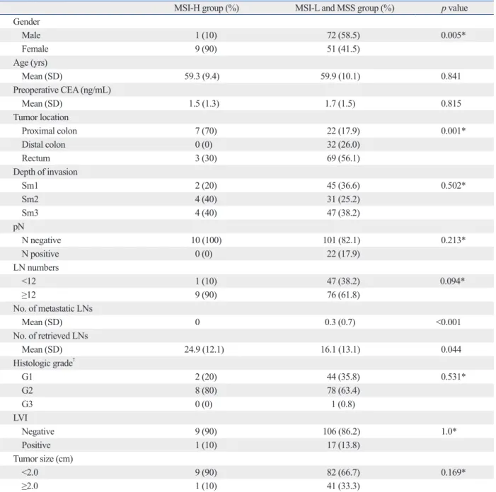
together and denoted as MSI-L/MSS. Comparisons of clin- icopathological characteristics between the MSI-H and MSI-L/MSS groups are summarized in Table 2. There was a female predominance in the MSI-H group (p=0.005). The predominant location in the MSI-H group was the proximal colon (p=0.001). There was no significant difference be- tween the two groups with regard to the depth of tumor in- vasion, histologic grade, presence of lymphovascular inva- sion, or tumor size. In contrast, the number of total retrieved lymph nodes was higher in the MSI-H group (p=0.044). The number of metastatic lymph nodes in the MSI-H group was significantly lower in comparison to the MSI-L/MSS group (p<0.001).
end of the forward primer.
All samples were prepared for fragment separation on an ABI Prism 3100 Genetic Analyzer using 0.7 μL of the am- plified samples combined with 0.3 μL of GeneScan 500 Size Standard and 9 μL of HiDi Formamide.
MSI was diagnosed when there were aberrant peaks or peak shifts compared to the normal control. A case was cate- gorized as MSI-H if MSI was present at two or more mark- ers, MSI-low (MSI-L) if only one of the five markers showed instability, and microsatellite stable (MSS) if no marker had evidence of MSI.26 In all of the analyses, MSI-L, and MSS tumors were grouped together and denoted as MSI-L/MSS.
Statistical analysis
All calculations and analyses were performed using SPSS version 20.0 (SPSS Inc., Chicago, IL, USA). The associa- tion of clinicopathological features with MSI status was an- alyzed using the two-sided Pearson’s chi-square test or Fisher’s exact test for categorical variables and Student’s t- test for continuous variables. Factors associated with LNM were analyzed by logistic regression with forward stepwise selection of variables. Disease-free survival (DFS) was de- fined as the time from the date of operation to the date of tumor recurrence or last follow-up. Survival analysis was performed with the Kaplan-Meier method. The log-rank test was used to compare survival outcome between groups.
A p-value <0.05 was considered to indicate significance.
RESULTS
Patient characteristics
Patients’ characteristics are summarized in Table 1. The median age was 60 years (range: 32‒84 years). The distri- bution of tumor locations was as follows: 29 in the proxi- mal colon, 32 in the distal colon and 72 in the rectum.
Twenty-two patients (16.5%) had regional LNM. Histolog- ic grade examination revealed only one patient (0.8%) with poorly differentiated adenocarcinoma. The distribution of invasion was 47 sm1, 35 sm2, and 51 sm3. Lymphovascu- lar invasion was detected in 18 patients (13.5%). The medi- an tumor size was 1.9 cm (range: 0.2‒8.0 cm).
Clinical characteristics of patients with MSI
An evaluation of tumor MSI status revealed MSS in 115 patients (86.5%), MSI-L in 8 patients (6.0%) and MSI-H in 10 patients (7.5%). MSI-L and MSS tumors were grouped
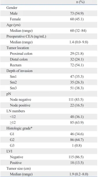
Table 1. Patient Baseline Characteristics
n (%) Gender
Male 73 (54.9)
Female 60 (45.1)
Age (yrs)
Median (range) 60 (32–84)
Preoperative CEA (ng/mL)
Median (range) 1.4 (0.0–9.8)
Tumor location
Proximal colon 29 (21.8)
Distal colon 32 (24.1)
Rectum 72 (54.1)
Depth of invasion
Sm1 47 (35.3)
Sm2 35 (26.3)
Sm3 51 (38.3)
pN
Node negative 111 (83.5)
Node positive 22 (16.5)
LN numbers
<12 48 (36.1)
≥12 85 (63.9)
Histologic grade*
G1 46 (34.6)
G2 86 (64.7)
G3 1 (0.8)
LVI
Negative 115 (86.5)
Positive 18 (13.5)
Tumor size (cm)
Median (range) 1.9 (0.2–8.0)
CEA, carcinoembryonic antigen; LN, lymph node; LVI, lymphovascular inva- sion.
*Histologic grade: G1, well differentiated; G2, moderately differentiated;
G3, poorly differentiated.
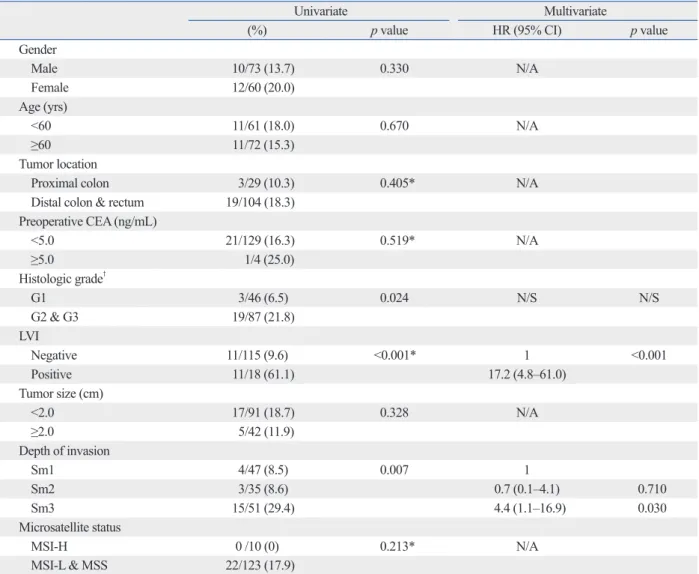
er’s exact test). However, there was no LNM in the MSI-H group (Table 3).
Survival analysis
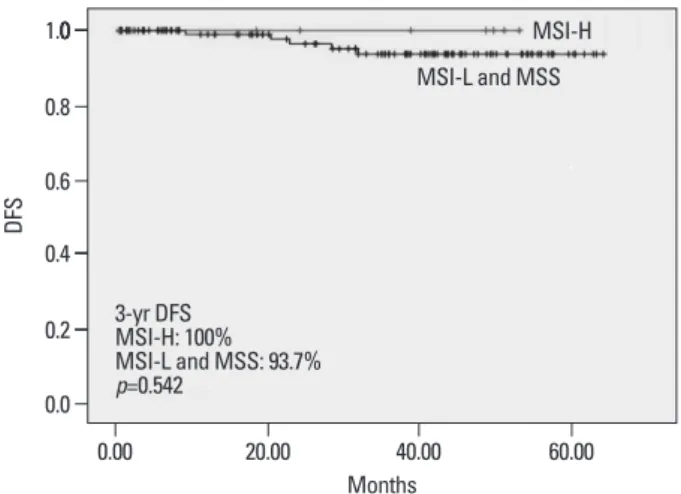
We compared disease-free survival between patients with MSI-H and those with MSI-L/MSS (Fig. 1). There was no significant difference in three-year DFS between the two groups (100% in the MSI-H group; 93.7% in the MSI-L/
MSS group; p=0.542) (Fig. 1).
Factors associated with LNM
Histologic grade, lymphovascular invasion, and depth of in- vasion were significantly associated with LNM by univariate analysis. Multivariate analysis indicated that lymphovascular invasion [hazard ratio (HR), 17.2; 95% confidence interval (CI), 4.8‒61.0; p<0.001] and depth of invasion (comparing sm1 to sm3: HR, 4.4; 95% CI, 1.1‒16.9; p=0.030) were in- dependent risk factors for LNM. MSI-H was not correlated with regional LNM by univariate analysis (p=0.213, Fish-
Table 2. Comparison of Clinicopathological Characteristics between MSI-H Group and MSI-L and MSS Group
MSI-H group (%) MSI-L and MSS group (%) p value
Gender
Male 1 (10) 72 (58.5) 0.005*
Female 9 (90) 51 (41.5)
Age (yrs)
Mean (SD) 59.3 (9.4) 59.9 (10.1) 0.841
Preoperative CEA (ng/mL)
Mean (SD) 1.5 (1.3) 1.7 (1.5) 0.815
Tumor location
Proximal colon 7 (70) 22 (17.9) 0.001*
Distal colon 0 (0) 32 (26.0)
Rectum 3 (30) 69 (56.1)
Depth of invasion
Sm1 2 (20) 45 (36.6) 0.502*
Sm2 4 (40) 31 (25.2)
Sm3 4 (40) 47 (38.2)
pN
N negative 10 (100) 101 (82.1) 0.213*
N positive 0 (0) 22 (17.9)
LN numbers
<12 1 (10) 47 (38.2) 0.094*
≥12 9 (90) 76 (61.8)
No. of metastatic LNs
Mean (SD) 0 0.3 (0.7) <0.001
No. of retrieved LNs
Mean (SD) 24.9 (12.1) 16.1 (13.1) 0.044
Histologic grade†
G1 2 (20) 44 (35.8) 0.531*
G2 8 (80) 78 (63.4)
G3 0 (0) 1 (0.8)
LVI
Negative 9 (90) 106 (86.2) 1.0*
Positive 1 (10) 17 (13.8)
Tumor size (cm)
<2.0 9 (90) 82 (66.7) 0.169*
≥2.0 1 (10) 41 (33.3)
CEA, carcinoembryonic antigen; LN, lymph node; LVI, lymphovascular invasion; SD, standard deviation; MSI-H, high-frequency microsatellite instability;
MSI-L, low-frequency microsatellite instability; MSS, microsatellite stable.
*Fisher’s exact test.
†Histologic grade: G1, well differentiated; G2, moderately differentiated; G3, poorly differentiated.
aging and histologic characteristics of the primary tumor such as depth of tumor invasion, tumor differentiation and lymphovascular invasion.8 In current practice, even after complete removal of the primary tumor, an additional radi- cal resection is recommended for patients found to have high LNM risk upon histologic examination. However, a large-scale multicenter study found that the actual rate of LNM was only 14‒23% according to respective high risk factors.1 For this reason, a great deal of effort has gone into reducing potential over-treatment by precise prediction of LNM risk.
In this study, the role of MSI was investigated as one of the predictive factors for LNM in T1 colorectal cancer, since we feel that in the prediction of early metastasis, biological
DISCUSSION
The major finding of this study was that MSI status could be used as a negative predictive marker in estimating LNM in T1 colorectal cancer given that there was no LNM in MSI- H patients.
The standard treatment for colorectal cancer is complete resection of the primary tumor with regional lymphadenec- tomy due to the potential risk of regional LNM. However, in T1 colorectal cancer, regional lymphadenectomy can be omitted without deterioration of oncologic outcomes for se- lected patients with little risk of regional LNM. The predic- tion of regional LNM was made based on preoperative im-
Table 3. Factors Associated with Lymph Node Metastasis in T1 Colorectal Carcinoma
Univariate Multivariate
(%) p value HR (95% CI) p value
Gender
Male 10/73 (13.7) 0.330 N/A
Female 12/60 (20.0)
Age (yrs)
<60 11/61 (18.0) 0.670 N/A
≥60 11/72 (15.3)
Tumor location
Proximal colon 3/29 (10.3) 0.405* N/A
Distal colon & rectum 19/104 (18.3) Preoperative CEA (ng/mL)
<5.0 21/129 (16.3) 0.519* N/A
≥5.0 1/4 (25.0)
Histologic grade†
G1 3/46 (6.5) 0.024 N/S N/S
G2 & G3 19/87 (21.8)
LVI
Negative 11/115 (9.6) <0.001* 1 <0.001
Positive 11/18 (61.1) 17.2 (4.8–61.0)
Tumor size (cm)
<2.0 17/91 (18.7) 0.328 N/A
≥2.0 5/42 (11.9)
Depth of invasion
Sm1 4/47 (8.5) 0.007 1
Sm2 3/35 (8.6) 0.7 (0.1–4.1) 0.710
Sm3 15/51 (29.4) 4.4 (1.1–16.9) 0.030
Microsatellite status
MSI-H 0 /10 (0) 0.213* N/A
MSI-L & MSS 22/123 (17.9)
CEA, carcinoembryonic antigen; LVI, lymphovascular invasion; N/A, not applicable; N/S: non significant; HR, hazard ratio; CI, confidence interval; MSI-H, high-frequency microsatellite instability; MSI-L, low-frequency microsatellite instability; MSS, microsatellite stable.
Factors with p value less than 0.2 in univariate analysis were entered into multivariate analysis.
*Fisher’s exact test.
†Histologic grade: G1, well differentiated; G2, moderately differentiated; G3, poorly differentiated.
colon cancer and not in stage II colon cancer, the associa- tion was not evident in stage III colorectal cancer in another series.33 It was reported that this discrepancy might have originated from different uses of panels in defining MSI-H, different incidences of MSI-H phenotype, and limited num- bers of MSI-H cases.26,34 In our study, although the frequency of MSI-H was relatively low (7.5%), a higher lymph node harvest was observed in MSI-H tumors (p=0.044), confirm- ing the positive correlation of MSI-H and retrieved lymph node numbers. In the sub-group analysis of stage I patients (n=111), there was still a trend toward increased total re- trieved lymph nodes in MSI-H tumors (MSI-H: mean of 24.9; MSI-L/MSS: mean of 16; p=0.062; data not shown).
The incidence of regional LNM was 16.5% in the current study, higher than in previous reports (6.3% to 13%).1-4 This difference may be the result of patient selection bias in this study. Most of the included patients were candidates for radical surgery because of the presence of risk factors for regional LNM or technical factors such as incomplete resection or difficulty in complete resection of the primary tumor. These factors could have contributed to the relative- ly high incidence of regional LNM; however, the fact that all included patients underwent radical surgery is a unique aspect of this study. For this reason, the presence of cancer metastasis to regional lymph nodes was confirmed by patho- logical examination. It is also noteworthy that there was no regional LNM in patients with MSI-H, even among high risk patients. In conclusion, given that there was no LNM in patients with MSI-H tumors, MSI status could serve as a negative predictive factor in estimating LNM in T1 colorec- tal cancer. Although this study showed the possibility of negative predictive power of MSI-H in LNM, the sample size is relatively small. Further large scale studies are re- quired to confirm our observation.
ACKNOWLEDGEMENTS
This study was supported by a faculty research grant from Yonsei University College of Medicine (6-2010-0134).
REFERENCES
1. Kitajima K, Fujimori T, Fujii S, Takeda J, Ohkura Y, Kawamata H, et al. Correlations between lymph node metastasis and depth of submucosal invasion in submucosal invasive colorectal carcinoma:
characteristics should be taken into account in addition to conventional pathologic examination. MSI-H cancers are re- ported to have a decreased likelihood of metastases to either regional LNs or distant organs.22,23,27 Interestingly, regional LNM was not identified in patients with MSI-H T1 colorec- tal cancers in this study. Although not statistically signifi- cant, the prominent difference in LNM between the MSI-H group (0%) and MSI-L/MSS group (17.9%) indicate the possibility that the lack of an observed association between MSI status and LNM may have been the result of a type II error related to insufficient sample size. In the studied co- hort, lymphovascular invasion, and depth of invasion were identified as independent risk factors for LNM by multivar- iate analysis, consistent with other reports.8 Although this study was not substantial enough to make definite conclu- sions, our results suggest that MSI has the potential to be used as a predictive factor for LNM in T1 colorectal cancer.
Colorectal cancers with MSI-H have distinct clinicopath- ological features. MSI-H cancers tend to be associated with a slight predominance in females, proximal tumor location, large tumor size, greater depth of tumor invasion, and poor histology.20,21,23,24 Our data also demonstrated that MSI-H T1 colorectal cancer showed a female predominance, prox- imal tumor location and more retrieved lymph nodes.
The total number of retrieved lymph nodes in colorectal cancer is known to be positively correlated with good prog- nosis.28-30 Many studies demonstrated an association be- tween MSI-H status and higher total lymph node counts.31 However, there are discrepancies among some studies.
While Belt, et al.32 reported that high lymph node retrieval was associated with MSI-H tumors especially in stage III
Fig. 1. Comparison of survival between MSI-H group and MSI-L/MSS group.
There was no difference of survival outcomes between “MSI-H group” and
“MSI-L/MSS group” (mean follow-up periods: 31 months). DFS, disease-free survival; MSI-H, high-frequency microsatellite instability; MSI-L, low-fre- quency microsatellite instability; MSS, microsatellite stable.
Months 0.0
0.2 0.4 000
0.6 0.8 1.0
DFS
0.00 20.00 40.00 60.00
MSI-L and MSS
3-yr DFS MSI-H: 100%
MSI-L and MSS: 93.7%
p=0.542
MSI-H
of gastrointestinal cancer. Yonsei Med J 2009;50:309-21.
19. Michel S, Benner A, Tariverdian M, Wentzensen N, Hoefler P, Pommerencke T, et al. High density of FOXP3-positive T cells in- filtrating colorectal cancers with microsatellite instability. Br J Cancer 2008;99:1867-73.
20. Gafà R, Maestri I, Matteuzzi M, Santini A, Ferretti S, Cavazzini L, et al. Sporadic colorectal adenocarcinomas with high-frequency microsatellite instability. Cancer 2000;89:2025-37.
21. Liang JT, Huang KC, Cheng AL, Jeng YM, Wu MS, Wang SM.
Clinicopathological and molecular biological features of colorec- tal cancer in patients less than 40 years of age. Br J Surg 2003;90:
205-14.
22. Gryfe R, Kim H, Hsieh ET, Aronson MD, Holowaty EJ, Bull SB, et al. Tumor microsatellite instability and clinical outcome in young patients with colorectal cancer. N Engl J Med 2000;342:69-77.
23. Lim SB, Jeong SY, Lee MR, Ku JL, Shin YK, Kim WH, et al.
Prognostic significance of microsatellite instability in sporadic colorectal cancer. Int J Colorectal Dis 2004;19:533-7.
24. Malesci A, Laghi L, Bianchi P, Delconte G, Randolph A, Torri V, et al. Reduced likelihood of metastases in patients with microsat- ellite-unstable colorectal cancer. Clin Cancer Res 2007;13:3831-9.
25. Loukola A, Eklin K, Laiho P, Salovaara R, Kristo P, Järvinen H, et al. Microsatellite marker analysis in screening for hereditary non- polyposis colorectal cancer (HNPCC). Cancer Res 2001;61:4545-9.
26. Kim H, An JY, Noh SH, Shin SK, Lee YC, Kim H. High micro- satellite instability predicts good prognosis in intestinal-type gas- tric cancers. J Gastroenterol Hepatol 2011;26:585-92.
27. Huddy SP, Husband EM, Cook MG, Gibbs NM, Marks CG, Heald RJ. Lymph node metastases in early rectal cancer. Br J Surg 1993;80:1457-8.
28. Tepper JE, O’Connell MJ, Niedzwiecki D, Hollis D, Compton C, Benson AB 3rd, et al. Impact of number of nodes retrieved on out- come in patients with rectal cancer. J Clin Oncol 2001;19:157-63.
29. Chang GJ, Rodriguez-Bigas MA, Skibber JM, Moyer VA. Lymph node evaluation and survival after curative resection of colon can- cer: systematic review. J Natl Cancer Inst 2007;99:433-41.
30. Le Voyer TE, Sigurdson ER, Hanlon AL, Mayer RJ, Macdonald JS, Catalano PJ, et al. Colon cancer survival is associated with in- creasing number of lymph nodes analyzed: a secondary survey of intergroup trial INT-0089. J Clin Oncol 2003;21:2912-9.
31. Ogino S, Nosho K, Irahara N, Shima K, Baba Y, Kirkner GJ, et al.
Negative lymph node count is associated with survival of colorec- tal cancer patients, independent of tumoral molecular alterations and lymphocytic reaction. Am J Gastroenterol 2010;105:420-33.
32. Belt EJ, te Velde EA, Krijgsman O, Brosens RP, Tijssen M, van Essen HF, et al. High lymph node yield is related to microsatellite instability in colon cancer. Ann Surg Oncol 2012;19:1222-30.
33. MacQuarrie E, Arnason T, Gruchy J, Yan S, Drucker A, Huang WY. Microsatellite instability status does not predict total lymph node or negative lymph node retrieval in stage III colon cancer.
Hum Pathol 2012;43:1258-64.
34. Søreide K, Ogino S. Microsatellite instability and retrieval of lymph nodes in stage III colon cancer: harbinger or hermit? Hum Pathol 2012;43:1785-6.
a Japanese collaborative study. J Gastroenterol 2004;39:534-43.
2. Tominaga K, Nakanishi Y, Nimura S, Yoshimura K, Sakai Y, Shi- moda T. Predictive histopathologic factors for lymph node metas- tasis in patients with nonpedunculated submucosal invasive colorectal carcinoma. Dis Colon Rectum 2005;48:92-100.
3. Yamamoto S, Watanabe M, Hasegawa H, Baba H, Yoshinare K, Shiraishi J, et al. The risk of lymph node metastasis in T1 colorec- tal carcinoma. Hepatogastroenterology 2004;51:998-1000.
4. Nascimbeni R, Burgart LJ, Nivatvongs S, Larson DR. Risk of lymph node metastasis in T1 carcinoma of the colon and rectum.
Dis Colon Rectum 2002;45:200-6.
5. Minsky BD, Rich T, Recht A, Harvey W, Mies C. Selection crite- ria for local excision with or without adjuvant radiation therapy for rectal cancer. Cancer 1989;63:1421-9.
6. Takano S, Kato J, Yamamoto H, Shiode J, Nasu J, Kawamoto H, et al. Identification of risk factors for lymph node metastasis of colorectal cancer. Hepatogastroenterology 2007;54:746-50.
7. Wang H, Wei XZ, Fu CG, Zhao RH, Cao FA. Patterns of lymph node metastasis are different in colon and rectal carcinomas.
World J Gastroenterol 2010;16:5375-9.
8. Mou S, Soetikno R, Shimoda T, Rouse R, Kaltenbach T. Patho- logic predictive factors for lymph node metastasis in submucosal invasive (T1) colorectal cancer: a systematic review and meta- analysis. Surg Endosc 2013;27:2692-703.
9. Shibata D, Peinado MA, Ionov Y, Malkhosyan S, Perucho M. Ge- nomic instability in repeated sequences is an early somatic event in colorectal tumorigenesis that persists after transformation. Nat Genet 1994;6:273-81.
10. Yamashita K, Dai T, Dai Y, Yamamoto F, Perucho M. Genetics su- persedes epigenetics in colon cancer phenotype. Cancer Cell 2003;4:121-31.
11. Rajagopalan H, Nowak MA, Vogelstein B, Lengauer C. The sig- nificance of unstable chromosomes in colorectal cancer. Nat Rev Cancer 2003;3:695-701.
12. Nasu T, Oku Y, Takifuji K, Hotta T, Yokoyama S, Matsuda K, et al. Predicting lymph node metastasis in early colorectal cancer us- ing the CITED1 expression. J Surg Res 2013;185:136-42.
13. Chung DC, Rustgi AK. DNA mismatch repair and cancer. Gastro- enterology 1995;109:1685-99.
14. Rhyu MS. Molecular mechanisms underlying hereditary nonpol- yposis colorectal carcinoma. J Natl Cancer Inst 1996;88:240-51.
15. Thibodeau SN, Bren G, Schaid D. Microsatellite instability in cancer of the proximal colon. Science 1993;260:816-9.
16. Boland CR, Thibodeau SN, Hamilton SR, Sidransky D, Eshleman JR, Burt RW, et al. A National Cancer Institute Workshop on Mic- rosatellite Instability for cancer detection and familial predisposi- tion: development of international criteria for the determination of microsatellite instability in colorectal cancer. Cancer Res 1998;58:
5248-57.
17. Aaltonen LA, Salovaara R, Kristo P, Canzian F, Hemminki A, Peltomäki P, et al. Incidence of hereditary nonpolyposis colorectal cancer and the feasibility of molecular screening for the disease. N Engl J Med 1998;338:1481-7.
18. Boland CR, Shin SK, Goel A. Promoter methylation in the genesis