https://doi.org/10.4174/astr.2017.93.5.266 Annals of Surgical Treatment and Research
Histopathologic risk factors for lymph node metastasis in patients with T1 colorectal cancer
Ryun Kyong Ha1, Kyung Su Han1,2, Dae Kyung Sohn1,2, Byung Chang Kim1,2, Chang Won Hong1,2, Hee Jin Chang1, Jong Hee Hyun1, Min Jung Kim1, Sung Chan Park1, Jae Hwan Oh1
1Center for Colorectal Cancer, Research Institute and Hospital, National Cancer Center, Goyang, 2Center for Cancer Prevention and Early Detection, Research Institute and Hospital, National Cancer Center, Goyang, Korea
INTRODUCTION
Recently, with the increasing implementation of endoscopic screening programs for colorectal cancer (CRC), the incidence of early CRC has been increasing [1]. Although intraepithelial or intramucosal (Tis) CRC carries no risk of lymph node metastasis (LNM) [2,3], LNM has been observed in 7%–15% of patients with T1 CRC, defined as tumors invading the submucosa [4- 7]. Endoscopic resection of Tis CRC is therefore considered standard therapy, whereas endoscopic resection of T1 CRC is limited to selected patients.
Thus, evaluation of the risk of LNM in the latter patients after endoscopic resection is critical for determining whether these patients should undergo additional surgery or be monitored regularly.
According to the current guidelines, additional surgery is preferable when the resection margin is positive or any of risk factors for LNM are present. The risk factors for LNM include deep submucosal invasion, histologic high grade, budding, and vascular invasion [7-13].
Our previous study, involving 435 patients with T1 CRC treated in 2001–2010, found that histologic high grade, budding, Purpose: Evaluating the risk of lymph node metastasis (LNM) is critical for determining subsequent treatments following endoscopic resection of T1 colorectal cancer (CRC). This study analyzed histopathologic risk factors for LNM in patients with T1 CRC.
Methods: This study involved 745 patients with T1 CRC who underwent endoscopic (n = 97) or surgical (n = 648) resection between January 2001 and December 2015 at the National Cancer Center, Korea. LNM in endoscopically resected patients, which could not be evaluated directly, was estimated indirectly based on follow-up results and histopathologic reports of salvage surgery. The relationships of depth of submucosal invasion, histologic grade, budding, vascular invasion, and background adenoma with LNM were evaluated statistically.
Results: Of the 745 patients, 91 (12.2%) were found to be positive for LNM. Univariate and multivariate analyses identified deep submucosal invasion (P = 0.010), histologic high grade (P < 0.001), budding (P = 0.034), and vascular invasion (P < 0.001) as risk factors for LNM. Among the patients with one, two, three, and four risk factors, 6.0%, 18.7%, 36.4%, and 100%, respectively, were positive for LNM.
Conclusion: Deep submucosal invasion, histologic high grade, budding, and vascular invasion are risk factors for LNM in patients with T1 colorectal cancer. If any of these risk factors are present, additional surgery following endoscopic resection should be determined after considering the potential risk of LNM and each patient’s situation.
[Ann Surg Treat Res 2017;93(5):266-271]
Key Words: Colorectal neoplasm, Colonoscopy, Lymphatic metastasis
Reviewed January February March April May June July August September October November December
Received January 19, 2017, Revised April 19, 2017, Accepted April 28, 2017 Corresponding Author: Kyung Su Han
Center for Colorectal Cancer, Center for Cancer Prevention and Early Detection, Research Institute and Hospital, National Cancer Center, 323 Ilsan-ro, Ilsandong-gu, Goyang 10408, Korea
Tel: +82-31-920-2626, Fax: +82-31-920-0002 E-mail: [email protected]
Copyright ⓒ 2017, the Korean Surgical Society
cc Annals of Surgical Treatment and Research is an Open Access Journal. All articles are distributed under the terms of the Creative Commons Attribution Non- Commercial License (http://creativecommons.org/licenses/by-nc/4.0/) which permits unrestricted non-commercial use, distribution, and reproduction in any medium, provided the original work is properly cited.
vascular invasion, and the absence of background adenoma were associated with LNM in patients with T1 CRC [14].
This study updates our previous findings, assessing the factors associated with LNM in patients with T1 CRC treated in 2001–2015.
METHODS
Between January 2001 and December 2015, 937 patients with T1 CRC underwent endoscopic or surgical resection at the National Cancer Center, Korea, excluding those with ypT1 pathology who received preoperative chemoradiation treatment.
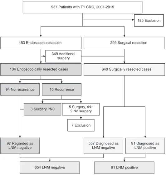
Among these 937 patients, those with familial adenomatous polyposis (n = 16) or synchronous advanced CRC (n = 75) were excluded, as were patients endoscopically resected but followed up for <24 months (n = 94). Of the endoscopically resected patients, patients with recurrence who were found to be positive for LNM on salvage surgery (rpN1 or 2) (n = 5) and those with multiple metastases who did not undergo salvage surgery (n = 2) were excluded because of the uncertainty in timing of LNM and the possibility of skip metastases, respectively. Thus, this study
included 745 patients, 97 who underwent endoscopic resection and 648 who underwent surgical resection (Fig. 1). The database of the National Cancer Center and patients’ clinical charts were reviewed retrospectively.
Based on the Paris classification, endoscopically resected tumors were classified into 4 types: pedunculated, sessile, flat, and depressed [15].
Tumor location was classified into 3 groups: right colon (defined as cecum – splenic flexure), left colon (defined as splenic flexure – rectosigmoid junction), and rectum (defined as rectosigmoid junction – anal verge).
The depth of submucosal invasion was evaluated using Kudo’s classification as infiltration into the upper third (sm1), middle third (sm2), or lower third (sm3) of the submucosal layer in surgically resected specimens [16]. For endoscopically resected sessile and flat tumors, the cut-off between sm1 and sm2 was 1,000 μm according to the Paris classification, with submucosal invasion >2,000 μm defined as sm3 [15].
For endoscopically resected pedunculated tumors, the cut- off between sm1 and sm2 was at the level of the neck [2], and submucosal invasion >3,000 μm from the neck was defined as
937 Patients with T1 CRC, 2001-2015
453 Endoscopic resection 299 Surgical resection
104 Endoscopically resected cases 648 Surgically resected cases
94 No recurrence 10 Recurrence
7 Exclusion
97 Regarded as LNM negative
557 Diagnosed as LNM negative
91 Diagnosed as LNM positive
654 LNM negative 91 LNM positive
Exclusion 185
349 Additional surgery
3 Surgery, rN0 5 2
Surgery, rN+
No surgery
Fig. 1. Pathway for estimating the status of lymph node metastasis (LNM) in endoscopically resected patients. CRC, colorectal cancer.
sm3. Submucosal invasion depth ≥sm2 was defined as deep submucosal invasion.
Differentiation of adenocarcinomas was classified according to World Health Organization criteria: grade 1 (well differ- entiated), grade 2 (moderately differentiated), or grade 3 (poorly differentiated) [17]. Grades 1 and 2 were defined as histologic low grade, and grade 3, mucinous carcinoma, signet ring cell carcinoma, and carcinoma with neuroendocrine differentiation were defined as histologic high grade.
An isolated cell or a small cluster of <5 tumor cells in the invasive front was defined as a “budding” focus, and >10 budding foci viewed at ×200 magnification was defined as budding positive [18].
Vascular invasion was defined as the presence of cancer cells within endothelial-lined channels, including angiolymphatic invasion and venous invasion. Vascular invasion of small vessels without a vascular smooth muscle layer was defined as angiolymphatic invasion, and vascular invasion of large vessels with a vascular smooth muscle layer was defined as venous invasion.
Background, or preexisting, adenoma was defined as benign adenomatous tissue contiguous to a resected carcinoma.
Estimation of the status of LNM in endoscopically resected patients
The 97 endoscopically resected patients were followed up by colonoscopy, performed 3–6 months after resection and annually thereafter, and by annual CT scan of the abdomen and chest and annual measurement of serum carcinoembryonic antigen (CEA) concentration. LNM status in these patients
was determined indirectly, based on follow-up results and/or histopathologic reports of salvage surgery.
Patients were followed up for a minimum of 24 months.
Patients with no evidence of recurrence during follow-up and those with recurrence who had no evidence of LNM on salvage surgery (rpN0) were regarded as negative for LNM.
Statistical analyses
The relationship between histopathologic factors and LNM was evaluated using the chi-squared test and Fisher’s exact test.
Univariate and multivariate logistic regression analyses were performed to identify the risk factors associated with LNM. A P-value < 0.05 was defined as statistically significant.
This study was approved by the Institutional Review Board of the National Cancer Center, Korea (NCC2017-0189), and informed consent was obtained from all patients.
RESULTS
Characteristics of patients
The 745 patients enrolled in this study included 471 men (63.2%) and 274 women (36.8%), of mean age 61.4 years (range, 26–91 years). Of these 745 patients, 123 (16.5%) had right colon tumors, 355 (47.7%) had left colon tumors, and 267 (35.8%) had rectal tumors. Tumors were pedunculated in 45 patients (6.0%), sessile in 540 (72.5%), flat in 92 (12.4%), and depressed in 68 (9.1%). Of these 745 patients, 97 (13.0%) underwent only endoscopic resection and 648 (87.0%) underwent surgical resection. Mean tumor size was 17.2 ± 11.0 mm (range, 1.5–115 mm) (Table 1).
Follow-up results and estimation of LNM status of endoscopically resected patients
The mean follow-up period was 59.5 months (range, 24–142 months). Of the 97 endoscopically resected patients, 94 (96.9%) showed no evidence of recurrence during the follow-up period, and they were regarded as negative for LNM. The remaining 3 patients (3.1%) with recurrence were negative for LNM (rpN0) on salvage surgery. Details of 3 patients with recurrence are shown in Table 2.
Table 1. Demographic and clinical characteristics of study pa tients (n = 745)
Variable Value
Age (yr) 61.4 ± 10.6 (26–91)
Tumor size (mm) 17.2 ± 11.0 (1.5–115) Sex
Male 471 (63.2)
Female 274 (36.8)
Location
Right colon 123 (16.5)
Left colon 355 (47.7)
Rectum 267 (35.8)
Endoscopic type
Pedunculated 45 (6.0)
Sessile 540 (72.5)
Flat 92 (12.3)
Depressed 68 (9.1)
Resection type
Endoscopic 97 (13.0)
Surgical 648 (87.0)
Values are presented as mean ± standard deviation (range) or number (%).
Table 2. Details of 3 patients with recurrence after endo- scopic resection
Sex/
age (yr) Risk
factors Recurrence
site Salvage
surgery rpTNM
M/49 Positive margin Local AR rpT3N0M0
F/63 Positive margin Local RHC rpT1N0M0
F/65 Positive margin Local LAR rpTisN0M0 AR, anterior resection; RHC, right hemicolectomy; LAR, low anterior resection.
Relationship between histopathologic factors and LNM
Of the 745 patients, 91 (12.2%) were found to be LNM positive. Both univariate (Table 3) and multivariate (Table 4) analyses indicated that histologic high grade (P < 0.001), vascular invasion (P < 0.001), deep submucosal invasion (P = 0.010), and budding (P = 0.034) were significantly associated with LNM.Incidence of LNM in patients subgrouped by number of risk factors
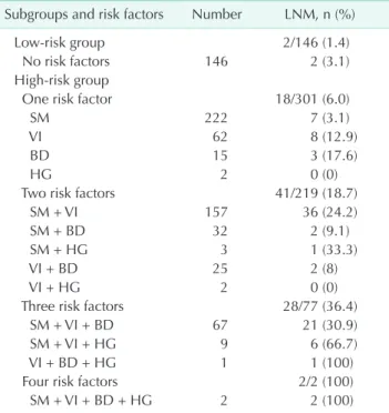
Four risk factors for LNM were identified in patients with T1 CRC: deep submucosal invasion, histologic high grade, budding, and vascular invasion. The relationship between the number of risk factors and LNM was analyzed (Table 5).
Among the patients with one, two, three, and four risk factors, 6.0%, 18.7%, 36.4%, and 100%, respectively, were positive for LNM.
DISCUSSION
The current guidelines recommend additional surgery for patients at high risk of LNM. In agreement with the current guidelines, this study identified deep submucosal invasion, histologic high grade, budding, and vascular invasion as risk factors predicting LNM in patients with T1 CRC.
Our previous study excluded patients who underwent endo- scopic resection from the statistical analyses [14], because their LNM status could not be evaluated directly. Because most T1 CRCs without risk factors undergo endoscopic resection, exclusion of endoscopically resected patients may cause a selection bias, affecting the study results. Therefore, in the current study, LNM status of endoscopically resected patients was estimated indirectly based on follow-up results and/or histopathologic reports of salvage surgery, with these patients included in statistical analyses to overcome any possible selec- tion bias.
Consistent with previous findings, the current study showed that deep submucosal invasion was significantly associated with LNM. Several other studies, however, have evaluated the area, not the depth, of submucosal invasion [19]. According to one of those studies, lymphatic vessels were reported to be significantly more numerous within sm1 than within sm3, suggesting that tumors invading sm1 alone, but on a broader front, may be able to gain greater access to the lymphatic system Table 3. Relationships between histopathologic factors and
lymph node metastasis (LNM)
Variable Number LNM, n (%) P-value
Submucosal invasion depth <0.001
sm1 253 16 (6.3)
sm2 232 28 (12.1)
sm3 260 47 (18.1)
Histologic grade <0.001
Low 726 81 (11.2)
High 19 10 (52.6)
Budding <0.001
Negative 603 60 (10.0)
Positive 142 31 (21.8)
Vascular invasion <0.001
Negative 420 15 (3.6)
Positive 325 76 (23.4)
Background adenoma 0.013
Present 518 53 (10.2)
Absent 227 38 (16.7)
Table 4. Multivariate analysis of histopathologic factors asso ciated with lymph node metastasis
Variables P-value OR 95% CI
Deep submucosal invasion 0.010 2.193 1.207–3.984 Histologic high grade <0.001 7.340 2.623–20.535
Budding 0.034 1.757 1.044–2.955
Vascular invasion <0.001 6.631 3.671–11.979 Absence of
background adenoma 0.066 1.580 0.971–2.571 OR, odds ratio; CI, confidence interval.
Table 5. Relationship between lymph node metastasis (LNM) and number of risk factors
Subgroups and risk factors Number LNM, n (%)
Low-risk group 2/146 (1.4)
No risk factors 146 2 (3.1)
High-risk group
One risk factor 18/301 (6.0)
SM 222 7 (3.1)
VI 62 8 (12.9)
BD 15 3 (17.6)
HG 2 0 (0)
Two risk factors 41/219 (18.7)
SM + VI 157 36 (24.2)
SM + BD 32 2 (9.1)
SM + HG 3 1 (33.3)
VI + BD 25 2 (8)
VI + HG 2 0 (0)
Three risk factors 28/77 (36.4)
SM + VI + BD 67 21 (30.9)
SM + VI + HG 9 6 (66.7)
VI + BD + HG 1 1 (100)
Four risk factors 2/2 (100)
SM + VI + BD + HG 2 2 (100)
SM, deep submucosal invasion; VI, vascular invasion; BD, budding; HG, histologic high grade.
than narrow but deeply invading sm3 tumors [20].
The current study also found that histologic high grade and vascular invasion were the most relevant risk factors for LNM in patients with T1 CRC, in good agreement with many earlier studies.
Budding is a histologic feature thought to correspond to the initial phase of tumor invasion and reported to be associated with metastatic activity [21]. Japanese guidelines accept budding as a risk factor for LNM in T1 CRC, but not western guidelines.
The current study showed that budding was significantly associated with LNM in T1 CRC.
Most CRCs develop from adenomatous polyps through the adenoma-carcinoma sequence, although some of these tumors develop de novo [22,23]. Although the relationship between the absence of background adenoma and LNM in T1 CRC has not been clearly determined, the absence of background adenoma may indicate more aggressive biologic behavior. In contrast to our previous study, which showed that the absence of background adenoma was significantly associated with LNM on both univariate and multivariate analyses, the current study found that the absence of background adenoma was significant on univariate analysis alone.
The current study showed that the incidence of LNM ranges widely according to the kind and number of risk factors in T1 CRC (6.0%–100%) (Table 5). Therefore, when determining subsequent treatments, the potential risk of LNM should be
considered, instead of dichotomous decisions depending on the presence or absence of risk factors.
This study had several limitations. Most importantly, LNM status in endoscopically resected patients was estimated in- directly, but these patients were included in statistical analyses.
However, this may help overcome a possible selection bias, as described above. Other limitations included the retrospective design of the study and its relatively short follow-up period.
In conclusion, this study showed that deep submucosal invasion, histologic high grade, budding, and vascular invasion are independent risk factors for LNM in patients with T1 CRC, and the incidence of LNM ranges widely according to the type and number of risk factors. If any of these risk factors are present, additional surgery following endoscopic resection should be determined after considering the potential risk of LNM and each patient’s situation.
CONFLICTS OF INTEREST
No potential conflict of interest relevant to this article was reported.
ACKNOWLEDGEMENTS
This work was supported by a National Cancer Center Grant, Korea (NCC-1510150).
REFERENCES
1. Gupta AK, Melton LJ 3rd, Petersen GM, Timmons LJ, Vege SS, Harmsen WS, et al.
Changing trends in the incidence, stage, survival, and screen-detection of colo- rectal cancer: a population-based study.
Clin Gastroenterol Hepatol 2005;3:150-8.
2. Haggitt RC, Glotzbach RE, Soffer EE, Wruble LD. Prognostic factors in colo rectal car cinomas arising in ade no mas: im pli ca- tions for lesions re moved by endo sco pic poly pectomy. Gastroenterology 1985;89:
328-36.
3. Kyzer S, Begin LR, Gordon PH, Mitmaker B.
The care of patients with colorectal polyps that contain invasive adenocarcinoma.
Endo scopic polypectomy or colectomy?
Cancer 1992;70:2044-50.
4. Kikuchi R, Takano M, Takagi K, Fujimoto N, Nozaki R, Fujiyoshi T, et al. Man age-
ment of early invasive colo rec tal cancer.
Risk of recurrence and clini cal guidelines.
Dis Colon Rectum 1995;38:1286-95.
5. Nascimbeni R, Burgart LJ, Nivatvongs S, Larson DR. Risk of lymph node metastasis in T1 carcinoma of the colon and rectum.
Dis Colon Rectum 2002;45:200-6.
6. Kitajima K, Fujimori T, Fujii S, Takeda J, Ohkura Y, Kawamata H, et al. Correlations bet ween lymph node metastasis and depth of submucosal invasion in sub mu cosal invasive colorectal car ci no ma: a Japanese collaborative study. J Gastroenterol 2004;
39:534-43.
7. Sohn DK, Chang HJ, Park JW, Choi DH, Han KS, Hong CW, et al. Histopathological risk factors for lymph node metastasis in submucosal invasive colorectal carcinoma of pedunculated or semipedunculated
type. J Clin Pathol 2007;60:912-5.
8. National Comprehensive Cancer Network clinical practice guidelines in oncology (NCCN Guidelines): colon cancer. Version 2. 2016. Fort Wathington (PA): National Comprehensive Cancer Network; c2017 [cited 2017 Jan 5]. Available from: http://
www.nccn.org/professionals/physician_
gls/f_guidelines.asp.
9. National Comprehensive Cancer Network clinical practice guidelines in oncology (NCCN Guidelines): rectal cancer. Version 1. 2016. Fort Wathington (PA): National Comprehensive Cancer Network; c2017 [cited 2017 Jan 5]. Available from: http://
www.nccn.org/professionals/physician_
gls/f_guidelines.asp.
10. Watanabe T, Itabashi M, Shimada Y, Tanaka S, Ito Y, Ajioka Y, et al. Japanese
So ciety for Cancer of the Colon and Rec- tum (JSCCR) Guidelines 2014 for treat- ment of colorectal cancer. Int J Clin Oncol 2015;20:207-39.
11. Ueno H, Mochizuki H, Hashiguchi Y, Shimazaki H, Aida S, Hase K, et al.
Risk factors for an adverse outcome in early invasive colorectal carcinoma.
Gastroenterology 2004;127:385-94.
12. Choi DH, Sohn DK, Chang HJ, Lim SB, Choi HS, Jeong SY. Indications for sub- sequent surgery after endoscopic resec- tion of submucosally invasive colorectal carcinomas: a prospective cohort study.
Dis Colon Rectum 2009;52:438-45.
13. Tateishi Y, Nakanishi Y, Taniguchi H, Shimoda T, Umemura S. Pathological prog nostic factors predicting lymph node meta stasis in submucosal invasive (T1) colorectal carcinoma. Mod Pathol 2010;
23:1068-72.
14. Suh JH, Han KS, Kim BC, Hong CW, Sohn DK, Chang HJ, et al. Predictors for lymph
node metastasis in T1 colorectal cancer.
Endoscopy 2012;44:590-5.
15. The Paris endoscopic classification of super ficial neoplastic lesions: esophagus, stomach, and colon: November 30 to December 1, 2002. Gastrointest Endosc 2003;58(6 Suppl):S3-43.
16. Kudo S. Endoscopic mucosal resection of flat and depressed types of early colo- rectal cancer. Endoscopy 1993;25:455-61.
17. Hamilton S, Aaltonen L. WHO Classi fi ca- tion of Tumours. Pathology and genetics of tumours of the digestive system. Lyon:
IARC press; 2000. (World Health Orga- nization classification of tumours; v. 2).
18. Ueno H, Price AB, Wilkinson KH, Jass JR, Mochizuki H, Talbot IC. A new prognostic staging system for rectal cancer. Ann Surg 2004;240:832-9.
19. Toh EW, Brown P, Morris E, Botterill I, Quirke P. Area of submucosal invasion and width of invasion predicts lymph node metastasis in pT1 colorectal cancers.
Dis Colon Rectum 2015;58:393-400.
20. Smith KJ, Jones PF, Burke DA, Treanor D, Finan PJ, Quirke P. Lymphatic vessel distribution in the mucosa and submu- cosa and potential implications for T1 colo rectal tumors. Dis Colon Rectum 2011;54:35-40.
21. Hase K, Shatney C, Johnson D, Trollope M, Vierra M. Prognostic value of tumor
“budding” in patients with colorectal cancer. Dis Colon Rectum 1993;36:627-35.
22. Vogelstein B, Fearon ER, Hamilton SR, Kern SE, Preisinger AC, Leppert M, et al. Genetic alterations during colorectal- tumor development. N Engl J Med 1988;
319:525-32.
23. Shimoda T, Ikegami M, Fujisaki J, Matsui T, Aizawa S, Ishikawa E. Early colorectal carcinoma with special reference to its development de novo. Cancer 1989;64:
1138-46.