INTRODUCTION
Sentinel lymph node biopsy (SLNB) has been validated in early breast cancer patients and is the accepted standard of care for axillary staging in patients with clinically node-nega- tive axilla [1-3]. Although patients undergoing axillary lymph node dissection (ALND) have higher morbidities including seroma, lymphedema, and arm weakness than SLNB [4], it is still a valid procedure for patients with a positive SLN. Never- theless, 40% to 70% of patients with positive SLNs on the final histology have no metastases in the nonsentinel lymph nodes (NSLN) [5-7]. For this reason, most investigators beleive that ALND has minimal therapeutic effect for patients with only SLN involvement [8,9].
Currently, several risk factors are defined for the probability
of metastasis in NSLNs, such as the histological primary tumor size, size of the SLN metastasis, number of positive SLNs, the ratio of positive SLNs to all removed SLNs, lymphovascular in- vasion, and extracapsular extension of the positive SLN [8,10, 11]. None of these parameters alone is sufficient to identify which patients need ALND. Furthermore, although some mathematical models have been developed to predict the NSLN status in these patients [11-14], the predictive value of these models does not always reach a sufficiently high accuracy.
In the last decade, breast cancer has been classified into dis- tinct molecular subtypes based on the prognostic significance of gene expression profiles [15,16]. For practical use in hospi- tal setting, it has been suggested that immunohistochemical surrogate panels, composed of the estrogen receptor (ER), progesterone receptor (PR), and oncogene erbB-2/human epidermal growth factor receptor 2 (HER2), be used to define breast cancer subtypes rather than gene expression profiling [17-19]. It has been reported that different breast cancer sub- types have variable responses to treatments and oncologic outcomes [20,21]. Additionally, it has been demonstrated that there is a close relationship between subtype and risk of me-
Subtype Is a Predictive Factor of Nonsentinel Lymph Node Involvement in Sentinel Node-Positive Breast Cancer Patients
Kaptan Gülben, Uğur Berberoğlu, Ogün Aydoğan, Volkan Kınaş1
Department of Surgery, Ankara Oncology Training and Research Hospital, Ankara; 1Department of Surgery, Samsun Training and Research Hospital, Samsun, Turkey
ORIGINAL ARTICLE
Purpose: This study aimed to identify the effect of breast cancer subtype on nonsentinel lymph node (NSLN) metastasis in pa- tients with a positive sentinel lymph node (SLN). Methods: The records of 104 early breast cancer patients with a positive SLN between April 2009 and September 2013 were retrospectively evaluated. All patients underwent axillary lymph node dissection.
The effects of the tumor subtype (luminal A, luminal/HER2+, hu- man epidermal growth factor receptor 2 [HER2] overexpression, and triple-negative) and other clinicopathological factors on NSLN metastasis were examined by univariate and multivariate statistical analyses. Results: Fifty of 104 patients (48%) exhibited NSLN metastasis. Univariate and multivariate analyses revealed that tumor size and the ratio of positive SLNs were significant risk factors of NSLN metastasis in patients with a positive SLN.
The rate of NSLN metastasis was higher in patients with luminal/
HER2+ and HER2 overexpression subtypes than that in patients with other subtypes in the univariate analysis (p<0.001). In the multivariate analysis, both patients with luminal/HER2+ (p<
0.006) and patients with HER2 overexpression (p<0.031) sub- types had a higher risk of NSLN metastasis than patients with the luminal A subtype. Conclusion: Subtype classification should be considered as an independent factor when evaluating the risk of NSLN metastasis in patients with a positive SLN. This result supports the development of new nomograms including breast cancer subtype to increase predictive accuracy.
Key Words: Breast neoplasms, Molecular subtypes, Predictive value of tests, Sentinel lymph node biopsy
Correspondence to: Kaptan Gülben
Department of Surgery, Ankara Oncology Training and Research Hospital, Kardelen mah., 2040. sok., B/27, Ankara 06370, Turkey
Tel: +90-312-3360909, Fax: +90-312-3454979 E-mail: [email protected]
Received: July 9, 2014 Accepted: September 25, 2014
Cancer
tastasis to one or more axillary SLNs [22,23]. However, data on which subtype has a higher risk of NSLN metastasis are limited. Here, we determined if the breast cancer subtype in- fluences NSLN metastasis independently compared to other clinicopathological factors in 104 patients with a positive SLN.
METHODS
We examined the records of 480 patients with invasive breast cancer who underwent SLNB between April 2009 and September 2013 at Ankara Oncology Training and Research Hospital. Institutional Review Board approval was obtained with approval number 2014/352, for the study. Of these pa- tients, 104 women with a positive SLN who underwent subse- quent ALND were included in the study. The inclusion crite- ria were early infiltrating breast carcinoma based on clinical and radiological findings, normal physical examination of the axilla, and initial treatment of mastectomy or lumpectomy plus SLNB. Patients who received neoadjuvant chemotherapy and/or radiotherapy were excluded from the study.
The SLNB procedure was performed using a blue dye and radioisotope combination. SLN imaging was done 2 to 18 hours before the operation by superficial injection of a radio- isotope followed by gamma camera imaging. On the day of surgery, blue dye was administered and manual massage was applied. All blue and/or radioactive lymph nodes were consid- ered as SLNs and examined by frozen section intraoperatively.
Any firm or enlarged node encountered during the SLNB pro- cedure was also considered as an SLN [24]. Standard ALND was performed during the same procedure when metastases were detected in SLNs. If the definitive diagnosis with either hematoxylin and eosin staining or immunohistochemistry (IHC) revealed metastasis in an SLN postoperatively, when the frozen section had been negative, a second operation for ALND was performed.
Pathological examinations were performed by the same team for all patients. SLN materials were reviewed by the pa- thologist following serial sectioning and staining with hema- toxylin and eosin. Positive SLNs were classified into two groups according to the size of the metastasis: micrometasta- sis (≤2 mm) and macrometastasis (>2 mm). Patients who had isolated tumor cells in NSLN were not included in this study. For analysis of NSLNs, routine hematoxylin and eosin staining was used. For hormone receptor status determina- tion, tumor cells that showed at least 1% immunohistochemi- cal staining in paraffin blocks were accepted as receptor posi- tive [25]. HER2 overexpression status was determined accord- ing to the American Society of Clinical Oncology guidelines [26]. Patients were classified into four distinct subtypes based
on the different possible IHC combinations of ER, PR, and HER2 status: luminal A (ER-positive and/or PR-positive, HER2-negative), luminal/HER2+, HER2 overexpression (ER- negative, PR-negative, HER2-positive), and triple-negative (ER-negative, PR-negative, HER2-negative) [17-19].
Clinicopathological factors including age, menopausal sta- tus, histological primary tumor size, histological grade, lym- phovascular invasion (LVI), number of SLNs removed, size of the largest SLN metastasis, extracapsular extension status of the positive SLNs, the ratio of positive SLNs to all removed SLNs, and subtype of the tumor were assessed as predictors of NSLN metastasis using chi-square or Fisher exact tests in the univariate analysis. Statistically significant risk factors for NSLN metastasis in the univariate analysis were included in a multivariate analysis using the logistic regression model. All tests of significance were two-sided and a p-value of <0.05 was considered statistically significant. Statistical tests were carried out using the SPSS version 15.0 for Windows (SPSS Inc., Chicago, USA).
RESULTS
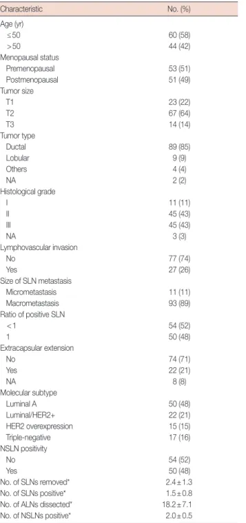
Clinicopathological characteristics of 104 early-stage breast cancer patients with a positive SLN who underwent ALND are summarized in Table 1. The median age of these patients was 49 years (range, 26–75 years) and the median pathological tu- mor size was 3 cm (range, 0.5–6.5 cm). The mean number of SLNs removed was 2.4±1.3 (mean±SD) and the mean num- ber of positive SLNs was 1.5±0.8. Moreover, the mean number of ALNs dissected was 18.2±7.1. The median number of NSLNs dissected in luminal A, luminal/HER2+, HER2 over- expression, and triple-negative cases was 18, 16, 19, and 19, re- spectively. The mean number of positive NSLNs was 2.0±0.5 and the rate of the patients with a positive NSLN was 48%. In this retrospective cohort, the proportion of the patients who had the luminal A subtype was 48%, while the proportion of patients with the luminal/HER2+, HER2 overexpression and triple-negative subtypes was 21%, 15%, and 16%, respectively.
Univariate analysis results are presented in Table 2. Age, menopausal status, number of SLNs removed, size of SLN me- tastasis, and extracapsular extension were not significant risk factors for NSLN metastasis (p>0.166). However, tumor size, histological grade, LVI, ratio of positive SLNs, and subtype classification were significant risk factors for NSLN metastsis (p<0.012). With respect to the breast cancer subtypes, patients with the luminal/HER2+ and HER2 overexpression subtypes had a significantly higher rate of NSLN metastasis than pa- tients with the luminal A and triple negatif subtypes (p<
0.001). The rate of NSLN metastasis in both patients with lu-
minal/HER2+ and HER2 overexpression subtypes was 73%.
In the multivariate analysis (Table 3), a large tumor size and a high ratio of positive SLN were significant predictors of
NSLN metastasis (p<0.001 and p<0.002, respectively). Fur- thermore, a significant relationship was revealed between sub- type classification and NSLN metastasis in the multivariate Table 1. Clinical and pathological characteristics of breast cancer pa-
tients with positive sentinel lymph nodes
Characteristic No. (%)
Age (yr)
≤50 60 (58)
>50 44 (42)
Menopausal status
Premenopausal 53 (51)
Postmenopausal 51 (49)
Tumor size
T1 23 (22)
T2 67 (64)
T3 14 (14)
Tumor type
Ductal 89 (85)
Lobular 9 (9)
Others 4 (4)
NA 2 (2)
Histological grade
I 11 (11)
II 45 (43)
III 45 (43)
NA 3 (3)
Lymphovascular invasion
No 77 (74)
Yes 27 (26)
Size of SLN metastasis
Micrometastasis 11 (11)
Macrometastasis 93 (89)
Ratio of positive SLN
<1 54 (52)
1 50 (48)
Extracapsular extension
No 74 (71)
Yes 22 (21)
NA 8 (8)
Molecular subtype
Luminal A 50 (48)
Luminal/HER2+ 22 (21)
HER2 overexpression 15 (15)
Triple-negative 17 (16)
NSLN positivity
No 54 (52)
Yes 50 (48)
No. of SLNs removed* 2.4±1.3
No. of SLNs positive* 1.5±0.8
No. of ALNs dissected* 18.2±7.1
No. of NSLNs positive* 2.0±0.5
NA =not available; SLN =sentinel lymph node; HER2 =human epidermal growth factor receptor 2; NSLN =nonsentinel lymph node; ALN =axillary lymph node.
*Mean±SD.
Table 2. Univariate analysis of factors affecting nonsentinel lymph node positivity
Variable Negative NSLN
No. (%)
Positive NSLN
No. (%) p-value
Age (yr) 0.356
≤50 29 (48) 31 (52)
>50 23 (52) 21 (48)
Menopausal status 0.410
Premenopausal 25 (47) 28 (53)
Postmenopausal 28 (55) 23 (45)
Tumor size 0.001
T1 20 (87) 3 (13)
T2/T3 32 (40) 49 (60)
Histological grade 0.008
I/II 36 (64) 20 (36)
III 14 (31) 31 (69)
LVI 0.003
No 45 (58) 32 (42)
Yes 7 (26) 20 (74)
No. of SLN removed 0.294
<3 26 (46) 31 (54)
≥3 27 (57) 20 (43)
Size of SLN metastasis 0.166
Micro 8 (73) 3 (27)
Macro 46 (49) 47 (51)
Extracapsular extension 0.191
No 39 (53) 35 (47)
Yes 7 (32) 15 (68)
Ratio of positive SLN 0.012
<1 35 (65) 19 (35)
1 16 (32) 34 (68)
Molecular subtype 0.001
Luminal A 36 (72) 14 (28)
Luminal/HER2+ 6 (27) 16 (73)
HER2 overexpression 4 (27) 11 (73)
Triple-negative 8 (47) 9 (53)
NSLN=nonsentinel lymph node; LVI=lymphovascular invasion; SLN=sentinel lymph node; HER2=human epidermal growth factor receptor 2.
Table 3. Multivariate analysis of factors affecting nonsentinel lymph node positivity
Variable p-value HR 95% CI
Tumor size (T1 vs. T2/T3) 0.001 8.4 1.4–25.6
Histological grade (I/II vs. III) 0.243
LVI (no vs. yes) 0.127
Ratio of positive SLN (<1 vs. 1) 0.002 5.5 1.9–16.1 Molecular subtype
Luminal A - 1.0
Luminal/HER2+ 0.006 6.1 1.6–22.2
HER2 overexpression 0.031 5.4 1.1–26.0
Triple-negative 0.442
HR=hazard ratio; CI=confidence interval; LVI=lymphovascular invasion; SLN=
sentinel lymph node; HER2=human epidermal growth factor receptor 2.
model. Among patients with a positive SLN, both patients with the luminal/HER2+ and HER2 overexpression subtypes had a higher risk of NSLN metastasis than patients with the luminal A subtype (p<0.006; hazard ratio [HR], 6.1; 95%
confidence interval [95% CI], 1.6–22.2; and p<0.03; HR, 5.4;
95% CI, 1.1–26.0, respectively). However, patients with the triple-negative subtype did not have a higher risk than patients with the luminal A subtype (p<0.442).
DISCUSSION
SLNB has become the standard of care to assess axillary sta- tus, particularly in patients with early-stage invasive breast cancer. Generally, ALND is still the standard surgical interven- tion for patients with a positive SLN. However, the therapeutic efficacy of this procedure is not clear.
Various risk factors for likelihood of NSLN metastasis, such as the primary tumor size, grade of primary tumor, size of the SLN metastasis, number of positive SLNs, ratio of positive SLNs, and LVI have been identified [8,10]. In addition, al- though some nomograms using various combinations of these factors have been developed to predict NSLN status [11- 14], the predictive value of these models does not always reach the intended high accuracy level.
Our study shows that clinicopathological factors including the primary tumor size and the ratio of positive SLNs predict the presence of NSLN metastasis in patients with a positive SLN. Patients with a tumor size >2 cm had a significantly higher risk of NSLN metastasis than those with tumors ≤2 cm (60% vs. 13%). Similarly, patients with the ratio of positive SLNs to all removed SLNs equal to 1, that is, patients with me- tastases in all SLNs, had a higher risk of NSLN metastasis than those with a ratio <1 (68% vs. 35%). All groups with a ratio of positive SLN lower than 1 (e.g., values of 0.5, values from 0.5 to <1) had a significantly lower risk of NSLN metastasis than patients with a ratio of 1. Thus, we divided the ratio of positive SLN into two groups (<1 and 1) in this study. The size of the SLN metastasis was not a risk factor for NSLN metastasis.
However, the rate of NSLN metastasis in patients with macro- metastatic SLN (51%) was higher than that in patients with micrometastatic SLN (27%). Sample size and the small num- ber of patients with a micrometastatic SLN may have contrib- uted to this result. Similarly, although histological grade and LVI were significant risk factors for NSLN metastasis in the univariate analysis, they were not of significant predictive val- ue in the multivariable models. Although the effect of extra- capsular extension of the positive SLN on the risk of NSLN metastasis did not reach statistical significance, patients with extracapsular extension had a higher rate of NSLN metastasis
(68%) than the patients without extracapsular extension (47%). This difference might reach statistical significance in a larger study population.
ER, PR, and HER2 status have been investigated in several studies in terms of the effects on NSLN metastasis in patients with a positive SLN, but they have not been identified as risk factors [7,27,28]. Conversely, a limited number of studies have been published regarding the effect of breast cancer subtypes on NSLN metastasis. We tested whether the subtype of breast cancer, classified based on different IHC combinations of ER, PR, and HER2 status had a significant influence on NSLN metastasis in patients with a positive SLN.
Certain luminal subtypes of breast cancer have been report- ed to have a greater risk of metastasis to axillary lymph nodes than other subtypes [22,23]. One study suggested that subtype classification was a predictor of SLN positivity and found that patients with the ER-negative and HER2-negative subtype had a lower risk of SLN metastasis than patients with other sub- types [23]. Conversely, the association between breast cancer subtype and NSLN metastasis in patients with a positive SLN is not well defined. A recent study analyzed how breast cancer subtypes interact with the NSLN status of patients with an SLN metastasis [29]. In order to validate their hypothesis the au- thors also tested the performance of the Tenon score and Me- morial Sloan-Kettering Cancer Center nomogram designed to predict NSLN status in patients with a positive SLN. Significant differences between the four molecular subtypes in terms of percentage of NSLN metastasis were identified. Although the performance of the two predictors was high in patients with ER-positive and HER2-negative subtype, at least 200 samples in each breast cancer molecular subgroup were required to ob- tain a definitive result. In another recent study consisting of 130 patients with a positive SLN, Zhou et al. [30] evaluated the relationship between subtype and NSLN metastasis, as well as, if breast cancer subtype increased the predictive accuracy of the Cambridge model. This study reported that patients with the luminal subtypes had a higher risk of NSLN metastasis than those with triple-negative subtype breast cancer. Patients with the HER2 overexpression subtype also had a higher risk of NSLN metastasis, but this difference was not significant.
Additionally, the authors emphasized that their new version of the Cambridge model had a more accurate predictive perfor- mance when the molecular subtype was added to the model.
Our study showed a statistically significant association between the subtype classification and NSLN metastasis in patients with a positive SLN. In this retrospective series, the luminal A sub- type of breast cancer had the lowest risk of NSLN metastasis.
Patients with the luminal/HER2+ subtype had a higher risk of NSLN metastasis than those with the luminal A subtype. In
addition, the HER2 overexpression subtype was significantly associated with NSLN metastasis, in contrast to the study find- ings of Zhou et al. [30]. However, we found no relationship be- tween the triple-negative breast cancer and NSLN metastasis.
Although triple-negative breast cancer is generally accepted as a more aggressive subtype of breast cancer, both the present study and those mentioned above have observed that it has a smaller effect on NSLN metastasis.
The limitations of this study include a small sample size, the retrospective nature of the study, and the limited number of patients with an SLN micrometastatis. However, the conclu- sions of this study are quite interesting and are useful when planning additional studies. Future prospective studies with a large sample size are needed to validate our findings.
In conclusion, the results of this study suggest that, in addition to a large tumor size and a high ratio of positive SLNs, the sub- type of breast cancer should be considered as an independent predictor of NSLN involvement in patients with a positive SLN.
These results support the development of new nomograms including breast cancer subtypes to increase predictive accuracy.
CONFLICT OF INTEREST
The authors declare that they have no competing interests.
REFERENCES
1. Keshtgar MR, Ell PJ. Clinical role of sentinel-lymph-node biopsy in breast cancer. Lancet Oncol 2002;3:105-10.
2. Veronesi U, Paganelli G, Viale G, Luini A, Zurrida S, Galimberti V, et al.
A randomized comparison of sentinel-node biopsy with routine axil- lary dissection in breast cancer. N Engl J Med 2003;349:546-53.
3. Zhou WB, Liu XA, Dai JC, Wang S. Meta-analysis of sentinel lymph node biopsy at the time of prophylactic mastectomy of the breast. Can J Surg 2011;54:300-6.
4. Veronesi U, Paganelli G, Galimberti V, Viale G, Zurrida S, Bedoni M, et al. Sentinel-node biopsy to avoid axillary dissection in breast cancer with clinically negative lymph-nodes. Lancet 1997;349:1864-7.
5. Hwang RF, Krishnamurthy S, Hunt KK, Mirza N, Ames FC, Feig B, et al. Clinicopathologic factors predicting involvement of nonsentinel ax- illary nodes in women with breast cancer. Ann Surg Oncol 2003;10:
248-54.
6. Chu KU, Turner RR, Hansen NM, Brennan MB, Bilchik A, Giuliano AE. Do all patients with sentinel node metastasis from breast carcino- ma need complete axillary node dissection? Ann Surg 1999;229:536- 41.
7. Nos C, Harding-MacKean C, Fréneaux P, Trie A, Falcou MC, Sastre- Garau X, et al. Prediction of tumour involvement in remaining axillary lymph nodes when the sentinel node in a woman with breast cancer contains metastases. Br J Surg 2003;90:1354-60.
8. Moghaddam Y, Falzon M, Fulford L, Williams NR, Keshtgar MR.
Comparison of three mathematical models for predicting the risk of
additional axillary nodal metastases after positive sentinel lymph node biopsy in early breast cancer. Br J Surg 2010;97:1646-52.
9. Cserni G, Burzykowski T, Vinh-Hung V, Kocsis L, Boross G, Sinkó M, et al. Axillary sentinel node and tumour-related factors associated with non-sentinel node involvement in breast cancer. Jpn J Clin Oncol 2004;
34:519-24.
10. Reynolds C, Mick R, Donohue JH, Grant CS, Farley DR, Callans LS, et al. Sentinel lymph node biopsy with metastasis: can axillary dissection be avoided in some patients with breast cancer? J Clin Oncol 1999;17:
1720-6.
11. Degnim AC, Reynolds C, Pantvaidya G, Zakaria S, Hoskin T, Barnes S, et al. Nonsentinel node metastasis in breast cancer patients: assessment of an existing and a new predictive nomogram. Am J Surg 2005;190:
543-50.
12. Van Zee KJ, Manasseh DM, Bevilacqua JL, Boolbol SK, Fey JV, Tan LK, et al. A nomogram for predicting the likelihood of additional nodal metastases in breast cancer patients with a positive sentinel node biopsy.
Ann Surg Oncol 2003;10:1140-51.
13. Kohrt HE, Olshen RA, Bermas HR, Goodson WH, Wood DJ, Henry S, et al. New models and online calculator for predicting non-sentinel lymph node status in sentinel lymph node positive breast cancer pa- tients. BMC Cancer 2008;8:66.
14. Pal A, Provenzano E, Duffy SW, Pinder SE, Purushotham AD. A model for predicting non-sentinel lymph node metastatic disease when the sentinel lymph node is positive. Br J Surg 2008;95:302-9.
15. Perou CM, Sørlie T, Eisen MB, van de Rijn M, Jeffrey SS, Rees CA, et al.
Molecular portraits of human breast tumours. Nature 2000;406:747-52.
16. Sørlie T, Perou CM, Tibshirani R, Aas T, Geisler S, Johnsen H, et al.
Gene expression patterns of breast carcinomas distinguish tumor sub- classes with clinical implications. Proc Natl Acad Sci U S A 2001;98:
10869-74.
17. Cheang MC, Voduc D, Bajdik C, Leung S, McKinney S, Chia SK, et al.
Basal-like breast cancer defined by five biomarkers has superior prog- nostic value than triple-negative phenotype. Clin Cancer Res 2008;14:
1368-76.
18. Carey LA, Dees EC, Sawyer L, Gatti L, Moore DT, Collichio F, et al. The triple negative paradox: primary tumor chemosensitivity of breast can- cer subtypes. Clin Cancer Res 2007;13:2329-34.
19. Cheang MC, Chia SK, Voduc D, Gao D, Leung S, Snider J, et al. Ki67 Index, HER2 Status, and Prognosis of Patients With Luminal B Breast Cancer. J Natl Cancer Inst 2009;101:736-50
20. Weigelt B, Mackay A, A’hern R, Natrajan R, Tan DS, Dowsett M, et al.
Breast cancer molecular profiling with single sample predictors: a retro- spective analysis. Lancet Oncol 2010;11:339-49.
21. Gabos Z, Sinha R, Hanson J, Chauhan N, Hugh J, Mackey JR, et al.
Prognostic significance of human epidermal growth factor receptor positivity for the development of brain metastasis after newly diagnosed breast cancer. J Clin Oncol 2006;24:5658-63.
22. Yin WJ, Lu JS, Di GH, Lin YP, Zhou LH, Liu GY, et al. Clinicopathologi- cal features of the triple-negative tumors in Chinese breast cancer pa- tients. Breast Cancer Res Treat 2009;115:325-33.
23. Reyal F, Rouzier R, Depont-Hazelzet B, Bollet MA, Pierga JY, Alran S, et al. The molecular subtype classification is a determinant of sentinel node positivity in early breast carcinoma. PLoS One 2011;6:e20297.
24. Lyman GH, Giuliano AE, Somerfield MR, Benson AB 3rd, Bodurka
DC, Burstein HJ, et al. American Society of Clinical Oncology guide- line recommendations for sentinel lymph node biopsy in early-stage breast cancer. J Clin Oncol 2005;23:7703-20.
25. Hammond ME, Hayes DF, Dowsett M, Allred DC, Hagerty KL, Badve S, et al. American Society of Clinical Oncology/College Of American Pa- thologists guideline recommendations for immunohistochemical test- ing of estrogen and progesterone receptors in breast cancer. J Clin Oncol 2010;28:2784-95.
26. Wolff AC, Hammond ME, Schwartz JN, Hagerty KL, Allred DC, Cote RJ, et al. American Society of Clinical Oncology/College of American Pathologists guideline recommendations for human epidermal growth factor receptor 2 testing in breast cancer. J Clin Oncol 2007;25:118-45.
27. Rahusen FD, Torrenga H, van Diest PJ, Pijpers R, van der Wall E, Licht J,
et al. Predictive factors for metastatic involvement of nonsentinel nodes in patients with breast cancer. Arch Surg 2001;136:1059-63.
28. Schrenk P, Konstantiniuk P, Wölfl S, Bogner S, Haid A, Nemes C, et al.
Prediction of non-sentinel lymph node status in breast cancer with a micrometastatic sentinel node. Br J Surg 2005;92:707-13.
29. Reyal F, Belichard C, Rouzier R, de Gournay E, Senechal C, Bidard FC, et al. Non-sentinel lymph node metastasis prediction in breast cancer with metastatic sentinel lymph node: impact of molecular subtypes classification. PLoS One 2012;7:e47390.
30. Zhou W, He Z, Xue J, Wang M, Zha X, Ling L, et al. Molecular subtype classification is a determinant of non-sentinel lymph node metastasis in breast cancer patients with positive sentinel lymph nodes. PLoS One 2012;7:e35881.